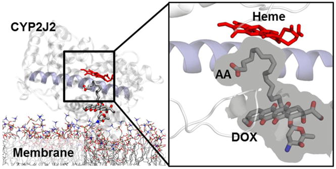
Figure 7.

Representative snapshot (i.e., most often observed configuration) of DOX docked pose obtained from docking to membrane-bound CYP2J2-AA structures, with AA located close to the heme (see Methods). The volume available is shown as a grey surface, surrounding the two ligands. The docked poses reveal that DOX cannot be accommodated within the active site when AA is present, and is located away from the heme and at the entrance of the substrate access channel (with center of mass distance ~17 Å).
