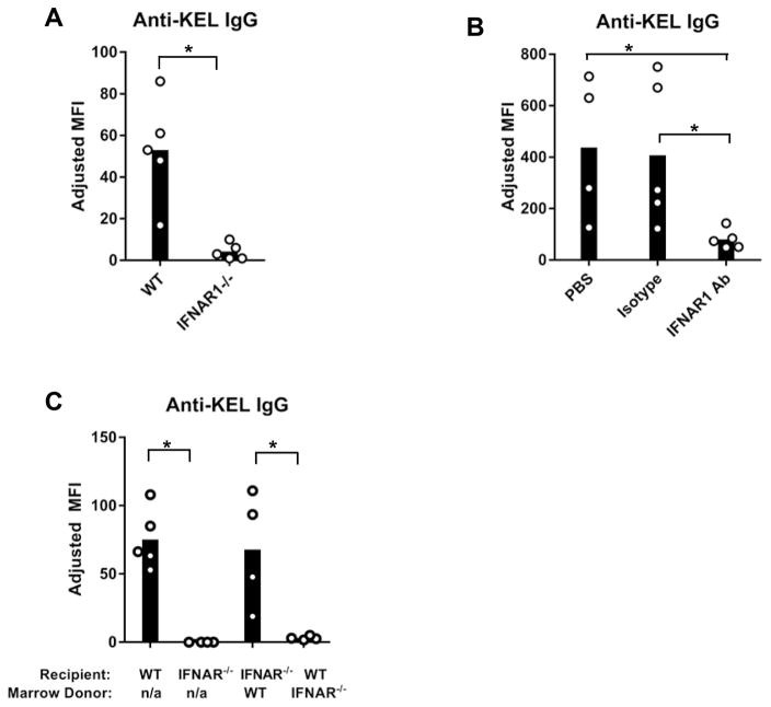
Figure 1. IFNα/β promotes alloimmunization to KEL RBCs.
Peripheral blood of KEL-expressing transgenic mice was leuko-reduced and transfused into recipients on Day 0. Serum anti-KEL IgG was measured by flow cytometric crossmatch. The adjusted MFI was calculated by subtracting the reactivity of serum with WT RBCs from serum reactivity with KEL RBCs. (A) Anti-KEL IgG in serum of WT or IFNAR1−/− mice 28 days after transfusion with KEL RBCs. (B) Serum anti-KEL IgG of transfused WT mice injected i.p. with anti-IFNAR1 blocking antibody (MAR1-5A3), an isotype control IgG1 antibody (MOPC-21), or PBS on Day −1, +2, and +7. (C) Anti-KEL IgG of transfused WT, IFNAR1−/−, and bone marrow chimeric mice. Recipients were irradiated and reconstituted with donor bone marrow cells, 8 weeks prior to transfusion. “n/a”; non applicable. One of 2 (B–C) or 3 (A) independent experiements with 4–5 mice/group. Data from repeated experiments are shown in Supplemental Figure 1. Bars on graphs indicate the mean of the data, and the circles represent individual data points from one mouse.*p<0.05 by (A, C) Mann Whitney U test and (B) Kruskal-Wallis test with a Dunn’s post-test.

