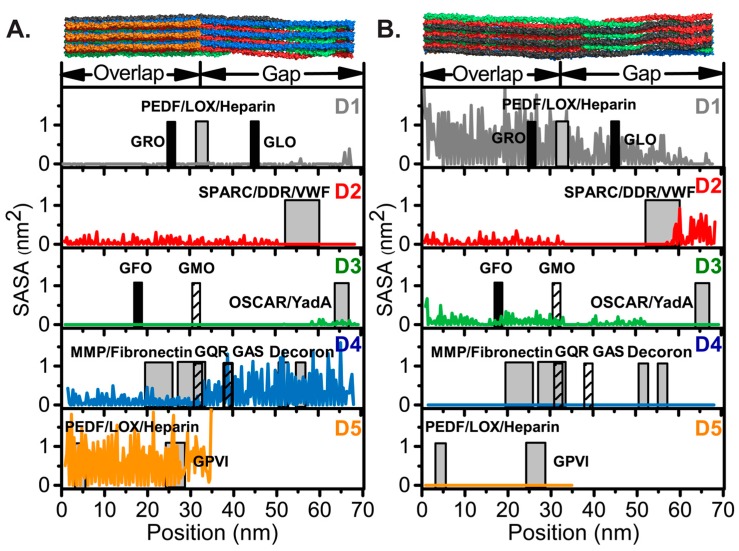
Figure 3.
Residue-specific modified 5 Å SASA calculations with respect to surfaces (A) A [98] and (B) B [58]. The D-segments, D1–D5, are indicated and colored as in Figure 1 and Figure 2. The locations of collagen ligands are shown by gray boxes with heparin [104], PEDF [105] and LOX [57] in D1 and D5; SPARC [86], DDRs [64,65] and VWF [78] in D2; OSCAR [84,85] and YadA [87] in D3; MMPs [68], fibronectin [70], and the decorin core protein, decoron [106], in D4; and GPVI [80] in D5. Integrin high and low affinity motifs are represented by black and hashed boxes, respectively, and labeled by the first three residues of the indicated six-residue binding motif. The width of the boxes corresponds to the length of the identified recognition sequence. Abbreviations: PEDF, pigment epithelium-derived factor; LOX, lysyl oxidase; SPARC, secreted protein acidic and rich in cysteine; DDRs, discoidin domain receptors; VWF, von Willebrand factor; OSCAR, osteoclast-associated immunoglobulin-like receptor; Yad A, Yersinia adhesin A; MMPs, matrix metalloproteinases; GPVI, glycoprotein VI.

