Abstract
The Morel-Lavallée lesion is a closed degloving injury that usually occurs following high-energy trauma. We present a case demonstrating endoscopic management of this lesion. A 44-year-old man fell from scaffolding. Initial assessment demonstrated no significant injury. An ultrasound scan 2 days post injury revealed a large fluid collection along the lateral right thigh. This subsequently became infected and did not respond to antibiotic therapy.
Due to the extent of the lesion, we were reluctant to perform a traditional open drainage. An endoscopic probe was inserted at the proximal and distal poles of the lesion and the wound debrided.
This resulted in a rapid improvement in symptoms and a complete resolution of the lesion at 1 year postsurgery, with no wound-associated morbidity.
This is only the second description of endoscopic debridement of a large, acute Morel-Lavallée lesion, with an excellent outcome.
Keywords: Trauma, Orthopaedics, Orthopaedic and trauma surgery
Background
The Morel-Lavallée lesion (MLL) is a well established but rare finding first described by the French physician Maurice Morel-Lavallée in 1853.1 It is a closed internal degloving injury that usually occurs following high-energy trauma due to a shearing force or crush injury. There is traumatic separation of the skin and hypodermis from underlying fascia. Disruption of the perforating arteries and lymphatics cause formation of a haematoma or seroma within this potential space. The compromise of the skin’s blood supply can lead to necrosis.2
The lesion classically occurs over the greater trochanter of the femur1 due to the relative mobility of the overlying skin compared with the underlying fascia. It can be identified in the acute clinical period as a fluctuant mass overlying an injured area, often with skin contusions and abrasions. They are easily missed in the acute period.3 Investigation is commonly by ultrasound, CT and MRI.4
Although a closed injury, it is thought these collections can become colonised with circulating bacteria.5 A number of treatment methods have been described including compression bandaging, sclerotherapy and open incision, drainage and debridement.6
Case presentation
A 44-year-old male scaffolder presented to the emergency department following a fall from 15 feet. His initial assessment and management was according to Advanced Trauma and Life Support guidelines. A primary survey revealed no systemic injuries. His primary complaint was pain in the right thigh. A plain X-ray demonstrated no fracture and he was discharged with analgesia.
The patient re-presented to the emergency department 3 days later with increasing pain, rigours and a swelling over the posterolateral aspect of his right thigh.
Investigations
An ultrasound scan of the area was performed and showed a large subcutaneous fluid collection extending over the posterolateral aspect of the thigh from the level of the gluteus maximus down to the lateral femoral condyle. The maximum depth was 4.6 cm and transverse diameter 10 cm. There were a few fine loculations and echogenicities within the collection.
It was felt that the ultrasound appearance was in keeping with a Morel-Lavallée closed degloving injury with seroma formation.
Haemoglobin: 13.1 g/dL
White cell count (WBC): 13.2×10⁹/L
C-reactive protein (CRP): 47 mg/dL
Aspiration of the seroma under sterile conditions grew Staphylococcus aureus sensitive to flucloxacillin.
Differential diagnosis
MLL
Necrotising fasciitis
Cellulitis
Haematoma
Thigh compartment syndrome
Treatment
The patient was admitted for intravenous antibiotics and a period of observation.
Despite treatment the patient continued to have elevated temperatures and his CRP climbed to 195 within 4 days. Clinical examination revealed that his collection appeared to have increased in size and remained exquisitely tender to palpate.
Treatment options were discussed with the patient. It was explained that despite the trial of conservative treatment, his condition had continued to deteriorate. Surgical options were discussed. We explained that due to the extent of the lesion, an open incision and debridement to adequately drain the lesion and break up any loculations would require an extensive incision with potential wound complications and an unsightly scar.
On further discussion it was felt that the introduction of endoscopic probes at the proximal and distal ends of the wounds would allow adequate access to both drain the wound and break up loculations without the need for an extensive approach.
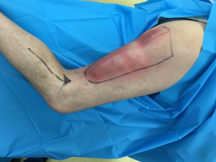
The patient was positioned and draped in lateral decubitus position (figure 1). Two 1 cm incisions were placed at the proximal and distal aspects of the collection. Eight hundred millilitres of blood-stained fluid was evacuated via the incisions. The incisions were then used as entrance ports for endoscopic equipment. An endoscopic camera (as used in knee arthroscopy) allowed visualisation of the interior of the collection. A probe and sterile brush were then guided under direct vision to break down loculations and debride the interior of the collection. Six litres of normal saline was used to lavage the tissues with removal by active suction. Two ¼ inch fenestrated negative pressure Redivac drains were left in-situ to prevent reaccumulation of fluid. Pressure dressings were placed tight across the lateral thigh to close the dead space.
Figure 1.
Patient in lateral decubitus position with lesion marked.
Outcome and follow-up
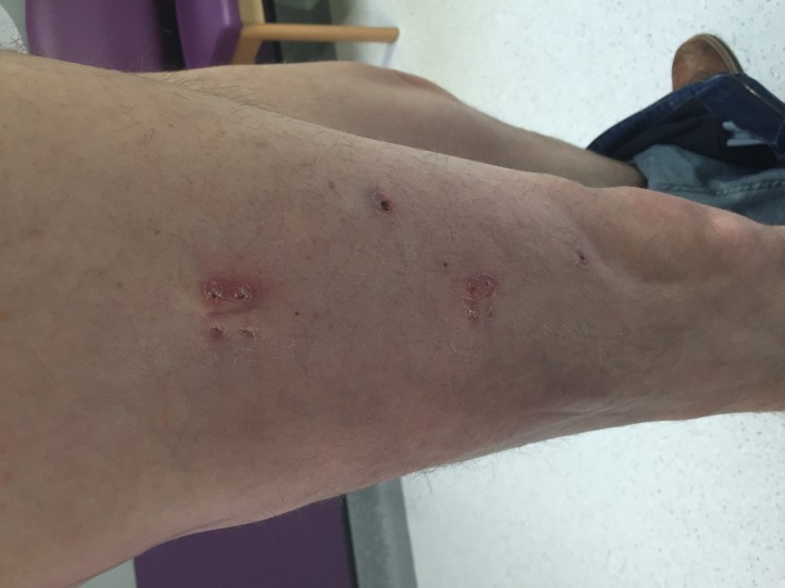
Postoperatively the drains were closely observed with 100 and 40 mL in the two containers on day 1, respectively. The drains were withdrawn 1 inch each day until the fenestrations appeared and the suction was lost on day 5 with prompt removal. With the drains removed and wounds satisfactory, the patient was instructed to wear tight fitting cycling shorts, which provided uniform compression over the involved area. Intravenous antibiotics were converted to oral therapy 3 days following surgery. The patient’s CRP and WBC returned to normal parameters within a week of surgery. Two weeks following surgical drainage, the patient attended clinic for assessment and the picture shows well-healed wounds with no collection (figure 2). Longer term follow-up of the patient confirmed complete resolution.
Figure 2.
Two weeks following surgical drainage, the patient attended clinic for assessment and the picture shows well-healed wounds with no collection.
Discussion
The use of endoscopic equipment to perform an endoscopic drainage and debridement of a MLL has been described once before.7 In this case, it was used to drain and debride a lesion that had been present for 3 months and was aided by adjuvant sclerotherapy. They reported a successful outcome with no recurrence of the seroma. There are no other reports of the use of endoscopic techniques to drain these lesions.
Both open incision and drainage and percutaneous drain were considered. However, in view of the patient’s impending sepsis and the presence of loculations within the lesion on ultrasound scan, the endoscopic probe provided several benefits. It allowed accurate debridement of necrotic tissue and loculations, visualisation to ensure complete drainage and creation of a healing tissue bed, as well as targeted lavage.
Incision and drainage in such an extensive lesion would require a large incision. This would result in a large scar and has the potential to require repeated debridement with a high chance of developing a chronic infection.8
Percutaneous drainage is described by Tseng and Tornetta.5 They describe a technique whereby two 2 cm incisions are made at the proximal and distal ends of the lesion, a plastic brush is used to debride injured fatty tissue and pulsed lavage to wash the wound. They describe this technique in the early management of these wounds. We felt that the direct visualisation of the interior of the lesion allowed more accurate debridement of all areas back to healthy tissue and to ensure that all loculations were divided. This is particularly pertinent due to the extent of the lesion in this case.
Timely removal of drains coupled with direct compression with tight fitting cycling shorts in the postoperative period prevents reaccumulation of the haemolymphatic mass. Cycling shorts are particularly effective for lesions at the greater trochanter and proximal thigh, as this is a difficult place to accurately place traditional compression dressings. Clinicians must be aware that this carries a risk of compartment syndrome particularly if the lesion is misdiagnosed and is in fact a haematoma.
This report demonstrates that endoscopic debridement, followed by compression dressing with cycling shorts and without sclerotherapy is a safe and effective means of treating an acute MLL.
Learning points.
The Morel-Lavallée lesion should be suspected in high-energy trauma especially around greater trochanter and thigh. It is often underestimated for a haematoma and not monitored or treated adequately.
Ultrasound and MRI are the imaging modalities of choice. They can also be identified on CT scan.
These lesions require operative intervention for adequate resolution. Our described arthroscopic method achieves the end goal of a more radical open debridement but minimises the surgical scar and associated morbidity.
Direct compression of the involved area for a prolonged period postoperatively closes the dead space. Cycling shorts provide an effective means of providing compression to the greater trochanter and proximal thigh.
Footnotes
Contributors: AW: concept of case report, initial write up and editing. SEM: literature search, write up of report and editing. JM: literature search, write up and editing. JB: concept, overall supervision and editing of report.
Competing interests: None declared.
Patient consent: Obtained.
Provenance and peer review: Not commissioned; externally peer reviewed.
References
- 1.Morel-Lavalle ´e M. [Epanchements traumatique de derosite]. Arch Gen Med 1853;31:691Y731. [Google Scholar]
- 2.Nickerson TP, Zielinski Martin D, Donald H, et al. . The Mayo Clinic experience with Morel-Lavalle’e lesions:Establishment of a practice management guideline. [DOI] [PubMed]
- 3.Hudson DA. Missed closed degloving injuries: late presentation as a contour deformity. Plast Reconstr Surg 1996;98:334–7. 10.1097/00006534-199608000-00020 [DOI] [PubMed] [Google Scholar]
- 4.Puig J, Pelaez I, Banos J, et al. . Longstanding Morel-Lavallée lesion in the proximal thigh: ultrasound and MR findings with surgical and histopathological correlation. Australas Radiol 2006;50:594Y597 10.1111/j.1440-1673.2006.01640.x [DOI] [PubMed] [Google Scholar]
- 5.Tseng S, Tornetta P. Percutaneous management of Morel-Lavallee lesions. J Bone Joint Surg Am 2006;88:92–6. 10.2106/JBJS.E.00021 [DOI] [PubMed] [Google Scholar]
- 6.Greenhill D, Haydel C, Rehman S. Management of the Morel-Lavallée lesion. Orthop Clin North Am 2016;47:115–25. 10.1016/j.ocl.2015.08.012 [DOI] [PubMed] [Google Scholar]
- 7.Kim S. Endoscopic treatment of Morel-Lavallee lesion. Injury 2016;47:1064–6. 10.1016/j.injury.2016.01.029 [DOI] [PubMed] [Google Scholar]
- 8.Hak DJ, Olson SA, Matta JM. Diagnosis and management of closed internal degloving injuries associated with pelvic and acetabular fractures: the Morel-Lavallée lesion. J Trauma 1997;42:1046–51. 10.1097/00005373-199706000-00010 [DOI] [PubMed] [Google Scholar]


