Abstract
Lipoma is a common subcutaneous tumour composed of fat tissue. It may occur as a sporadic solitary lesion or as multiple lesions. They often grow very slowly. They vary between 2 and 10 cm in size. There are rarely any subjective symptoms. Lipomas do not usually require treatment unless they are big enough to be symptomatic. We reported a 75-year-old man with a rapidly enlarging and ulcerated mass on his right upper thigh.
Keywords: dermatology, general surgery
Background
The presentation of lipoma in this case is unusually. Herein, it is rapidly enlarging with surface ulceration likely due to mass effect. The clinical appearance seen here is not of typical lipoma.
Case presentation
A 74-year-old male presented with a 4-month history of painless, enlarging and ulcerated mass on his right upper thigh. There is no known precipitating factor such as trauma. He has a history of type II diabetes mellitus, hypothyroidism, hyperlipidaemia and right nephrectomy for renal cell carcinoma in 1992.
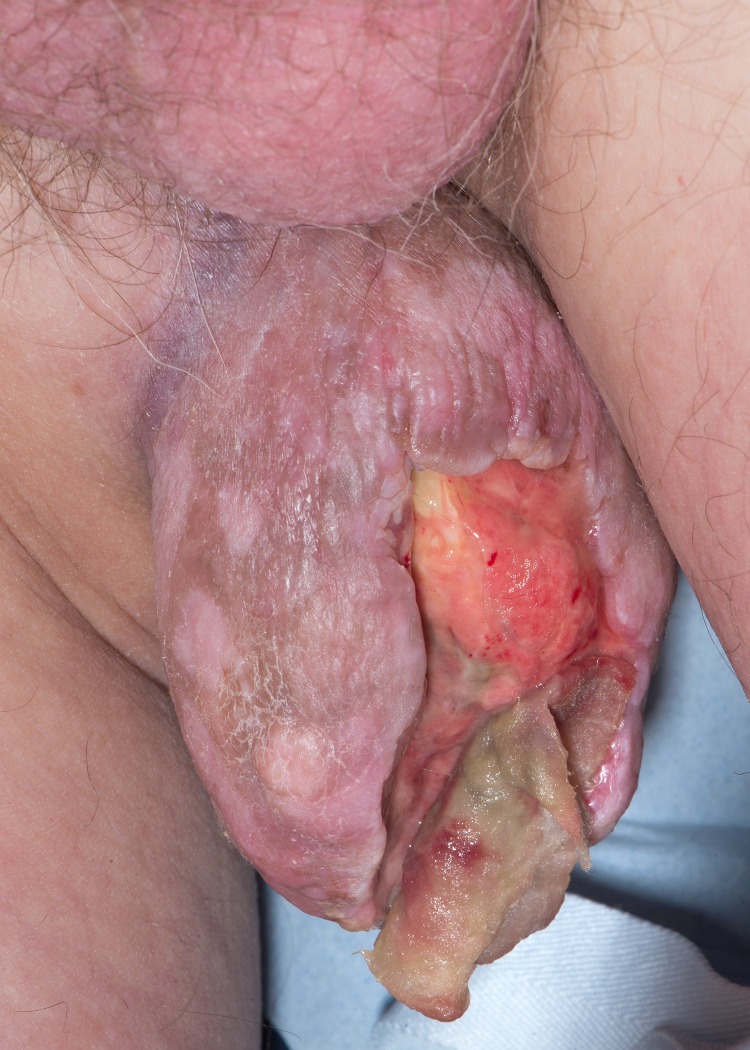
Clinical examination revealed a 10×8 cm, firm, ulcerated and pedunculated nodule on the inner aspect of his right upper thigh (figure 1). There is no lymphadenopathy palpable and the rest of the examination was normal. Given the clinical findings, MRI, staging CT of chest, lung and pelvis were performed. The images had shown a pedunculated lesion with central ulceration on the inner aspect of right thigh just inferior to the scrotum. No other abnormality was detected. Excision was performed and histology was consistent with a lipoma with ulceration.
Figure 1.
A 10×8 cm, firm, ulcerated and pedunculated nodule on the inner aspect of right upper thigh.
Investigations
MRI, staging CT of chest, lung and pelvis.
Differential diagnosis
Secondary metastasis from renal cell carcinoma, lipomatosis, Dercum’s disease and cutaneous sarcomas.
Treatment
Surgical excision.
Outcome and follow-up
No recurrence 6 months later.
Discussion
A lipoma is a common, benign, slow-growing tumour of adipose tissue. Lipomas are commonly found in the subcutaneous tissue and to a lesser extent in internal organs. Lesions are solitary or multiple and usually of no significance. They rarely reach a size larger than 2 cm. Lesions larger than 10 cm are called giant lipoma. Giant lipoma usually affects the upper torso such as back and to a lesser extent upper and lower extremities. Lipomas can occur at any age. However, they tend to arise in adulthood. Solitary lipomas are more common in women, while multiple lipomas occur more frequently in men. Lesions are often asymptomatic unless large in which case they can cause pain through pressure on various structures. Differential diagnosis includes lipomatosis, Dercum’s disease and cutaneous sarcomas.1 2
The exact aetiology of lipoma is not known. Subcutaneous lipomas are associated with hypercholesterolaemia, obesity and trauma. Radiological evaluation of lipoma such as ultrasound is useful for diagnosis and delineating the extent of the lesion and to assist in operative planning. Surgical excision remains the definitive diagnosis (to exclude malignancy) and treatment.
Lipoma can be conservatively observed unless patients experience symptoms due to physical limitation of the mass such as lymphoedema, pain or nerve compression. This case is unusual due to its rapid growth and surface ulceration. The surface ulceration could be due to pressure effect. He was successfully treated with surgical excision and remains in remission 6 months later.
Learning points.
Lipoma may occur as a sporadic solitary lesion or as multiple lesions.
Exclude any conditions associated with a familial component such as Gardner syndrome.
Consider soft tissue sarcoma such as liposarcoma when a fatty tumour becomes larger than 10 cm in size.
Footnotes
Contributors: TK and TNS contribute to the writing of this case.
Competing interests: None declared.
Patient consent: Obtained.
Provenance and peer review: Not commissioned; externally peer reviewed.
References
- 1.Rubinstein A, Goor Y, Gazit E, et al. . Non-symmetric subcutaneous lipomatosis associated with familial combined hyperlipidaemia. Br J Dermatol 1989;120:689–94. doi:10.1111/j.1365-2133.1989.tb01357.x [DOI] [PubMed] [Google Scholar]
- 2.Self TH, Akins D. Dramatic reduction in lipoma associated with statin therapy. J Am Acad Dermatol 2008;58:S30–S31. doi:10.1016/j.jaad.2007.08.034 [DOI] [PubMed] [Google Scholar]


