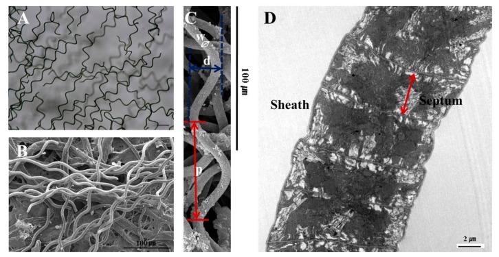
Figure A1.
Morphology of Spirulina maxima. (A) Light micrograph. Original magnification, 100×; (B) Scanning electron micrograph. Original magnification, 350×; (C) The width (w) of the trichomes is 6–12 m, the pitch (p) is 12–72 m, and the diameter (d) is 30–70 m. Original magnification, 500×; (D) Longitudinal section along the trichome, illustrating the thin, diffluent, fibrillar, net-like sheath enveloping the trichome. The regularly spaced cross walls form a septum that divide the trichome into cells. Original magnification, 8600×.

