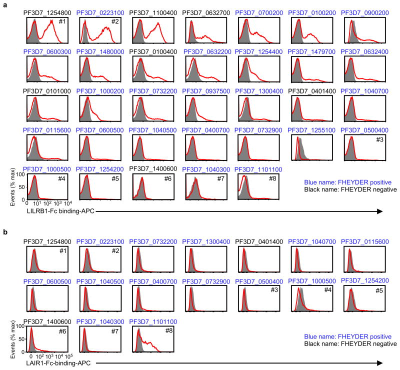
Extended Data Figure 4. Screening of RIFINs that bound the LILRB1-Fc or LAIR1-Fc fusion protein.
a, IEs of 3D7 carrying RIFIN-transgenes were stained with the LILRB1-Fc fusion protein. RIFIN-transgenes are indicated in the figure. Red and shaded histograms indicate staining with LILRB1-Fc and control-Fc fusion proteins, respectively. b, IEs of 3D7 carrying RIFIN-transgenes were stained with the LAIR1-Fc fusion protein. RIFIN-transgenes are indicated in the figure. Red and shaded histograms indicate staining with LAIR1-Fc and control-Fc fusion proteins, respectively. Presence of the FHEYDER sequence in each RIFIN is indicated in the figure. Representative data from independent analyses are shown. Therefore, the proportions of IEs bound to Fc-fusion proteins and the levels of Fc-fusion protein binding IEs may not be comparable among different RIFINs. All experiments were replicated twice.

