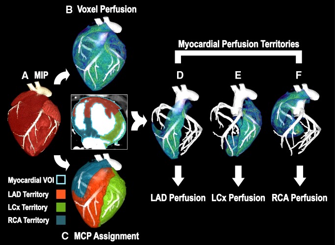
Figure 4:
Image processing scheme for FPA perfusion measurement. A, The entire heart is segmented from the V1 and V2 maximum-intensity projection (MIP), resulting in a whole-heart VOI (axial view; light blue outline = whole-heart VOI). B, Voxel-by-voxel perfusion is computed within the whole-heart VOI. C, Minimum-cost-path (MCP) myocardial assignment is performed, yielding three vessel-specific sub-VOIs (red = LAD sub-VOI, green = LCx sub-VOI, blue = RCA sub-VOI). D–F, Vessel-specific perfusion is derived by averaging the voxel-by-voxel perfusion within each sub-VOI.

