Abstract
Adrenal insufficiency is divided into three types based on the etiology of its development. In primary adrenal insufficiency, pathology resides in end-organ failure at the level of the adrenal cortex, while in secondary and tertiary adrenal insufficiency, impairment rests in the pituitary gland and hypothalamus, respectively. Regardless of etiology, adrenal insufficiency results in a hypocortisolemic condition. While the relationship between neuropsychiatric symptoms, especially psychosis, and hypercortisolemia has been extensively documented, the development of hypocortisolemia-induced psychosis is less common. We present a case of secondary adrenal insufficiency caused by a pituitary tumor. During the course of evaluation and treatment, the patient developed a psychotic episode. We will briefly review the condition of adrenal insufficiency and propose how hypocortisolemia can result in psychosis.
Keywords: Psychosis, secondary adrenal insufficiency, hypocortisolemia, proinflammatory cytokines
Incidentally found lesions of the pituitary gland, referred to as “pituitary incidentalomas,” are present in approximately 10 percent of individuals undergoing brain magnetic resonance imaging (MRI). Up to 40 percent of individuals with these tumors will have evidence for hypopituitarism, including hypoadrenalism in up to 18 percent of patients.1 A variety of diseases might cause hypopituitarism, and accordingly, this disorder can be divided into two types depending on its cause. Primary hypopituitarism is caused by disorders of the pituitary gland itself and might be due to the loss, damage, or dysfunction of pituitary hormone-secreting cells. On the other hand, secondary hypopituitarism results from diseases of the hypothalamus or pituitary stalk interrupting the nerve or vascular connections to the pituitary gland, thereby reducing the secretion of the pituitary hormones.1
Adrenal insufficiency is a potentially life-threatening disease resulting from primary (adrenal glands) or secondary and tertiary (hypothalamic-pituitary axis) failure of glucocorticoid action or production with or without impairment also in mineralocorticoids and adrenal androgens.2 The most frequent cause of primary adrenal insufficiency in western countries is autoimmune adrenalitis, whereas secondary adrenal insufficiency is most often caused by pituitary tumors and their treatment (e.g., surgery).3
While various psychiatric disturbances have been described in association with pituitary and adrenal dysfunction, many physicians, including psychiatrists and endocrinologists, are not aware of the correlation between these disorders. This is partially secondary to the well-known correlation between corticoid excess disorders, such as Cushing’s syndrome and neuropsychiatric presentations.4 It is, therefore, less intuitive that disorders of corticoid deficiency (i.e., adrenal insufficiency) would result in similar psychiatric changes. Although the mechanism responsible for this change in adrenal insufficiency is not currently known, a postulated mechanism is the upregulation and increased sensitivity of both glucocorticoid and mineralocorticoid receptors in the hippocampus during periods of deficiency.5–9 Once exogenous glucocorticoids aimed at treating the insufficiency are introduced, they might overstimulate these receptors, creating a Cushing’s-like effect, even in the presence of decreased serum cortisol, and therefore producing psychiatric symptoms. In this case, we present a report of secondary adrenal insufficiency (SAI) because of compression of the pituitary stalk by an adenoma, manifested by paranoid delusions.
CASE PRESENTATION
A 43-year-old man with a past medical history significant for hypertension and hyperlipidemia presented to the emergency room with progressively worsening fatigue and confusion over a two-month period. One week prior to admission, he developed flu-like symptoms accompanied by vomiting, decreased oral intake, and two falls, preceded by near-syncopal episodes. His family had noted slowing of his speech and felt that his thinking “wasn’t as clear as usual.” In the emergency room, he was noted to have a severely low sodium of 105mmol/L (normal 133–145mmol/L) and a significantly low glucose of 37mg/dL. He had no history of either hyponatremia or hypoglycemia and had not started any new medications. Per his family, he had not complained of any changes in vision or headache prior to his recent fall and had no history of seizures. His hypertension was chronic and well-controlled with lisinopril. He denied any changes in ring or shoe size, although he had noted a decrease in his libido and an increased thirst.
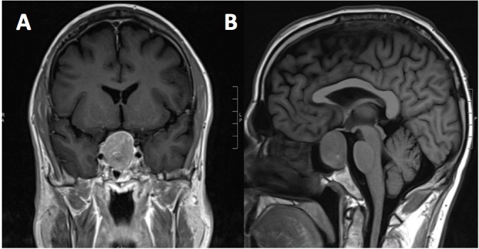
Brain MRI demonstrated an incidental 3-cm sellar/suprasellar mass impinging on the optic chiasm consistent with a pituitary macroadenoma (Figure 1). His hyponatremia and mild hypotension initially improved with normal saline hydration, but hypoglycemia persisted despite frequent intravenous dextrose administration. Given his sellar mass and concerns for hypoadrenalism, the neurosurgery and endocrine services were consulted and pituitary testing was performed (Table 1).
Figure 1.

Brain MRI demonstrating a 3-cm sellar/suprasellar mass on coronal (A) and sagittal (B) imaging.
Table 1.
Additional pertinent laboratory tests on admission to the hospital
| LABORATORY TEST | RESULT | REFERENCE RANGE |
|---|---|---|
| Adrenocorticotrophic hormone (ACTH) | 20.7 | 7.2–63.3pg/mL |
| Cortisol (at 4 a.m.) | 7.22 | 6.2–19.4mcg/dL |
| Growth hormone (GH) | 0.6 | 0.0–10.0ng/mL |
| Insulin-like growth factor-1 (IGF-1) | 75–216ng/mL | |
| Luteinizing hormone (LH) | 5.5 | 1.7–8.6mIU/mL |
| Testosterone | 348–1197ng/dL | |
| Thyroid stimulating hormone (TSH) | 1.29 | 0.27–4.20mcU/mL |
| Free thyroxine (fT4) | 1.0 | 0.9–1.8ng/dL |
| Prolactin (PRL) | 32.8 | 4.0–15.2ng/mL |
| Serum osmolality | 242 | |
| Serum uric acid | 4.1 | 3.9–9.0mg/dL |
| Urine osmolality | 431 | 200–1200mOs/kg |
| Urine sodium | <20 | <20mmol/L |
His cortisol was felt to be inappropriately low for his degree of illness, and given his significant hyponatremia, he was diagnosed with SAI and was treated with high-dose hydrocortisone (75mg daily of prednisone equivalent). With this treatment, his blood pressure improved, his hypoglycemia resolved, and his sodium began to slowly improve but did not normalize. After euvolemia was achieved, fluid restriction was implemented, and his sodium gradually rose to 130 at a rate of 8 to 10mmol/L/day. Initially, his mentation improved and was nearly back to baseline. However, on hospital Day 5, he developed paranoid delusions, agitation, and violent outbursts. Sodium at that time was 128mmol/L. An electroencephalogram (EEG) demonstrated diffuse mild background slowing, and an MRI showed no acute changes when compared to imaging on admission. Osmotic demyelination syndrome was thought unlikely. Subsequently, the patient began refusing serial metabolic panels as well as medications. Given the potential morbidity associated with his psychotic-driven behavior, the psychiatry service was consulted.
The patient’s mental status was remarkable for tangential speech but of normal rate and volume. His affect was constricted but without lability and appropriate to thought content. His thought content was remarkable for paranoid delusions (i.e., “you are playing games with me;” “I’m sure you want me to believe that”), but he denied auditory or visual hallucinations. He also denied suicidal or homicidal ideation, intent, or plan. The patient was oriented to person, place, and time and without fluctuation of mentation. He scored a 30/30 on the mini Mental State Examination. Yet, his insight into his medical condition and judgment in the necessity of treatment and assessment of basic metabolic profile was impaired. He was started on risperidone 0.25mg twice daily. On the day of our psychiatric consultation, the patient’s hydrocortisone had been lowered to 25mg daily of prednisone equivalent.
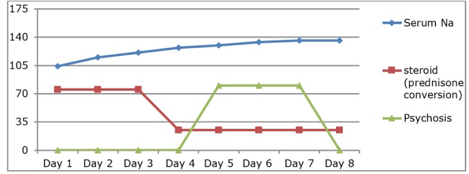
Over the course of the next three days, the patient’s paranoid delusions attenuated (Table 2), but he developed an increased thirst despite a dilute urine (urine osmolarity 140mOs/kg, sodium <13mmol/L) and serum sodium of 134mmol/L. This was thought to be secondary to primary polydipsia from tumor impingement on his anterior hypothalamus. Fluids were restricted with appropriate concentration of urine. Once he was stable with an appropriate serum sodium, the patient underwent transsphenoidal resection of his pituitary macroadenoma. Pathology was consistent with a pituitary adenoma with focal positive staining for LH, positive for follicle stimulating hormone (FSH), and negative for prolactin, ACTH, TSH, and GH. He tolerated surgery well and is currently managed on replacement low-dose corticosteroids, levothyroxine, and testosterone. Notably, risperidone was discontinued one month after transsphenoidal resection of his pituitary macroadenoma, and he has not had a recurrence of psychosis or any psychiatric phenomenology.
Table 2:

Time-Line: Psychosis, Serum Sodium, and (Relative) Corticosteroid Dosing
DISCUSSION
While beyond the scope or our article, we refer the readers to Tables 3 and 4 for a review of symptomatic differences between PAI-SAI and hypothalamic-pituitary-adrenal axis hormone differences between PAI-SAI-TAI, respectively. While not as common as Cushing’s disease, primary, secondary, and tertiary adrenal insufficiency (PAI, SAI, and TAI, respectively) can produce neuropsychiatric symptoms such as psychosis. We postulate that the symptoms can develop because of the direct effects of hypercortisolemia, upstream effects from hypernatremia, and/or increased production of proinflammatory cytokines due to disruption of the hypothalamic-pituitary-adrenal (HPA) axis.
Table 3.
| CLINICAL SYMPTOM | PRIMARY ADRENAL INSUFFICIENCY | SECONDARY ADRENAL INSUFFICIENCY | MECHANISM OF SYMPTOM PRODUCTION |
|---|---|---|---|
| Hyper-pigmentation | (+) | (-) | Lack of glucocorticoid negative feedback increases the release of ACTH and other POMC-peptides; these POMC-peptides are responsible for hyperpigmentation by acting on the MSH receptors in the skin |
| Hyperkalemia | (+) | (-) | Due to mineralocorticoid deficiency |
| Signs of other pituitary hormone deficiencies | (-) | (+) | Pan-hypopituitarism from lesions directly affecting pituitary gland |
| Headaches; visual field deficits | (-) | (+) | From direct effect of pituitary lesion |
| Hyponatremia | (+) | (+) | Due to mineralocorticoid deficiency, GC deficiency (leading to SIADH) in primary adrenal insufficiency, and dilutional in secondary adrenal insufficiency |
| Hypoglycemia | (+) | (++) | Glucocorticoid deficiency |
| Orthostatic hypotension | (+) | (+) | Mineralocorticoid deficiency is not seen in secondary adrenal insufficiency, as mineralocorticoids are principally regulated by the plasma renin-angiotensin system. Hypotension in secondary adrenal insuffiency occurs due to decreased vascular tone as a result of reduced vascular responsiveness to angiotensin II and norepinephrine |
| Anorexia; weight loss; fatigue; generalized malaise; decreased libido | (+) | (+) | Glucocorticoid deficiency |
| Loss of axillary/pubic hair in women | (+) | (+) | Loss of adrenal androgens |
Table 4.
Serum levels of corticotropin-releasing hormone (CRH), Adrenocorticotrophic hormone (ACTH), cortisol, and aldosterone in primary, secondary, and tertiary adrenal insufficiency
| PATHOLOGY MEASURE | PRIMARY ADRENAL INSUFFICIENCY | SECONDARY ADRENAL INSUFFICIENCY | TERTIARY ADRENAL INSUFFICIENCY |
|---|---|---|---|
| CRH (released from 1 hypothalamus) | |||
| ACTH (released from pituitary gland) | |||
| Cortisol (released from adrenal glands) | |||
| Aldosterone (released from adrenal glands) |
First, the etiology of our patient’s hyponatremia was multifactorial with contributions from hypovolemia, adrenal insufficiency, and primary polydipsia from the mass effect of his tumor. Hyponatremia has also been associated with sellar masses in general, including pituitary macroadenomas, Rathke’s cleft, and arachnoid cysts.10 Each aspect had to be corrected in turn before resolution of his condition.
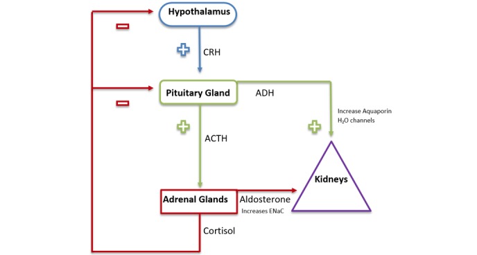
Urine sodium was low and not consistent with cerebral salt wasting. Extracellular volume depletion secondary to sodium loss through vomiting and decreased oral intake was suspected, resulting in persistent antidiuretic hormone (ADH) secretion, even though serum osmolality was low.11 The initial undetectable urine sodium is also explained by this loss of solute in the days leading up to presentation. ADH production occurs in the supraoptic nucleus and paraventricular nucleus of the hypothalamus, triggered in response to increases in serum osmolarity (osmoreceptors) and decreases in circulating volume (baroreceptors). It binds to the V2 receptor in the distal collecting duct of the kidney, which translocates aquaporins to the apical membrane to reabsorb water from the urine. Cortisol provides an important inhibitory signal for ADH and is required for removal of these aquaporins and excretion of free water.12 The absence of cortisol results in excess ADH, causing increased urine osmolality and urine sodium. This presents in a similar manner to the sydrome of inappropriate antidiuretic horome (SIADH) but is an appropriate bodily response to glucocorticoid deficiency. Our patient’s ADH value should have been suppressed in the setting of euvolemia and plasma hypoosmolarity but was within normal limits. The hyponatremia resulting from adrenal insufficiency is largely a result of this lack of ADH suppression (see Figure 2).13
Figure 2:

Mechanism of Vasopressln/ADH-induced Hyponatremia”
Hyponatremia occurs due to loss of epithelial Na+ channels (ENaC) involved in sodium reabsorption and the Increase in aquaporin channels that freely absorb water.
Hypotension can be seen in SAI as a result of decreased glucocorticoid-mediated expression of catecholamine receptors in the vasculature.12 Cortisol contributes to vascular resistance in this manner. Hypoglycemia is more common in SAI rather than PAI, as there are four counterregulatory hormones that guard against low blood glucose (glucagon, growth hormone, cortisol, and epinephrine). In our patient’s case, there is evidence of both GH and cortisol deficiency. Poor oral intake in the days prior to admission also might have diminished his glycogen stores and lessened the effect of glucagon.
The diagnosis of SAI was made given his clinical symptoms (hyponatremia, hypotension, and hypoglycemia), his history, and his inappropriately normal lab values. In the setting of a sodium of 112mmol/L (with serum osmolarity of 224mOs/kg) and a blood glucose of 45mg/dL, serum cortisol would be expected to be elevated in a normally functioning HPA axis. His simultaneously drawn cortisol of 7.22mcg/dL was inappropriately normal, as hypoglycemia is a potent stimulator of the HPA axis. Secondary adrenal insufficiency was differentiated from PAI by the presence of an ACTH value within the normal range. In PAI, ACTH would be expected to be markedly elevated given his low cortisol. This value was inappropriately normal as well. The hyponatremia associated with PAI is related to mineralocorticoid deficiency and increased renal excretion of sodium.12 The ACTH stimulation test was not performed given this information, and he was promptly given hydrocortisone, which immediately resolved the hypoglycemia and hypotension and improved the hyponatremia.
Although uncommon, neuropsychiatric manifestations such as depressive symptoms, irritability, sleep disorders, apathy, cognitive impairment, delusions, and hallucinations can develop secondary to AI, and in our patient’s case, paranoid delusions significantly interfered with the treatment of his hyponatremia and SAI and the resection of his pituitary adenoma.9
While the pathophysiology of psychosis in SAI is still unclear, it has been proposed that a decrease in glucocorticoids results in an increase in neural excitability, leading to enhanced ability to detect sensory input, including taste, olfaction, audition, and proprioception. It has also suggested that decreased glucocorticoids might increase conduction velocity along peripheral axons while prolonging conduction across synapses. This is felt to change the timing of the arrival of signals from the periphery to the central nervous system, resulting in a loss of perceptual ability and decreased integration of sensory inputs. It is reasonable to postulate that if patients are receiving abnormally high sensory signals but are unable to process and integrate these signals appropriately, there might be a tendency to develop hallucinations and a lower threshold for psychosis.13
We posit a possible explanation of our patient’s psychosis due to SAI (or TAI) based on the inflammatory hypothesis of schizophrenia. First, we reiterate that (chronic) stress exerts effects on the paraventricular nucleus of the hypothalamus. In response, the hypothalamus secretes corticotropin-releasing factor (CRF) and arginine vasopressin (AVP), which activate the HPA axis and ultimately upregulate glutocorticoids (GCs) from the adrenal cortex. GCs act on glucocorticoid receptors (GR) on the surfaces or in the cytoplasm of immune cells; monocytes and neutrophils are mainly included, which in turn inhibit the secretion of the proinflammatory cytokines (PIC) such as interleukin (IL)-1β, IL-6, tumor necrosis factor alpha (TNF-α), and interferon (INF)-γ14
In regard to our patient and through the above-discussed mechanism, his underlying medical conditions/”chronic stress” (pituitary macroadenoma with compression of the pituitary stalk resulting in SAI and subsequent metabolic derangements) would typically result in a decrease in PIC such as IL-1β, IL-6, and TNF-α. In those with SAI (as in our patient), deficiency in cortisol production could subsequently lead to an increase, rather than the expected decrease, in PIC.15
A possible association between schizophrenia (the prototypical psychotic illness) and the immune system was postulated over a century ago and is supported by epidemiological and genetic studies pointing to links with infection and inflammation. Meta-analyses of many cross-sectional studies show that schizophrenia is associated with disruption of the cytokine milieu and the propensity for the production of PIC. Antipsychotic-naive first-episode psychosis16 and acute psychotic relapse17 are also associated with increased serum concentrations of IL-6 and other PIC such as TNF-α, IL-1β, and INF-γ and decreased serum concentrations of anti-inflammatory cytokine IL-10, which are normalized after remission of symptoms with antipsychotic treatment.18
Evidence has accumulated to suggest that neuroinflammation might be an early pathology of schizophrenia (i.e., the prototypical primary psychotic disorder) that later leads to neurodegeneration. Its exact role in the etiology as well as the source of neuroinflammation are still unknown.19 However, one recent study provided significant evidence for strong epistatic interactions among PIC genes IL-6 and INF-G in the development of schizophrenia. This study suggested that associated risk variants were indicative of altered transcriptional activity with higher production of IL1α, IL-6, TNF-α, and IFN-γ cytokines.20
Our theory needs to be considered in light of evidence, which supports overactivation of the HPA axis and higher baseline cortisol levels in patients with psychosis, especially non-medicated patients. Additionally, a causal role for HPA activity in triggering or exacerbating psychotic symptoms is supported by research on hypercortisolemia. For example, hypercortisolemia induced by administration of exogenous corticosteroids in high doses can trigger psychotic symptoms. Symptoms of hypercortisolemia-induced psychosis include pressured speech, hallucinations, delusions, and disorganized thought, which are often indistinguishable from the symptoms of psychotic disorders. Indeed, disorders characterized by hypercortisolemia, such as Cushing’s syndrome, often involve psychotic symptoms that remit in conjunction with cortisol levels in response to treatments such as etomidate, an adrenal suppressant, mifepristone, and other treatments.21
Thus, we propose that both an endogenous hypercortisolemic and hypocortisolemic state can result in the same phenotype, i.e., psychosis. There are two types of GR, mineralocorticoid receptors (MRs) and GRs, which are present throughout the limbic and paralimbic regions, including the hippocampus, amygdala, and prefrontal cortex. At rest, the majority of MRs are occupied, while only about 10 percent of GRs are bound to glucocorticoids. However, during stress, MRs are saturated, and increasing proportions of GRs become occupied. Both animal and human studies suggest that the relative proportion of MR to GR activation might be an important moderating factor in multiple brain processes, with the relation of receptor activation to brain function constituting an “inverted U” such that too much or too little can impair cognitive processes. Finally, research has revealed pervasive effects of glucocorticoids on brain structure and function, including hippocampal abnormalities that have been linked with psychosis.21 Thus, this seems to provide further evidence that elevated levels of PIC, independent of their downstream effect on glucocorticoids, might be partly responsible for the development of psychosis.
Our patient’s delusions attenuated, although it remains obscure as to whether this was due to lowering of the corticosteroid dosage, gradual normalization of serum sodium level, initiation of risperidone, and/or ultimately, transsphenoidal resection of his pituitary macroadenoma.22 With regard to the former, the Boston Collaborative Drug Surveillance Program found that of patients receiving more than 80mg/d prednisone, 18 percent had severe psychiatric presentations that included psychosis. Of those receiving 41 to 80mg/d, 4.6 percent had such symptoms, whereas only 2 percent receiving less than this amount had psychotic symptoms.23 Notably, psychotic symptoms have not recurred one month after transsphenoidal resection of his pituitary macroadenoma and without risperidone therapy.
CONCLUSION
Our patient presented with nonspecific symptoms, including fatigue and confusion, and was found to have a pituitary macroadenoma with associated hypopituitarism.
Specifically, he had adrenal insufficiency with resulting hypotension and hypoglycemia. Treatment with glucocorticoids improved these signs, but he subsequently developed symptoms of psychosis.
A multidisciplinary team involving psychiatrists, endocrinologists, neurosurgeons, and primary care physicians is necessary to help care for these often complicated patients. Communication between the different specialists is key and makes for an easier experience for the patient.
REFERENCES
- 1.Kim SY. Diagnosis and treatment of hypopituitarism. Endocrinol Metab (Seoul). 2015;30(4):443–455. doi: 10.3803/EnM.2015.30.4.443. [DOI] [PMC free article] [PubMed] [Google Scholar]
- 2.Diederich S, Franzen NF, Bähr V, Oelkers W. Severe hyponatremia due to hypopituitarism with adrenal insufficiency: report on 28 cases. Eur J Endocrinol. 2003;148(6):609–617. doi: 10.1530/eje.0.1480609. [DOI] [PubMed] [Google Scholar]
- 3.Hahner S, Allolio B. Management of adrenal insufficiency in different clinical settings. Expert Opin Pharmacother. 2005;6(14):2407–2417. doi: 10.1517/14656566.6.14.2407. [DOI] [PubMed] [Google Scholar]
- 4.Pivonello R, Simeoli C, De Martino MC, et al. Neuropsychiatric disorders in Cushing’s syndrome. Frontiers in Neuroscience. 2015;9:129. doi: 10.3389/fnins.2015.00129. [DOI] [PMC free article] [PubMed] [Google Scholar]
- 5.Berardelli R, Karamouzis I, D’Angelo V, et al. The acute effect of a mineralocorticoid receptor agonist on corticotrope secretion in Addison’s disease. J Endocrinol Invest. 2016;39(5):537–542. doi: 10.1007/s40618-015-0393-5. [DOI] [PubMed] [Google Scholar]
- 6.Prigent H, Maxime V, Annane D. Science review: mechanisms of impaired adrenal function in sepsis and molecular actions of glucocorticoids. Crit Care. 2004;8:243–252. doi: 10.1186/cc2878. [DOI] [PMC free article] [PubMed] [Google Scholar]
- 7.Jacobson L, Sapolsky R. The role of the hippocampus in feedback regulation of the hypothalamic-pituitary-adrenocortical axis. Endocr Rev. 1991;12:118–134. doi: 10.1210/edrv-12-2-118. [DOI] [PubMed] [Google Scholar]
- 8.Herman JP, McKlveen JM, Ghosal S, et al. Regulation of the hypothalamic-pituitary-adrenocortical stress response. Comprehensive Physiology. 2016;6(2):603–621. doi: 10.1002/cphy.c150015. [DOI] [PMC free article] [PubMed] [Google Scholar]
- 9.Farah Jde L, Lauand CV, Chequi L, et al. Severe psychotic disorder as the main manifestation of adrenal insufficiency. Case Rep Psychiatry. 2015;2015:512430. doi: 10.1155/2015/512430. [DOI] [PMC free article] [PubMed] [Google Scholar]
- 10.Conn PM, Freeman ME. Neuroendocrinology in Physiology and Medicine. 1st ed. Humana Press; 2000. pp. 331–333. [Google Scholar]
- 11.Kalra S, Zargar AH, Jain SM, et al. Diabetes insipidus: The other diabetes. Indian J Endocrinol Metab. 2016;20(1):9–21. doi: 10.4103/2230-8210.172273. [DOI] [PMC free article] [PubMed] [Google Scholar]
- 12.Goldman L, Schaefer AI. Goldman-Cecil Medicine. 25th ed. Elsevier Health Sciences; 2015. pp. 1497–1498. [Google Scholar]
- 13.Fenske W, Allolio B. Current state and future perspectives in the diagnosis of diabetes insipidus: a clinical review. J Clin Endocrinol Metab. 2012;97(10):3426–3437. doi: 10.1210/jc.2012-1981. [DOI] [PubMed] [Google Scholar]
- 14.Tian R, Hou G, Li D, et al. A possible change process of inflammatory cytokines in the prolonged chronic stress and its ultimate implications for health. Scientific World Journal. 2014;2014:780616. doi: 10.1155/2014/780616. [DOI] [PMC free article] [PubMed] [Google Scholar]
- 15.Sannarangappa V, Jalleh R. Inhaled corticosteroids and secondary adrenal insufficiency. Open Respir Med J. 2014;8:93–100. doi: 10.2174/1874306401408010093. [DOI] [PMC free article] [PubMed] [Google Scholar]
- 16.Upthegrove R, Manzanares-Teson N, Barnes NM. Cytokine function in medication-naive first episode psychosis: a systematic review and meta-analysis. Schizophr Res. 2014;155:101–108. doi: 10.1016/j.schres.2014.03.005. [DOI] [PubMed] [Google Scholar]
- 17.Miller BJ, Buckley P, Seabolt W, et al. Meta-analysis of cytokine alterations in schizophrenia: clinical status and antipsychotic effects. Biol Psychiatry. 2011;70:663–671. doi: 10.1016/j.biopsych.2011.04.013. [DOI] [PMC free article] [PubMed] [Google Scholar]
- 18.Khandaker GM, Cousins L, Deakin J, et al. Inflammation and immunity in schizophrenia: implications for pathophysiology and treatment. Lancet Psychiatry. 2015;2(3):258–270. doi: 10.1016/S2215-0366(14)00122-9. [DOI] [PMC free article] [PubMed] [Google Scholar]
- 19.Pasternak O, Kubicki M, Shenton ME. In vivo imaging of neuroinflammation in schizophrenia. Schizophr Res. 2016;173(3):200–212. doi: 10.1016/j.schres.2015.05.034. [DOI] [PMC free article] [PubMed] [Google Scholar]
- 20.Srinivas L, Vellichirammal NN, Alex AM, et al. Pro-inflammatory cytokines and their epistatic interactions in genetic susceptibility to schizophrenia. J Neuroinflammation. 2016;13(1):105. doi: 10.1186/s12974-016-0569-8. [DOI] [PMC free article] [PubMed] [Google Scholar]
- 21.Holtzman CW, Trotman HD, Goulding SM, et al. Stress and neurodevelopmental processes in the emergence of psychosis. Neuroscience. 2013;249:172–191. doi: 10.1016/j.neuroscience.2012.12.017. [DOI] [PMC free article] [PubMed] [Google Scholar]
- 22.Bordo G, Kelly K, McLaughlin N, et al. Sellar masses that present with severe hyponatremia. Endocr Pract. 2014;20(11):1178–1186. doi: 10.4158/EP13370.OR. [DOI] [PubMed] [Google Scholar]
- 23.Bornstein SR, Allolio B, Arlt W, et al. Diagnosis and treatment of primary adrenal insufficiency: an Endocrine Society clinical practice guideline. J Clin Endocrinol Metab. 2016;101(2):364–389. doi: 10.1210/jc.2015-1710. [DOI] [PMC free article] [PubMed] [Google Scholar]
- 24.Naziat A, Grossman A. Adrenal insufficiency. In: De Groot LJ, Beck-Peccoz P, Chrousos G, et al., editors. South Dartmouth, MA: MDText.com, Inc.; 2000. 2015. Endotext [Internet] Apr 12. [Google Scholar]
- 25.Malikova J, Flück CE. Novel insight into etiology, diagnosis and management of primary adrenal insufficiency. Horm Res Paediatr. 2014;82(3):145–157. doi: 10.1159/000363107. [DOI] [PubMed] [Google Scholar]
- 26.Reddy P. Clinical approach to adrenal insufficiency in hospitalised patients. Int J Clin Pract. 2011;65(10):1059–1066. doi: 10.1111/j.1742-1241.2011.02718.x. [DOI] [PubMed] [Google Scholar]
- 27.Oelkers W. Hyponatremia and inappropriate secretion of vasopressin (antidiuretic hormone) in patients with hypopituitarism. N Engl J Med. 1989;321(8):492–496. doi: 10.1056/NEJM198908243210802. [DOI] [PubMed] [Google Scholar]
- 28.Oelkers W. Adrenal insufficiency. N Engl J Med. 1996;335(16):1206–1212. doi: 10.1056/NEJM199610173351607. [DOI] [PubMed] [Google Scholar]


