Abstract
More than 50% of sepsis cases are associated with pneumonia. Sepsis is caused by infiltration of bacteria into the blood via inflammation, which is triggered by the release of cell wall components following lysis. However, the regulatory mechanism of lysis during infection is not well defined. Mice were infected with Streptococcus pneumoniae D39 wild-type (WT) and lipase mutant (ΔlipA) intranasally (pneumonia model) or intraperitoneally (sepsis model), and survival rate and pneumococcal colonization were determined. LipA and autolysin (LytA) levels were determined by qPCR and western blotting. S. pneumoniae Spd_1447 in the D39 (type 2) strain was identified as a lipase (LipA). In the sepsis model, but not in the pneumonia model, mice infected with the ΔlipA displayed higher mortality rates than did the D39 WT-infected mice. Treatment of pneumococci with serum induced LipA expression at both the mRNA and protein levels. In the presence of serum, the ΔlipA displayed faster lysis rates and higher LytA expression than the WT, both in vitro and in vivo. These results indicate that a pneumococcal lipase (LipA) represses autolysis via inhibition of LytA in a sepsis model.
Keywords: infection, LipA, LytA, sepsis, S. pneumoniae
INTRODUCTION
Sepsis is a medical emergency and requires immediate intervention to mitigate the various symptoms. Without treatment, the survival rate of sepsis patients lowers to close to zero at 36 h post-onset. Gram-positive bacterial sepsis accounts for 57% of all sepsis cases each year (Martin, 2012), and more than 50% of sepsis is associated with pneumonia (Munford and Suffredini, 2009). Streptococcus pneumoniae is a causative agent of pneumonia, bacteremia, meningitis, otitis media, and sinusitis (Liu et al., 2012), colonizes the human nasopharynx. (Berry et al., 1989; Lanie et al., 2007). S. pneumoniae contains many virulence factors that contribute to colonization and disease (Kadioglu et al., 2008; Orihuela et al., 2004). The autolysin LytA, an N-acetylmuramoyl-L-alanine amidase, cleaves the lactyl-amide bonds between the glycan strands of cell wall peptidoglycan (Berry et al., 1989). Furthermore, LytA is critical for S. pneumoniae pathogenesis during pneumonia and septicemia, as well as meningitis (Canvin et al., 1995; Hirst et al., 2008). However, the mechanism by which pneumococcal lysis is regulated in models of pneumonia and sepsis remains unclear. Therefore, an understanding of the mechanisms underlying sepsis and pneumonia, and the pathophysiology of sepsis by pneumococci would be required for the implementation of a new therapy for sepsis.
Lipases (triacylglycerol hydrolase, E.C. 3.1.1.3), with a consensus catalytic triad Gly-x-Ser-x-Gly around the active site serine, are defined as glycerol ester hydrolases that hydrolyze triglycerides to free fatty acids and glycerols (Joseph et al., 2008). In gram-negative Photorhabdus and Xenorhabdus spp., a putative lipase enhances the secretion of the toxin complex (Yang et al., 2012). Moreover, when Aeromonas lipase is secreted, it alters the host plasma membrane (Pemberton et al., 1997). In gram-positive Staphylococcus aureus and Staphylococcus epidermidis, extracellular lipases are considered to be virulence factors (Gill et al., 2005). In a study on S. aureus, strains isolated from patients with deep or subcutaneous infections (septicemia, furunculosis, and pyomyositis) displayed higher lipase expression than those from patients with superficial infections (Rollof et al., 1987). In addition, purified S. aureus lipase alters the surface structure of granulocytes, thereby impairing bacterial uptake by this cell type (Rollof et al., 1988).
In this study, the Spd_1447 protein was characterized as a lipase and named LipA. Unlike other pathogen lipases, the S. pneumoniae LipA was not detected in pneumococcal culture, suggesting that this lipase may play a different role in virulence. Furthermore, in a murine sepsis model, a ΔlipA displayed higher virulence under serum-rich conditions than the wild type (WT) protein because of higher LytA expression in the presence of serum in vitro and in vivo. Furthermore, serum can induce LipA expression, which suppresses pneumococcal autolysis. In summary, in the presence of serum, LipA inhibits LytA-mediated autolysis in S. pneumoniae, thus attenuating virulence.
MATERIALS AND METHODS
Animal study and ethics statement
Four-week-old male CD-1 (ICR) mice were purchased (Orient Bio Inc., Korea). All animal experiments (protocol PH-530518-06) were performed according to the animal care guidelines of the Korean Academy of Medical Sciences. Experimental procedures were approved and monitored by the Animal Care and Use Committee of Sungkyunkwan University (Korea).
Intranasal infection: S. pneumoniae was cultured to A550 = 0.3 (1.5 × 10⁸ CFU/ ml). 1 ml of pneumococcal culture was harvested by centrifugation (4,000 × g, 4°C, 10 min) followed by resuspension into 100 μl of phosphate buffer saline (PBS). Mice received 10 μl of suspension comprising 1.5 ×10⁷ CFUs into nares with nares up for 3 min to allow complete absorption of the inoculum.
Intraperitoneal infection: S. pneumoniae was cultured to A550 = 0.3 (1.5 × 10⁸ CFU/ ml). One ml of pneumococcal culture was obtained by centrifugation (4,000 × g, 4°C, 10 min) and then resuspended in 1 ml of phosphate buffer saline (PBS). The 100 μl of the suspension was serially diluted with PBS to make 1.5-3 × 10⁵ CFU/ ml. The 100 μl of the final suspension containing 1.5-3 × 10⁴ CFUs was injected into peritoneal cavity.
Bacterial strains and cell culture conditions
The bacterial strains and plasmids used in this study are displayed in Supplementary Table S1. All S. pneumoniae strains (WT and ΔlipA) were cultured in Todd Hewitt broth (THY) (Gibco, USA), as described previously (Luong et al., 2015).
The genes encoding D39 spd_1447 (GenBank ABJ55222.1) and WU2spd_1447 were cloned into E. coli BL21 using pET32b(+). E. coli strains were cultured in Luria broth.
The murine macrophage RAW 264.7 and lung carcinoma A549 epithelial cell lines were obtained from the American Type Culture Collection. Cells were cultured in Dulbecco’s modified Eagle’s medium with 10% fetal bovine serum (FBS; Gibco) and 1X penicillin/streptomycin (PAA Laboratories GmbH) (Nguyen et al., 2014).
Cloning and purification of recombinant D39 spd_1447 and WU2 spd_1447
The gene encoding spd_1447 (lipA) was amplified from the D39 or WU2 genome using primers (Supplementary Table S2) that contained HindIII and BamHI restriction enzyme sites. The PCR products were digested with HindIII and BamHI (New England BioLabs, USA) and cloned into the corresponding restriction sites in the pET32b(+) vector (Novagen, USA). E. coli BL21 (DE3) were transformed with the plasmids, and ampicillin (100 μg/ml) was used for selection of recombinants.
Because the recombinant Spd_1447 (LipA) formed an inclusion body, it was purified under denatured conditions and then refolded to a native form. At log phase, the protein was induced with 0.1 mM isopropyl-β-D-thiogalacto-pyranoside at 18°C for 18 h, followed by sonication in buffer A (50 mM NaH2PO4, 300 mM NaCl, and 1 mM PMSF at pH 8.0) containing 10 mM imidazole and 0.1% Triton X-100. The culture was centrifuged at 13,000 ×g at 4°C for 30 min, and subsequently, the pellet was dissolved in denaturing buffer B (buffer A containing 8 M Urea and 10 mM imidazole) with agitation for 15 min. Following centrifugation, the supernatant was subjected to nickel-nitrilotriacetic acid column (Invitrogen, USA) chromatography according to the manufacturer’s instructions. Following elution from the column, the fraction containing Spd_1447 was placed in a dialysis tube and refolded with a series of decreasing urea concentrations (4 M, 2 M, 1 M, and 0.5 M for 2 h each) in elution buffer at 4°C. Subsequently, the protein was further purified by HPLC using buffer A to obtain a pure fraction. The pure fraction was then concentrated using molecular sieve centrifugal filter units (Amicon Ultra-15; Millipore, USA). The protein was quantitated using the DC Protein assay (Bio-rad, USA). Unless otherwise specified, reagents were purchased from Sigma-Aldrich.
Construction of LipA S166A pneumococcal mutant strain
For construction of SPD_1447 (LipA) point mutant, spd_1447 (lipA) in pET32b(+) vector was isolated from E. coli BL21 (DE3) and amplified by LipA S166A primers (Supplementary Table S2). The PCR conditions comprised of 95°C for 30 s as an initial denaturation step and 12 cycles of 95°C for 30 s, 55°C for 1 min, 68°C for 7min, and 68°C for 5 min as extension step. The PCR products which contain LipA S166A mutation were treated with DpnI (New England BioLabs, USA) at 37°C for 2 h to remove the original un-mutagenized LipA, and then transformed into E. coli XL1 Blue, and ampicillin resistant (50 μg/ml) strain selected. The transformant containing LipA S166A and pCR 2.1 vector (ThermoFisher scientific, USA) were digested with NotI and XbaI (New England BioLabs, USA) and then ligated to clone the LipA S166A into pCR 2.1 to generate pCR 2.1Ω LipA S166A. Subsequently, kanamycin cassette was amplified by PCR using pDL 276 vector containing Kanr cassette as a template, and Kanr cassette-F and Kanr cassette-R as primers (Supplementary Table S2), and subsequently digested with EcoRI and NotI followed by cloning into the recombinant pCR 2.1Ω LipA S166A digested with the same restriction enzyme sites to generate pCR 2.1ΩLipA S166A Kanr. The final form of recombinant plasmid pCR 2.1ΩLipA S166AKanr-SPD_1447~8 containing the right flanking arm at the end of Kanr was amplified by PCR using D39 genomic DNA (serotype 2) as a template and SPD_1447~8 as primers (Supplementary Table S2) and digested with KpnI and Bam-HI and then ligated to the respective enzyme sites of pCR 2.1Ω LipA S166AΩ Kanr. Recombinant plasmid pCR 2.1ΩLipA S166A-Kanr-SPD_1447~8 was transformed into D39ΔlipA, and kanamycin (250 μg/ml) resistant colony was selected as recombinant. The transformants were confirmed by Western blot and DNA sequencing (CosmoGenetech) (data not shown).
Lipolytic activity assay
Lipolytic activity was determined by a colorimetric assay measuring the release of p-nitrophenol from a p-nitrophenyl laurate substrate (Pinsirodom and Parkin, 2001). Purified Spd_1447 (150 μg) was incubated with 400 μl of 420 μM p-nitrophenyl laurate and 400 μl of 0.1 M Tris-Cl (pH 8.2). Absorbance at 410 nm was recorded up to 8 h. The result was converted into the concentration of p-nitrophenol based on a p-nitrophenol solution standard curve (0–25 μM).
Purified Spd_1447 was incubated with p-nitrophenyl butyrate (C4), p-nitrophenyl dodecanoate (C12), or p-nitrophenyl palmitate (C16) for 2 h, and release of p-nitrophenol was determined as described above. All reagents were purchased from Sigma-Aldrich.
Tributyrin agar plate
The tributyrin agar plate was used to screen lipase-producing microbes (Jørgensen et al., 1991). THY agar with 1% (v/v) tributyrin (MB Cell, USA) was prepared by emulsifying by sonication followed by autoclaving. The S. pneumoniae D39 WT or ΔlipA strain or LipA S166A strain was plated on a THY tributyrin agar plate and incubated at 37°C for 12 h under anaerobic conditions. Clear halos formed around lipase-producing bacterial colonies.
For the lipase inhibition studies, the lipase inhibitor (−)-tetrahydrolipstatin (THL; Santa Cruz, USA) was added to THY tributyrin agar at 45°C to produce final concentrations of 0, 5, or 25 μg/ml. S. pneumoniae D39 culture (10 μl) was plated on the agar plates, as described above.
In vivo survival and colonization
For a pneumonia model, mice were infected intranasally (i.n.) with 1.5 × 10⁷ colony-forming units (CFU) of S. pneumoniae D39 and TIGR4 or 4.5 × 10⁷ CFU of WU2 (Luong et al., 2015). The survival rate was monitored daily. For a sepsis model, mice were infected intraperitoneally (i.p.) with 1.5 × 10⁴ CFU of D39, WU2, or TIGR4, and survival was recorded every day.
For the colonization experiment, mice were infected with 1.5 × 10⁷ CFU of S. pneumoniae i.n. or 1.5 × 10⁴ CFU i.p. At indicating time point, the organs were collected, homogenized, serially diluted, and plated on THY blood agar. Gentamycin (10 μg/ml) or erythromycin (2.5 μg/ml) was added to the blood agar to count the D39 WT or ΔlipA bacteria, respectively. The counted viable bacterial CFUs were divided by the number of injected bacterial CFUs.
Western blotting
S. pneumoniae lysate was collected, as described previously (Luong et al., 2015). Protein samples were prepared using DC™ protein assay (Bio-Rad) and separated on 10–12.5% sodium dodecyl sulfate-polyacrylamide gel, then transferred to polyvinylidene difluoride membranes (Millipore) using a Trans-Blot SD semi-dry transfer cell (Bio-Rad). The membrane was blocked to prevent non-specific binding with 5% skimmed milk (Difco) for 2 h at 25°C, and then incubated with the primary antibody overnight at 4°C. Later, the membrane was incubated with anti-mouse IgG antibody conjugated with horseradish peroxidase (Promega, USA). Bands were detected using WEST-ZOLTM Plus (iNtRON) and a chemiluminescence imaging system (Davinch-Chemi, Korea).
Cell cytotoxicity assay
S. pneumoniae [multiplicity of infection (MOI), 25] was infected to a confluent monolayer of RAW 264.7 or A549 cells for 2 h in the presence of 10% FBS. Cytotoxicity was determined by 3-(4,5-dimethylthiazole-2-yl)-2,5-dephenyl-tetrazolium bromide assay (Sigma-Aldrich), as previously described (Kang et al., 2002), and A540 was determined using an enzyme-linked immunosorbent assay reader (Softmax).
RNA isolation and quantitative real-time polymerase chain reaction (qPCR)
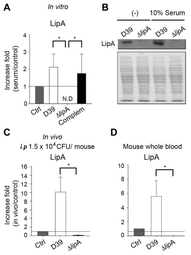
Bacterial RNA was isolated using the hot-phenol method, as previously described (Kwon et al., 2003). All RNA samples were treated with DNase I (TaKaRa, Japan). RNA (1 μg) was converted to cDNA by Easy Script Reverse Transcriptase (Abm, Canada) and random primers (TaKaRa). qPCR was performed using a Step-One-Plus Real-time PCR System (Applied Biosystems, USA) according to the manufacturer’s instructions, as previously described using the primers shown in Supplementary Table S2. The qPCR data was exported after dividing to 16s RNA level to control the loading amount. The value was displayed in Figs. 4A and 5C as increase fold of serum-induced pneumococci (serum) per no-serum pneumococci (control). A control bar (one fold value) was shown as a control level.
Fig. 4. LipA induction by serum.
(A, B) S. pneumoniae strains were cultured until log phase. Goat serum was added to a final concentration of 10% and then incubated for 5 min, followed by lipA mRNA quantitation by qPCR (A), or incubated for 10min, followed by western blot analysis (B). SDS-PAGE results following Coomasie Blue staining was displayed and served as a loading control. The experiment was performed three times independently. Statistical analyses were performed using one-way ANOVA (*p ≤ 0.05). (C) Mice (n = 3) were infected with 1.5 × 10⁴ CFU of S. pneumoniae D39 or ΔlipA intraperitoneally (i.p.). At the severely morbid stage, mouse blood was collected, and lipA mRNA expression was determined by qPCR. (D) Pneumococci at log phase in THY broth were incubated with whole mouse blood for 15min, and lipA mRNA expression was determined by qPCR. (C, D) Each mouse sample was analyzed in triplicate, and the Mann-Whitney rank sum test was used for statistical analysis. N.D: not detected. (*p ≤ 0.05). (A, C and D) Control bar (Ctrl) was displayed one fold value (control/ control).
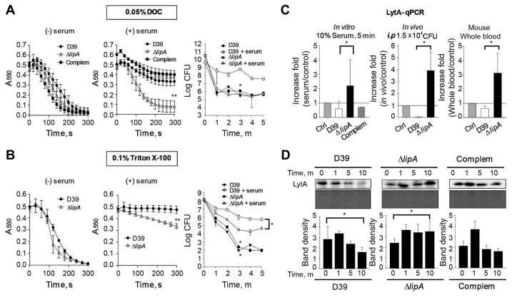
Fig. 5. Inhibition of autolysis and LytA expression by LipA in vitro and in vivo.
Pneumococcal strains cultured in THY broth until log phase were incubated with 10% goat serum for 10 min. (A, B) 0.05% DOC (A) and 0.1% Triton X-100 (B) were added to the bacterial culture and absorbance at 550 nm or viable cell number was determined. (C) (left) Pneumococcal strains cultured in THY broth to the log phase were incubated with 10% goat serum for 5 min, and lytA mRNA was analyzed by qPCR (middle). Mice were infected with 1.5 × 10⁴ CFU of pneumococcal strain intraperitoneally (i.p.). At the severely morbid stage, mouse blood was collected, and lytA RNA expression was analyzed by qPCR (right). The pneumococcal strain at the log phase was incubated with whole mouse blood for 15 min, and lytA mRNA was analyzed by qPCR. Each mouse sample was analyzed in triplicate, and the Mann–Whitney rank sum test was used for statistical analysis (*p ≤ 0.05). Control bar (Ctrl) was displayed one fold value (control/ control). (D) Pneumococcal strains cultured in THY broth to the log phase were incubated with 10% goat serum for 0, 1, 5, and 10 min. The LytA level was determined by western blotting. SDS-PAGE results following Coomasie Blue staining are displayed and served as a loading control. Band density was analyzed by ImageJ. (A, B, C-left, D) Representative data are displayed from three independent experiments and analyzed by one-way ANOVA (*p ≤ 0.05, **p ≤ 0.01).
Deoxycholate- and Triton X-100-induced lysis
Sodium deoxycholate (DOC) and Triton X-100 are known to induce pneumococcal autolysis (Mellroth et al., 2012). Goat serum (10%; Life Technologies) was added to the bacterial culture and incubated for 10 min. DOC or Triton X-100 was added to the bacterial culture at a final concentration of 0.05% or 0.1% (v/v), respectively, to induce bacterial lysis. For absorbance determination, A550 was monitored every 20 s for up to 5 min. For viable cell counting, the bacterial culture was serially diluted and plated onto THY blood agar plates.
Bacterial gene expression in a murine sepsis model
Mice were infected i.p with 1.5 × 10⁴ CFU of S. pneumoniae. Blood were collected at the most severely morbid stage (Hajaj et al., 2012) and subjected to centrifugation at 800 × g at 4°C for 10 min to remove red blood cells. Subsequently, the bacteria were harvested by centrifugation at 15,000 × g at 4°C for 10 min, and bacterial RNA was isolated by the hotphenol method, as previously described (Tran et al., 2011). RNA was treated with DNase I (TaKaRa) and converted to cDNA using random primers (TaKaRa). qPCR was performed using the Step One Plus Real-time PCR System (Applied Biosystems) according to the manufacturer’s instructions, as previously described (Tran et al., 2011). The value was displayed in Figs. 4C and 5C as increase fold of in vivo-collected pneumococci (in vivo) per non-infected pneumococci (control). A control bar (one fold value) was shown as a control level.
S. pneumoniae pellets at the log phase were harvested and incubated with whole mouse blood (CD-1, male) for 15 min. Subsequently, the bacteria in the whole blood were harvested by centrifugation at 15,000 × g at 4°C for 10 min. Total RNA was isolated and subjected to qPCR, as described above. The value was displayed in Figs. 4D and 5C as increase fold of whole blood-induced pneumococci (whole blood) per non-induced pneumococci (control). A control bar (one fold value) was shown as a control level.
Statistical analysis
Western blotting band density was analyzed by ImageJ 1.43 (NIH, USA). Animal studies were statistically analyzed by the log-rank test (survival) or the Mann–Whitney rank sum test (colonization). All other analyses were performed using one-way analysis of variance (ANOVA). p-values were denoted as follows: *p ≤ 0.05, **p ≤ 0.01, ***p ≤ 0.001.
RESULTS
Hypothetical protein Spd_1447 is a lipase
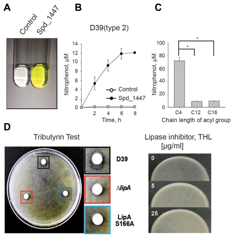
A hypothetical protein encoded by the spd_1447 locus in S. pneumoniae D39 was found to contain the conserved catalytic triad (Gly-His-Ser-Leu-Gly) (CDD: 238382) of the lipase family (NCBI). Sequence alignment also demonstrated that Spd_1447 is a lipase homolog of gram-positive Clostridium sp. or Lactobacillus sp. (Supplementary Fig. S1). For confirmation, the Spd_1447 proteins in S. pneumoniae serotype 2 D39 were purified and used for lipase activity determination (Pinsirodom and Parkin, 2001). After 2 h incubation, the purified Spd_1447 protein produced higher p-nitrophenol levels than the control (Fig. 1A). Furthermore, the D39 Spd_1447 protein displayed time-dependent lipase activity, and the product reached a plateau at 6 h post-incubation (Fig. 1B) indicating that this Spd_1447 protein, named LipA, can be considered a lipase.
Fig. 1. S. pneumoniae Spd_1447 (LipA) lipase activity.
(A) S. pneumoniae D39 (type 2) Spd_1447 hydrolyzed p-nitrophenyl laurate and released p-nitrophenol (yellow color) after a 2-h incubation. (B) S. pneumoniae D39 (type 2) Spd_1447 hydrolyzed p-nitrophenyl laurate to p-nitrophenol in a time-dependent manner. (C) S. pneumoniae D39 Spd_1447 hydrolyzed p-nitrophenyl butyrate (C4), p-nitrophenyl dodecanoate (C12), and p-nitrophenyl palmitate (C16), and released p-nitrophenol after a 2-h incubation. The values were calibrated with a blank control and expressed as released p-nitrophenol concentration. Statistical analyses were performed using one-way ANOVA (*p ≤ 0.05). (D) S. pneumoniae D39 WT (Black square) had a clearer zone on the tributyrin THY agar plate than the ΔlipA (Red square) or LipA S166A (Blue square). The lipase inhibitor THL impaired formation of the clear zone around the S. pneumoniae D39 WT colonies on tributyrin THY agar plates. The results displayed represent data from three independent experiments.
Next, lipase activity was analyzed using various lipid substrates, including p-nitrophenyl butyrate (C4), p-nitrophenyl dodecanoate (C12), and p-nitrophenyl palmitate (C16). The amount of p-nitrophenol released from the C4 substrate was dramatically higher than that from the C12 and C16 substrates (Fig. 1C), indicating that S. pneumoniae LipA preferentially hydrolyzes short-chain rather than long-chain lipid substrates.
A lipase-producing microbe can hydrolyze tributyrin and form a clear halo around the colony (Jørgensen et al., 1991). D39 WT displayed a larger clear zone around the colonies on a tributyrin agar plate than ΔlipA or LipA S166A, indicating LipA has lipase activity and serine residue at amino acid position 166 plays an important role (Fig. 1D). Although LipA was not secreted from D39 WT, our subcellular components fractionation experiment showed that substantial amount of LipA was localized at the cell wall and membrane (data not shown), thus allowing contact to substrate lipid and produce the clear zone in tributyrin assay. Moreover, when a lipase inhibitor (THL) was supplemented, the halo around the colony became fainter as lipase inhibitor concentrations increased, indicating inhibition of lipase activity (Fig. 1D). Thus, these data demonstrate that LipA possesses lipolytic activity.
LipA does not affect S. pneumoniae virulence in a pneumonia model
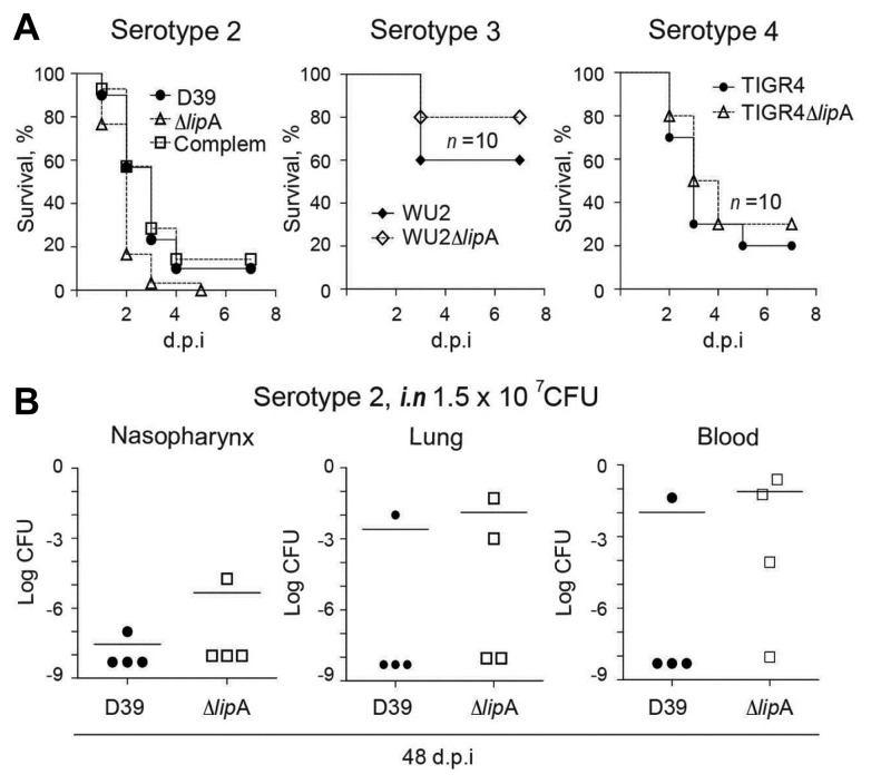
In some pathogens, lipases are considered to be secreted virulence factors (Gill et al., 2005; Stehr et al., 2004). However, LipA was not secreted from S. pneumoniae serotype 2 (D39; data not shown). For the pneumonia model, mice were infected i.n. with S. pneumoniae, and survival rates and bacterial colonization were determined. The survival rate of the mice infected with the ΔlipA was not significantly different from that in those infected with WT in three sero-types (Fig. 2A). In addition, the bacterial colonization in the lung, nasopharynx, and blood at 48 h post-infection did not differ between D39 WT and ΔlipA (Fig. 2B), demonstrating that LipA does not affect S. pneumoniae virulence following i.n. infection.
Fig. 2. LipA did not significantly attenuate virulence in a pneumonia model.
(A) Mice (n = 15) were infected i.n. with 1.5 × 10⁷ CFU of the type 2 and 4 strains or 4.5 × 10⁷ CFU of type 3, and survival time was recorded until 7 days post-infection (d.p.i). (B) Mice (n = 4) were infected with 1.5 × 10⁷ CFU of type 2 strain i.n., and viable cells in the nasopharynx, lung, and blood were determined by plating the homogenized samples on THY blood agar. The experiment was performed twice and representative data was shown. Significant differences were analyzed by the log-rank test (A) or the Mann-Whitney rank sum test (B).
LipA inhibits S. pneumoniae virulence during sepsis
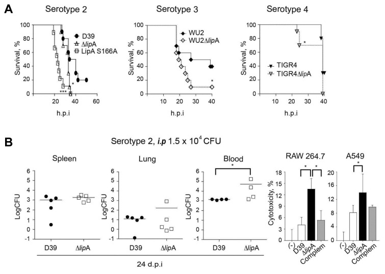
For the sepsis model, mice were infected i.p. with 3 × 10⁴ CFU of S. pneumoniae. The infection by ΔlipA or LipA S166A led to a dramatically higher mortality rate than D39 WT infection. Specifically, ΔlipA infection killed all the mice within 40 h post-infection, whereas 20% of D39 WT infected mice survived over 50 h post-infection (Fig. 3A). Furthermore, mice infected with WU2ΔlipA and TIGR4ΔlipA displayed a significantly higher mortality rate (90% and 30%, respectively) at 25 h post-infection than WT-infected mice (60% and 0% respectively; Fig. 3A), demonstrating the higher virulence of the ΔlipA with lack of lipase activity compared to the WT in a sepsis model.
Fig. 3. Virulence attenuation by LipA in a sepsis model.
(A) Mice (n = 10) were infected i.p. with 3 × 10⁴ CFU of type 2 strains or 1.5 × 10⁴ CFU of type 3 and 4 pneumococcal strains, and survival time was recorded until 60h post-infection (h.p.i). (B) Mice (n = 4–5) were infected with 1.5 × 10⁴ CFU of the type 2 strain i.p., and viable cells in spleen, lung, and blood were determined by plating the homogenized samples on THY blood agar. The experiment was performed in twice and representative data was from one experiment. (C) The ΔlipA displayed higher cytotoxicity in the presence of serum than that of D39 and the complemented strain. RAW 264.7 or A549 cells were infected with pneumococcal strains (MOI, 25) for 2h in the presence of 10% FBS, and cytotoxicity was determined by 3-(4,5-dimethylthiazole-2-yl)-2,5-dephenyl-tetrazolium bromide assay. Results represent data from three independent experiments. Significant differences were analyzed by the log-rank test (A), the Mann-Whitney rank sum test (B), or one-way ANOVA (C) (*p ≤ 0.05).
In the colonization experiments, after i.p. infection, ΔlipA infection produced higher bacterial loads in the blood at 24 h post-infection (p = 0.029; Fig. 3B) than D39 infection. Unlike i.p. infection, in the colonization experiments, after i.n. infection, ΔlipA infection did not produce any significant difference in bacterial loads from D39 infection (Fig. 2B).
Additionally, to determine effect of LipA on pneumococcal virulence in vitro, cells cytotoxicity was measured after pneumococcal infection at the indicated time. ΔlipA infection produced higher cytotoxicity in RAW 264.7 or A549 cell (MOI, 25) than D39 or the complemented strain infection (Fig. 3C) in the presence of 10% fetal bovine serum. These results demonstrate that ΔlipA possesses a higher virulence than WT in sepsis model.
Serum induces LipA
In the sepsis model, ΔlipA significantly increased virulence (Fig. 3). Therefore, the impact of serum on LipA expression was determined. The addition of serum to the S. pneumoniae culture significantly induced lipA mRNA expression in D39 WT and the complemented strain (Fig. 4A). Moreover, serum supplementation also induced LipA level increases in D39 WT (Fig. 4B), indicating that serum can induce increases in levels of both lipA mRNA and LipA protein in S. pneumoniae in vitro.
To check lipA induction by serum, mice were infected i.p. with S. pneumoniae (n = 3), and bacterial RNA was isolated from the blood and analyzed by qPCR. lipA expression in the D39-infected mice was significantly higher than in the non-infected control (Fig. 4C), and lipA was undetectable in the ΔlipA-infected samples. Moreover, when pneumococci were incubated with whole mouse blood for 15 min, lipA expression in D39 WT was significantly induced (Fig. 4D), indicating lipA induction by serum in the sepsis model.
Serum inhibits autolysis in a LipA-dependent manner
Human serum suppresses antibiotic-mediated autolysis in gram-positive S. aureus (Stratton et al., 1986). However, the role of serum in S. pneumoniae autolysis remains unknown. To identify whether serum can affect pneumococcal autolysis, DOC-induced autolysis were determined. Serum inhibited DOC-induced-autolysis of D39 WT and the complemented strain compared with ΔlipA (Fig. 5A). However, without serum, the autolysis rates of D39 WT, the complemented strain, and ΔlipA were similar (Fig. 5A). Moreover, in the presence of serum, after DOC treatment, the viability of the D39 WT was significantly higher than that of ΔlipA (Fig. 5A). However, without serum, the viability of the D39 WT was similar to that of ΔlipA (Fig. 5A).
Additionally, in the presence of serum, D39 WT Triton-X100-induced autolysis was abrogated compared with that of ΔlipA (Fig. 5B). In contrast, without serum, the autolysis rates of D39 WT and ΔlipA were not significantly different (Fig. 5B). Moreover, in the presence of serum, the viability of D39 WT was significantly higher than that of ΔlipA (Fig. 5B), while without serum, no significant difference in viability was observed (Fig. 5B). These data suggest that serum can inhibit S. pneumoniae autolysis via LipA.
Serum represses S. pneumoniae LytA levels via LipA
Among autolysins, LytA plays an important role in pneumococcal lysis (López and García, 2004). In the presence of serum, the lytA fold changes (serum/control) in D39 and the complemented strain were lower than 1, indicating that lytA expression after the addition of serum was lower than in the control (Fig. 5C). However, in the presence of serum, the lytA fold change in the ΔlipA mutant was approximately 2, indicating that lytA expression after the addition of serum was significantly increased relative to that in the control (Fig. 5C). Therefore, these data suggest that serum can inhibit lytA expression in S. pneumoniae D39 WT but not in the ΔlipA mutant.
Next, mice were infected i.p. with S. pneumoniae (n = 3), and lytA mRNA levels in the blood were determined. The lytA fold change (in vivo/control) in D39 was markedly lower than 1, indicating that lytA expression in a sepsis model was much lower than in the control. However, the lytA fold change in the ΔlipA mutant was approximately 4, suggesting that lytA expression in ΔlipA was significantly higher than in the control (Fig. 5C). Thus, in a sepsis model of infection, lytA expression was inhibited in D39 WT but not in the ΔlipA mutant.
In addition, when pneumococci were incubated with whole mouse blood, the lytA fold change (whole blood/control) in D39 was lower than 1, whereas the lytA fold change in the ΔlipA mutant was approximately 3 (Fig. 5C). These data indicate that lytA expression in D39 was lower than in the control, whereas lytA expression in ΔlipA was higher than in the control. Therefore, mouse blood inhibited lytA expression in D39 but not in the ΔlipA mutant.
At the protein level, pneumococcal culture at the log phase was supplemented with serum, and the total cell lysate was used for western blotting analysis. Serum inhibited LytA expression in D39 WT and the complemented strains at 5 and 10 min post-incubation, but did not inhibit in the ΔlipA mutant (Fig. 5D), suggesting that LipA suppresses LytA expression in the presence of serum.
DISCUSSION
Most studies on microbial lipase have focused on the broad applications of lipases in industrial biotechnology (Jaeger et al., 1994). In addition, the role of lipases in pathogenesis has been preferentially characterized in fungi rather than in bacteria. Deletion of lipase genes in Candida parapsilosis, a fungal pathogen, inhibited fungal growth and biofilm formation and resulted in virulence repression (Gácser et al., 2007). Furthermore, the lipase mutant in Fusarium graminearum increased mycotoxin production (Voigt et al., 2007). Bacterial lipase studies have been limited to the secretion of the lipase and damage to host cells during infection (Nawabi et al., 2008; Rollof et al., 1988). Gram-positive S. aureus lipase contains a signal peptide (295 amino acids), which is cleaved during secretion (Rollof and Normark, 1992). S. pneumoniae lipase (LipA) has no signal peptide sequence, and no LipA was detected in the bacterial culture supernatant (data not shown), indicating that pneumococcal LipA is not secreted. Additionally, the pneumococcal LipA did not affect virulence significantly following i.n. infection but inhibited virulence in a sepsis model of infection. These differences may be attributed to the presence of serum. Pneumococci in i.n route infection (pneumonia model) would experience transition from nasal cavity to pulmonary and to blood and might adapt slowly in due course of infection whereas i.p route infection (sepsis model) would directly be exposed to blood in such a short time and adapt to blood milieu and host immune system rapidly. Serum can induce LipA expression at both the mRNA and protein levels. Additionally, pneumococcal bacteremia patients displayed decreased levels of serum triglycerides, cholesterol, and high density lipoprotein cholesterol (Kerttula and Weber, 1987), indicating that serum and serum compounds play a role during pneumococcal sepsis. However, the correlation between the pneumococci and the host serum remains unclear. Here, we found that serum can affect bacterial lipase expression and pathogenesis.
Human serum inhibits antibiotic-mediated S. aureus autolysis (Stratton et al., 1986). Serum decreased DOC- or Triton X-100-induced pneumococcal autolysis. Moreover, serum impaired autolysis in the WT but not in the ΔlipA, suggesting that serum-dependent inhibition of pneumococcal autolysis is mediated by LipA.
In addition, higher lytA expression in ΔlipA seems to decrease CFUs than the WT, however, ΔlipA shows higher CFUs than the D39 WT in the blood (Fig. 3B). DOC- or Triton X-100-induced pneumococcal autolysis would occur only until a limited number of cells and seems to be subject to feedback regulation resulting in normal growth stage at 5 min of serum exposure (Figs. 5A and 5B). We assumed that immediately after serum exposure, the ΔlipA induces lytA and subsequently lysis and release of cell wall and other cellular components, which trigger both proinflammatory cytokines secretion from the host and nutrients procurement from the host resulting in inflammation (pneumonia and sepsis) as well as higher bacterial CFUs. Moreover, growth rate of the ΔlipA was recovered 5 h post-inoculation in the presence of serum (data not shown) suggesting that the ΔlipA seems to adapt to the serum environment and subject to feedback regulation resulting in repression of lytA expression. Therefore, the ΔlipA is more virulent immediately after the i.p infection.
Short-chain fatty acids (SFAs), such as butyrate, increase the chemotactic response of neutrophils and neutrophil migration (Vinolo et al., 2009) or acetate and propionate bound to the murine GPR43 receptor, and enhance calcium flux, reactive oxygen species release, and the phagocytic activity of neutrophils (Maslowski et al., 2009). Butyrate also suppresses cytokines such as TNF-α and IL-6, but increases IL-10 (Chakravortty et al., 2000; Park et al., 2007) by inhibition of histone deacetylase activity (Waldecker et al., 2008) resulting in an increase in histone acetylation and modulation of the expression of these genes. Thus, butyrate and SFAs might modulate host defense inflammation. S. pneumoniae LipA preferentially releases butyrate (C4), rather than dodecanoate (C10) or palmitate (C12). Therefore, LipA can modulate inflammation and host defense mechanisms by producing SFAs and other fatty acids during pneumococcal infection. How LipA affects virulence through preferential substrate hydrolysis, and how LipA-produced fatty acids modulate host immunity during infection requires further investigation.
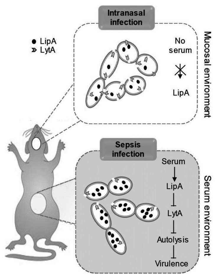
In summary, in a sepsis model, autolysis is inhibited to promote pneumococcal survival in the bloodstream prior to spreading into other niches. Pneumococcal autolysis suppression during septic infection is mediated by lipase LipA (Fig. 6). An investigation of pneumococcal LipA would provide a greater understanding of the mechanism by which S. pneumoniae modulates virulence to adapt to various host environments and maintain its survival until transfer to the next host.
Fig. 6. Schematic model of serum-dependent autolysis inhibition by LipA in S. pneumoniae during sepsis.
During intranasal infection, S. pneumoniae D39 WT and the ΔlipA did not display any significant differences in virulence. However, during sepsis infection, serum induced lipA expression, which subsequently inhibited LytA, leading to an inhibition of pneumococcal autolysis and virulence.
Supplementary data
ACKNOWLEDGMENTS
This work was supported by the National Research Foundation (NRF-2015R1 A2 A1 A10052511).
Footnotes
Note: Supplementary information is available on the Molecules and Cells website (www.molcells.org).
REFERENCES
- Berry A.M., Lock R.A., Hansman D., Paton J.C. Contribution of autolysin to virulence of Streptococcus pneumoniae. Infect Immun. 1989;57:2324–2330. doi: 10.1128/iai.57.8.2324-2330.1989. [DOI] [PMC free article] [PubMed] [Google Scholar]
- Canvin J.R., Marvin A.P., Sivakumaran M., Paton J.C., Boulnois G.J., Andrew P.W., Mitchell T.J. The role of pneumolysin and autolysin in the pathology of pneumonia and septicemia in mice infected with a type 2 pneumococcus. J Infect Dis. 1995;172:119–123. doi: 10.1093/infdis/172.1.119. [DOI] [PubMed] [Google Scholar]
- Chakravortty D., Koide N., Kato Y., Sugiyama T., Mu M.M., Yoshida T., Yokochi T. The inhibitory action of butyrate on lipopolysaccharide-induced nitric oxide production in RAW 264.7 murine macrophage cells. J Endotoxin Res. 2000;6:243–247. [PubMed] [Google Scholar]
- Gácser A., Trofa D., Schäfer W., Nosanchuk J.D. Targeted gene deletion in Candida parapsilosis demonstrates the role of secreted lipase in virulence. J Clin Invest. 2007;117:3049–3058. doi: 10.1172/JCI32294. [DOI] [PMC free article] [PubMed] [Google Scholar]
- Gill S.R., Fouts D.E., Archer G.L., Mongodin E.F., DeBoy R.T., Ravel J., Paulsen I.T., Kolonay J.F., Brinkac L., Beanan M., et al. Insights on evolution of virulence and resistance from the complete genome analysis of an early methicillin-resistant Staphylococcus aureus strain and a biofilm-producing methicillin-resistant Staphylococcus epidermidis strain. J Bacteriol. 2005;187:2426–2438. doi: 10.1128/JB.187.7.2426-2438.2005. [DOI] [PMC free article] [PubMed] [Google Scholar]
- Hajaj B., Yesilkaya H., Benisty R., David M., Andrew P.W., Porat N. Thiol peroxidase is an important component of Streptococcus pneumoniae in oxygenated environments. Infect Immun. 2012;80:4333–4343. doi: 10.1128/IAI.00126-12. [DOI] [PMC free article] [PubMed] [Google Scholar]
- Hirst R.A., Gosai B., Rutman A., Guerin C.J., Nicotera P., Andrew P.W., O’Callaghan C. Streptococcus pneumoniae deficient in pneumolysin or autolysin has reduced virulence in meningitis. J Infect Dis. 2008;197:744–751. doi: 10.1086/527322. [DOI] [PubMed] [Google Scholar]
- J⊘rgensen S., Skov K.W., Diderichsen B. Cloning, sequence, and expression of a lipase gene from Pseudomonas cepacia: lipase production in heterologous hosts requires two Pseudomonas genes. J Bacteriol. 1991;173:559–567. doi: 10.1128/jb.173.2.559-567.1991. [DOI] [PMC free article] [PubMed] [Google Scholar]
- Jaeger K.-E., Ransac S., Dijkstra B.W., Colson C., van Heuvel M., Misset O. Bacterial lipases. FEMS Microbiol Rev. 1994;15:29–63. doi: 10.1111/j.1574-6976.1994.tb00121.x. [DOI] [PubMed] [Google Scholar]
- Joseph B., Ramteke P.W., Thomas G. Cold active microbial lipases: some hot issues and recent developments. Biotechnol Adv. 2008;26:457–470. doi: 10.1016/j.biotechadv.2008.05.003. [DOI] [PubMed] [Google Scholar]
- Kadioglu A., Weiser J.N., Paton J.C., Andrew P.W. The role of Streptococcus pneumoniae virulence factors in host respiratory colonization and disease. Nat Rev Micro. 2008;6:288–301. doi: 10.1038/nrmicro1871. [DOI] [PubMed] [Google Scholar]
- Kang N.-S., Park S.-Y., Lee K.-R., Lee S.-M., Lee B.-G., Shin D.-H., Pyo S. Modulation of macrophage function activity by ethanolic extract of larvae of Holotrichia diomphalia. J Ethnopharmacol. 2002;79:89–94. doi: 10.1016/s0378-8741(01)00369-5. [DOI] [PubMed] [Google Scholar]
- Kerttula Y., Weber T. Serum lipids in pneumonia of different aetiology. Ann Clin Res. 1987;20:184–188. [PubMed] [Google Scholar]
- Kwon H.-Y., Kim S.-W., Choi M.-H., Ogunniyi A.D., Paton J.C., Park S.-H., Pyo S.-N., Rhee D.-K. Effect of heat shock and mutations in ClpL and ClpP on virulence gene expression in Streptococcus pneumoniae. Infect Immun. 2003;71:3757–3765. doi: 10.1128/IAI.71.7.3757-3765.2003. [DOI] [PMC free article] [PubMed] [Google Scholar]
- Lanie J.A., Ng W.-L., Kazmierczak K.M., Andrzejewski T.M., Davidsen T.M., Wayne K.J., Tettelin H., Glass J.I., Winkler M.E. Genome sequence of Avery’s virulent serotype 2 strain D39 of Streptococcus pneumoniae and comparison with that of unencapsulated laboratory strain R6. J Bacteriol. 2007;189:38–51. doi: 10.1128/JB.01148-06. [DOI] [PMC free article] [PubMed] [Google Scholar]
- Liu L., Johnson H.L., Cousens S., Perin J., Scott S., Lawn J.E., Rudan I., Campbell H., Cibulskis R., Li M. Global, regional, and national causes of child mortality: an updated systematic analysis for 2010 with time trends since 2000. The Lancet. 2012;379:2151–2161. doi: 10.1016/S0140-6736(12)60560-1. [DOI] [PubMed] [Google Scholar]
- López R., García E. Recent trends on the molecular biology of pneumococcal capsules, lytic enzymes, and bacteriophage. FEMS Microbiol Rev. 2004;28:553–580. doi: 10.1016/j.femsre.2004.05.002. [DOI] [PubMed] [Google Scholar]
- Luong T.T., Kim E.-H., Bak J.P., Nguyen C.T., Choi S., Briles D.E., Pyo S., Rhee D.-K. Ethanol-induced alcohol dehydrogenase E (AdhE). potentiates pneumolysin in Streptococcus pneumoniae. Infect Immun. 2015;83:108–119. doi: 10.1128/IAI.02434-14. [DOI] [PMC free article] [PubMed] [Google Scholar]
- Martin G.S. Sepsis, severe sepsis and septic shock: changes in incidence, pathogens and outcomes. Expert Rev Anti Infect Ther. 2012;10:701–706. doi: 10.1586/eri.12.50. [DOI] [PMC free article] [PubMed] [Google Scholar]
- Maslowski K.M., Vieira A.T., Ng A., Kranich J., Sierro F., Di Y., Schilter H.C., Rolph M.S., Mackay F., Artis D., et al. Regulation of inflammatory responses by gut microbiota and chemoattractant receptor GPR43. Nature. 2009;461:1282–1286. doi: 10.1038/nature08530. [DOI] [PMC free article] [PubMed] [Google Scholar]
- Mellroth P., Daniels R., Eberhardt A., Rönnlund D., Blom H., Widengren J., Normark S., Henriques-Normark B. LytA, major autolysin of Streptococcus pneumoniae, requires access to nascent peptidoglycan. J Biol Chem. 2012;287:11018–11029. doi: 10.1074/jbc.M111.318584. [DOI] [PMC free article] [PubMed] [Google Scholar]
- Munford R., Suffredini A. Principles and practice of infectious diseases. 7th ed. Philadelphia: Churchill Livingstone; 2009. Sepsis, severe sepsis, and septic shock. [Google Scholar]
- Nawabi P., Catron D.M., Haldar K. Esterification of cholesterol by a type III secretion effector during intracellular Salmonella infection. Mol Microbiol. 2008;68:173–185. doi: 10.1111/j.1365-2958.2008.06142.x. [DOI] [PubMed] [Google Scholar]
- Nguyen C.T., Kim E.-H., Luong T.T., Pyo S., Rhee D.-K. ATF3 confers resistance to pneumococcal infection through positive regulation of cytokine production. J Infect Dis. 2014;210:1745–1754. doi: 10.1093/infdis/jiu352. [DOI] [PubMed] [Google Scholar]
- Orihuela C.J., Gao G., Francis K.P., Yu J., Tuomanen E.I. Tissue-specific contributions of pneumococcal virulence factors to pathogenesis. J Infect Dis. 2004;190:1661–1669. doi: 10.1086/424596. [DOI] [PubMed] [Google Scholar]
- Park J.-S., Lee E.-J., Lee J.-C., Kim W.-K., Kim H.-S. Anti-inflammatory effects of short chain fatty acids in IFN-γ-stimulated RAW 264.7 murine macrophage cells: Involvement of NF-κB and ERK signaling pathways. Int Immunopharmacol. 2007;7:70–77. doi: 10.1016/j.intimp.2006.08.015. [DOI] [PubMed] [Google Scholar]
- Pemberton J.M., Kidd S.P., Schmidt R. Secreted enzymes of Aeromonas. FEMS Microbiol Lett. 1997;152:1–10. doi: 10.1111/j.1574-6968.1997.tb10401.x. [DOI] [PubMed] [Google Scholar]
- Pinsirodom P., Parkin K.L. Current protocols in food analytical chemistry. John Wiley & Sons, Inc.; 2001. Lipase Assays; pp. Unit C3.1.1–C3.1.13. [Google Scholar]
- Rollof J., Normark S. In vivo processing of Staphylococcus aureus lipase. J Bacteriol. 1992;174:1844–1847. doi: 10.1128/jb.174.6.1844-1847.1992. [DOI] [PMC free article] [PubMed] [Google Scholar]
- Rollof J., HedstrÖM SÅ, Nilsson-Ehle P. Lipolytic activity of Staphylococcus aureus strains from disseminated and localized infections. APMIS. 1987;95B:109–113. doi: 10.1111/j.1699-0463.1987.tb03096.x. [DOI] [PubMed] [Google Scholar]
- Rollof J., Braconier J.H., Söderström C., Nilsson-Ehle P. Interference of Staphylococcus aureus lipase with human granulocyte function. Eur J Clin Microbiol Infect Dis. 1988;7:505–510. doi: 10.1007/BF01962601. [DOI] [PubMed] [Google Scholar]
- Stehr F., Felk A., Gácser A., Kretschmar M., Mähnß B., Neuber K., Hube B., Schäfer W. Expression analysis of the Candida albicans lipase gene family during experimental infections and in patient samples1. FEMS Yeast Res. 2004;4:401–408. doi: 10.1016/S1567-1356(03)00205-8. [DOI] [PubMed] [Google Scholar]
- Stratton C.W., Evans M.E., Burch D.J., Hawley H.B., Horsman T.A., Tu K.K., Reller L.B. Effect of human serum on inhibition of growth of Staphylococcus aureus by antimicrobial agents. Eur J Clin Microbiol. 1986;5:351–353. doi: 10.1007/BF02017797. [DOI] [PubMed] [Google Scholar]
- Tran T.D.-H., Kwon H.-Y., Kim E.-H., Kim K.-W., Briles D.E., Pyo S., Rhee D.-K. Decrease in penicillin susceptibility due to heat shock protein ClpL in Streptococcus pneumoniae. Antimicrob Agents Chemother. 2011;55:2714–2728. doi: 10.1128/AAC.01383-10. [DOI] [PMC free article] [PubMed] [Google Scholar]
- Vinolo M., Rodrigues H., Hatanaka E., Hebeda C., Farsky S., Curi R. Short-chain fatty acids stimulate the migration of neutrophils to inflammatory sites. Clin Sci (Lond) 2009;117:331–338. doi: 10.1042/CS20080642. [DOI] [PubMed] [Google Scholar]
- Voigt C., von Scheidt B., Gácser A., Kassner H., Lieberei R., Schäfer W., Salomon S. Enhanced mycotoxin production of a lipase-deficient Fusarium graminearum mutant correlates to toxin-related gene expression. Eur J Plant Pathol. 2007;117:1–12. [Google Scholar]
- Waldecker M., Kautenburger T., Daumann H., Busch C., Schrenk D. Inhibition of histone-deacetylase activity by short-chain fatty acids and some polyphenol metabolites formed in the colon. J Nutr Biochem. 2008;19:587–593. doi: 10.1016/j.jnutbio.2007.08.002. [DOI] [PubMed] [Google Scholar]
- Yang G., Hernández-Rodríguez C.S., Beeton M.L., Wilkinson P., Waterfield N.R. Pdl1 is a putative lipase that enhances Photorhabdus toxin complex secretion. PLoS Pathog. 2012;8:e1002692. doi: 10.1371/journal.ppat.1002692. [DOI] [PMC free article] [PubMed] [Google Scholar]
Associated Data
This section collects any data citations, data availability statements, or supplementary materials included in this article.


