Abstract
Avulsion is defined as the complete displacement of the tooth out of its socket with disruption of the fibers of periodontal ligament, remaining some of them adhered to the cementum and the rest to the alveolar bone. This condition is more frequent in young permanent teeth, because the root development is still incomplete. Splints are used to immobilize traumatized teeth that suffered damage in their structures of support, preventing their constant movement. The literature has shown that after replantation, it is necessary to use splints in order to immobilize the teeth during the initial period, which is essential for the repair of periodontal ligament; the use of semi-rigid splint is more indicated than the rigid one, and long periods of splinting showed that substitutive resorption or ankylosis is an expected complication. Thus, the aim of this review is to describe the different types of splints; their time of permanency, and its influence on the process of healing and reparation on the occurrence of substitutive resorption or ankylosis. It is very important to keep gathering knowledge about this content, since it has been proved that the approaches and the protocols keep changing over time.
Keywords: Tooth replantation, tooth avulsion, ankylosis, substitutive resorption, splinting
Introduction
Traumatic dental injuries compromise the patient, in both functional and psychological aspects (1). Among dental traumas tooth avulsion has a prevalence from 0.5 to 16% in permanent dentition (2, 3, 4, 5), and it is defined as the complete displacement of the tooth out of its socket with disruption of the fibers of periodontal ligament, remaining some of them adhered to the cementum and the rest to the alveolar bone (6). Avulsion is more frequent in young permanent teeth, because the root development is still incomplete and periodontium is in formation as well. Consequently, light horizontal impacts may result in the total displacement of the tooth. Among the teeth, the upper central incisors are most involved while among children from seven to eleven years old, boys are more susceptible to this type of trauma than girls (7). Etiologically, it is associated with car accidents and practice of sports (8), protrusion of the anterior teeth; malocclusion class II, division 1; anterior open bite and mouth breathing as well. In such cases the severity of injury is higher as more pronounced is the dental protrusion (9, 10). Immediate treatment for avulsion is replantation (1), and splint is the device used to support, protect or immobilize in order to avoid possible damage to the pulp and periodontal tissue, which retards the repair of neurovascular bundle and reintegration of periodontal fibers broken by trauma (8). The goal of this study is to do a narrative review of the literature, describing the different types of dental splints used in replantation, as well as the immobilization time and its relation to substitutive resorption.
Historic progress of dental splints
The first experimental research regarding this content, according to Chelotti and Valentine (11) began with Wilkinson in 1918, who used monkeys to perform teeth replantation and subsequent histological evaluation, which led him to conclude that it was necessary the presence of the periodontal membrane so that fixation could happen. Chelotti and Valentine (11) also affirmed that years after that, in 1983, Tiley Bodecker confirmed Wilkinson’s thesis, stating that the replanted teeth should be treated and sealed, and once replanted they should be completely immobilized. In 1990, Chelotti and Valentine (11) reported that splints are used to immobilize the injured teeth, which by immobilization have better repair conditions. They stated that the most used splinting devices are: mouth guards, dental braces and splints made with composite resin. In 1993, after studying dental trauma and its consequences, Alvarez and Alvarez (12) concluded that once the tooth is positioned correctly, it should be splinted in order to be kept in place and to prevent constant movements, which would damage the organization of periodontal tissue. In situations when no bone or tooth fracture is detected, they suggested splinting for 2 to 3 weeks, on the contrary, the splint should be maintained for 6 to 8 weeks. In 2000, Trope et al. (13) indicated for avulsed teeth a semi-rigid splint for 7 to 10 days fixed with composite resin and steel wire with a diameter from 0.015 to 0.030. They recommended this type of splint and amount of time, because it allows physiological movements of the teeth during the healing process and it also results in a reduction of the incidence of ankylosis. In 2001, Vasconcelos et al. (10) also recommended this type of splint, in addition, they said that when a rigid fixation is placed, there is a higher degree of bone growth over the periodontal space with consequent ankylosis and replacement resorption. Also in 2001, a literature review made by Pereira et al. (14) reported a case that the avulsed teeth were replanted and fixed with a rigid wire (0.9) and composite resin for 2 weeks, and then the endodontic treatment was performed. One year and three months after the accident, by analyzing the radiographies, no evidence of resorption was found, thus they decided for root canal obturation. Two years later, a control radiography was taken, it was observed a suggestive image of external resorption. The authors concluded that maintaining the vitality of the cells and periodontal ligament fibers is an important factor that might affect the success, so does not matter how great is the splint technique if periodontal ligament cells became necrotic.
In 2002, Castro et al. (15) reported a clinical case about the consequences of dental replantation after avulsion. In an emergency care, the teeth were replanted, but was not placed any kind of splint. Two days later, the patient went to the Dental School of Araçatuba – UNESP - Brazil where was placed a splint with orthodontic wire and composite resin, then the patient was referred to endodontic treatment. The authors stated that the proper repositioning of the as: the organization of the blood clot, the presence of bone or tooth fragments, bone plate fracture or displacement of the socket wall (16, 17). Studying dental trauma, in 2004, Oliveira et al. (18) conducted a research and reported that an efficient splint is essential for the maintenance of the avulsed tooth; there are several forms and materials for splinting, such as: resin, by itself or with a flexible arch of nylon or metal wire, orthodontic brackets with malleable arch, and vestibular arches or bars. They reported that flexible and short-term splints allow the physiological mobility of the teeth, which has been proven to be in favor of periodontal healing and reduce the risk of ankylosis and external resorption. However, they highlighted that in cases of fractured bone plate, a rigid contention is necessary and suggests the use of Erich bars and braces. It was reported by Cobankara and Ungor (19), in 2007, a case of late replantation (1 week) of two incisors after a cycling accident. The teeth had their roots debrided, the root canals were treated endodontically and filled with calcium hydroxide, and then immediately were replanted and splinted with rigid contention for 5 weeks, ankylosis was observed after 10 months. They concluded that although the risk of substitutive root resorption and subsequent tooth loss is high, the technique appears to be advantageous not only for aesthetic, postponing a prosthetic treatment, but also for maintaining the height of the alveolar bone (19, 20).
In the same year, Westphalen et al. (21) conducted a study involving 250 general dentists in order to find the degree of knowledge regarding the treatment of dental avulsion. The results showed that the level of knowledge on the subject was sufficient; and that in relation to dental splints 73% of respondents used semi-rigid splint with nylon, 10% steel wire and 10% use restorative material. Regarding the amount of time, 36% use for 15 days, 38% for 3 days, 24% for 60 days, only 2% would use for 24 hours. Only 7% said they did not use any kind of splint, and could not explain their decision; only one respondent justified the decision of not placing the splint in case of a satisfactory stability after replantation of the tooth, but not mentioned which type of splint it would use if necessary.
In 2009, Granville-Garcia et al. (22) also assessed the knowledge of dental avulsion among dentists in a Program of Family Health Care, and the influence of professional experience, by interviewing 30 professionals. The results were similar to other studies (23) concluding that most dentists have adequate knowledge of dental avulsion, not being influenced by the time of experience. In this evaluation saline solution was the most appropriate media for storage (56.7%), the ideal extra-alveolar period of time was less than 30 minutes (60%), and 61.1% of respondents used Semi-rigid contention for up to 15 days (23). In 2012, de Jesus Soares et al. (24) described the use of multidisciplinary approaches to deal with an unsuccessful replantation. Both upper incisors of a 14-year-old female patient were replanted and splinted with a semi-rigid splint for 3 months. The patient reported that the extraalveolar period before first care lasted about 3 hours and 10 minutes, with the teeth being in dry storage for 10 minutes after being stored in saline solution. Due to presence of severe inflammatory root resorption, showing communication with periodontal tissue associated with enhanced tooth mobility, authors opted for extraction, use of a temporary prosthesis made by patient’s own crowns followed by adhesive prosthesis, and after 3 years follow up, implant surgery associated with porcelain crowns. Autotransplantation also has been pointed as an alternative to replace missing incisors (25, 26, 27, 28, 29). However, it has limitations as the root of the donor tooth has to be two thirds to three quarters formed, besides of anatomic concerns once about 60% of autotransplanted teeth were dissimilar in appearance with regard to an asymmetric gingival width or a color mismatch (26, 28).
In 2014, Sardana et al. (30) performed a replantation of avulsed maxillary central incisor with 15-hours extra-oral time. A 3-year follow-up was made in order to observe the consequences of delayed replantation. As expected, ankylosis and inflammatory resorption did happened, but clinically the tooth was asymptomatic. In addition, the authors concluded that it is very important perform a delayed replantation even after prolonged extra-oral time because it maintains the esthetics of the individual. It also works as a good alternative to prosthesis (implant or fixed partial denture) till the growth is completed due to preservation of the alveolar bone and psychological benefit to the patient. In 2015, Nagata et al. (31) described a case involving an immature maxillary left lateral incisor that was replanted and successfully treated with pulp revascularization technique. The same approach was also described by Lucisano at al. in the follow year to manage a similar case (32): in both cases a 8-year old boy had his teeth replanted after 30min and 1 day of avulsion, respectively. Besides dental splint, revascularization therapy was performed by irrigating the root canal and applying a calcium hydroxide paste and 2% chlorhexidine gel for 21 days. After that, the canal was cleaned and a blood clot was stimulated up to the cervical third of the root canal. Mineral trioxide aggregate (MTA) was placed at the entrance of the root canal and the crown was restored. In both cases, it was possible to notice periapical repair and apical closure (31, 32).
Pulp and periodontal regeneration were reported for mature teeth as well (33, 34, 35). In 2016, a study from Tambakad et al. (34), the use of Platelet-rich plasma (PRP) was evaluated for pulpal regeneration in an 11-year-old’s avulsed mature incisor after more than 8 hours extraoral dry time and delayed replantation. After disinfection and extraoral pulp extirpation, the tooth had its apex enlarged, and was placed in doxycycline solution for 20 minutes. After tooth replantation and splinting, PRP was injected up to the level of the cementoenamel junction and sealed with glass ionomer cement. Passed 6 months, it was noticed internal and external root resorption with periapical radiolucency and an apparent periodontal ligament space, which were treated by inserting antibiotics (minocycline and metronidazole) into the canal. Twelve months later, radiographs suggested resolution of periapical radiolucency as well as stagnation of internal resorption and positive response to thermal and electric pulp tests (34).
Usage of splinting
Several factors might influence in the success of replantation, such as: extension of the trauma, extraalveolar permanence period, means of preservation, contamination, manipulation and conditions of the avulsed tooth (36). Also, relevant factors such as type of splint used and time of permanence. Currently, it has been suggested to make a slight splint and immediately to establish an occlusal function that will generate physiological stimulus in the metabolism of periodontal tissues (8). A consensus among authors is that after replantation, it is necessary to use a splint. This device is used to immobilize the injured teeth when necessary. They believe that this stabilization helps the periodontal ligament to have better repair conditions, however those devices should be the least traumatic as possible (2, 7, 9, 10, 11, 12, 13, 14, 15, 17, 18, 36, 37, 38, 39, 40, 41, 42, 43, 44, 45, 46). Several types of splints are available, depending on the mobility degree, they are classified as: flexible, semi-rigid and rigid. The authors ideally recommend the use of semi-rigid splint in cases of dental avulsions when no bone fracture is detected (2, 7, 10, 13, 15, 17, 18, 37, 39, 40, 42, 45, 46). Non-rigid immobilization is the ideal device due to its passive, atraumatic and flexible features, which allows a certain functional movement, and thus a functional arrangement of the periodontal ligament fibers, reducing the risk of external resorption and ankyloses (2, 10, 17, 37, 44, 47, 48, 49). Some authors also advocate the use of flexible splint in avulsions (4, 36, 38, 43). Even though Pereira et al. used the rigid contention in their clinical case (14), this contention produces a higher degree of external bone growth along the periodontal space with consequent ankylosis or substitutive resorption (2, 10). However, the rigid splint is necessary in cases of fracture of the bone plate and late replantation (7, 9, 19, 46).
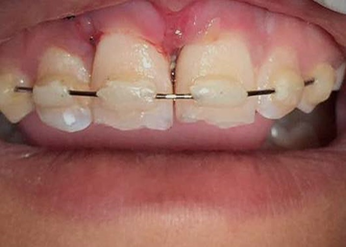
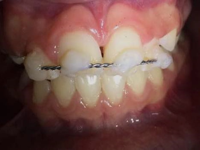
Most authors believe that the ideal semi-rigid splint type is the one made with composite resin and orthodontic wire or nylon thread. The variation regards the type of thread and material used. Ruellas et al. (44) and Prokopowitsch et al. (7), used a 0.018 inch wire twistflex; The orthodontic wire is utilized by McDonald and Avery (42), Castro et al. (16), Manfrim et al. (41) with smaller diameter than or equal to 0.5 mm; Prokopowitsch et al. (7) use the 0.20 mm to 0.40 mm in diameter; Andreasen et al. (37) the one with 0.3mm in diameter; Trope et al. (13) 0.015 to 0,030mm diameter. The nylon thread recommended by Andreasen et al. (37) is the fishing line type; McDonald and Avery (42), Soares and Goldberg (45), and Manfrim et al. (41) recommended the use of nylon monofilament 20 to 30 pounds (Figure 1). Authors like Prokopowitsch et al. (7), Isolan et al. (50) and Manfrin et al. (41) considered the splint with nylon composite resin as flexible and not as semirigid. On the other hand, Côrtes and Bastos (39) used as flexible splint the steel wire for osteosynthesis 0.12 or 0.25mm fixed with composite resin. Rigid immobilization can be made with composite and rigid wire 0.9 (14). On the other hand, Oliveira et al. (18) recommend Erich bars and orthodontic appliances. Prokopowitsch et al. (7) used steel wire 0.5mm and photo-polymerized resin, although Manfrim et al. (41) consider the wire of 0.5 mm as flexible (Figure 2). There are other types of alternative splints, such as the ones made with orthodontic brackets associated with a passive wire, sutures or vestibular bars (18, 40, 45, 47, 51). Regarding the time of immobilization, authors such as Isolan et al. (50), Trope et al. (13) and Baldissera et al. (38) agreed that long periods of splinting are factors that may contribute to the occurrence of substitutive resorption, therefore, over the years, it has been used in shorter periods of time. In cases of avulsion with no fracture in the bone plate, some authors advocate the use of splinting for just one week (2, 5, 36, 48, 49). Others, on the other hand, believe that 2 weeks are sufficient (14, 40, 41), however, Trope et al. (13) indicate for 7 to 10 days. McDonald and Avary (42) suggest for 7 to 14 days and Soares and Goldberg (45), for 7 to 15 days. According to Souza Neto et al. (46) and Prokopowitsch et al. (7) the ideal period should be 10 to 14 days and to Alvarez and Alvarez (12) would be 2 to 3 weeks. In cases of avulsion associated with fracture of the bone plate Alvarez and Alvarez (12) suggest the use of splints for 6 to 8 weeks. Ruellas et al. (44) recommended for 5 weeks. Trope et al. (13) use splints for 4 to 8 weeks and Prokopowitsch et al. (7) for 25 to 30 days. In late replantation, Prokopowitsch et al. (7) advocate the use of splints for a period of 45 days and Cobankara and Ungor (19) believe that it should be used for 5 weeks.
Figure 1.

Semi-rigid splint, placed after tooth avulsion of the central incisors.
Figure 2.

Rigid Splint placed after tooth intrusion of an upper left central incisor. At the moment of the consult, the patient reported to be using this splint for 2 months. It was revived immediately. After CBCT, it was possible to confirm the substitutive resorption on the root of the tooth.
Conclusion
Based on the literature, it can be concluded that after replantation the use of splint is compulsory for allowing immobilization of the teeth during the initial period, which is essential for the repair of periodontal ligament; the use of semi-rigid splint is more indicated than the rigid one, and that long periods of splinting showed that substitutive resorption or ankylosis is an expected complication.
Footnotes
Source of funding: None declared.
Conflict of interest: None declared.
References
- 1.Caldas AF Jr, Burgos ME. A retrospective study of traumatic dental injuries in a Brazilian dental trauma clinic. Dent Traumatol. 2001. December;17(6):250–3. 10.1034/j.1600-9657.2001.170602.x [DOI] [PubMed] [Google Scholar]
- 2.Andreasen JO, Andreasen FM. Texto e atlas colorido de traumatismo dental. São Paulo. Art Med. 2001;•••:383–453. [Google Scholar]
- 3. International Association of Dental Traumatology. Dental trauma guide [Internet]. [San Diego]: International Association of Dental Traumatology; 2016. August [revised 2016 Dec; cited 2017 Dec 2]. [about 20 p.] Available from: http://www.dentaltraumaguide.org. [Google Scholar]
- 4.Viegas CM, Scarpelli AC, Carvalho AC, Ferreira FM, Pordeus IA, Paiva SM. Predisposing factors for traumatic dental injuries in Brazilian preschool children. Eur J Paediatr Dent. 2010. June;11(2):59–65. [PubMed] [Google Scholar]
- 5.Wendt FP, Torriani DD, Assunção MC, Romano AR, Bonow ML, da Costa CT et al. Traumatic dental injuries in primary dentition: epidemiological study among preschool children in South Brazil. Dent Traumatol. 2010. April;26(2):168–73. 10.1111/j.1600-9657.2009.00852.x [DOI] [PubMed] [Google Scholar]
- 6.Vasconcellos RJ, Marzola C, Genu PR. Trauma dental aspectos clínicos e cirúrgicos. ATO. 2006;6(12):774–96. [Google Scholar]
- 7.Prokopowitsh I. Condutas endodônticas em dentes reimplantados. Espelho Clínico:São Caetano do Sul. 2001;4(24):8–9. [Google Scholar]
- 8.Busato AL, González PA, Camilo EM, De Deus MN. Alternative treatment for teeth with substitutive root resorption. ROBRAC. 2000;9(27):18–23. [Google Scholar]
- 9.Ceschin JR. Reimplante dental na época atual. São Paulo: Panamed; 1984. pp. 131–8. [Google Scholar]
- 10.Vasconcelos BC, Fernandes BC, Aguiar ER. Reimplante dental. Rev. Cir. Traumat Buco-Maxilo-Facial. 2001;1(2):45–51. [Google Scholar]
- 11.Chelotti A, Valentim C. Lesões traumáticas em dentes anteriores. Odontopediatria. São Paulo: Editora Santos; 1990. pp. 771–97. [Google Scholar]
- 12.Álvares S, Álvares S. Tratamento do traumatismo dentário e de suas seqüelas. São Paulo: Livraria Editora Santos; 1993. pp. 59–70. [Google Scholar]
- 13.Trope M, Chivian N, Sigurdsson A. Traumatismo dentário. Rio de Janeiro; 2000. pp. 520–64. [Google Scholar]
- 14.Pereira NR, Júnior JP, Ribeiro BL, Silva PG, Fukada MY. Replantation of avulsed permanent tooth. RGO. 2001;49(4):230–4. [Google Scholar]
- 15.Castro JC, Poi WR, Lucas LV, Ângelo LT. Correcting the consequences of tooth replantation. RGO. 2002;50(4):213–6. [Google Scholar]
- 16.Filippi A, Pohl Y, Kirschner H. Replantation of avulsed primary anterior teeth: treatment and limitations. ASDC J Dent Child. 1997. Jul-Aug;64(4):272–5. [PubMed] [Google Scholar]
- 17.Flores MT 1, Andreasen JO 2, Bakland LK 3, Feiglin B, Gutmann JL, Oikarinen K et al. ; International Association of Dental Traumatology . Guidelines for the evaluation and management of traumatic dental injuries. Dent Traumatol. 2001. October;17(5):193–8. 10.1034/j.1600-9657.2001.170501.x [DOI] [PubMed] [Google Scholar]
- 18.Oliveira FA, Oliveira MG, Orso VD, Oliveira VR. Dentoalveolar trauma: literature review. Rev. Cir. Traumat. Buco-Maxilo-Facial. 2004;4(1):15–21. [Google Scholar]
- 19.Cobankara FK, Ungor M. Replantation after extended dry storage of avulsed permanent incisors: report of a case. Dent Traumatol. 2007. August;23(4):251–6. 10.1111/j.1600-9657.2005.00425.x [DOI] [PubMed] [Google Scholar]
- 20.Gomes MC, Westphalen VP, Westphalen FH, Silva Neto UX, Fariniuk LF, Carneiro E. Study of storage media for avulsed teeth. Brazilian Journal of Dental Traumatology. 2009;1(2):69–76. [Google Scholar]
- 21.Westphalen VP, Martins WD, Deonizio MD, da Silva Neto UX, da Cunha CB, Fariniuk LF. Knowledge of general practitioners dentists about the emergency management of dental avulsion in Curitiba, Brazil. Dent Traumatol. 2007. February;23(1):6–8. 10.1111/j.1600-9657.2005.00392.x [DOI] [PubMed] [Google Scholar]
- 22.Granville-Garcia AF, de Menezes VA, de Lira PI. Dental trauma and associated factors in Brazilian preschoolers. Dent Traumatol. 2006. December;22(6):318–22. 10.1111/j.1600-9657.2005.00390.x [DOI] [PubMed] [Google Scholar]
- 23.Granville-Garcia AF, Menezes VA, Cavalcanti SD, Leonel MT, Cavalcanti AL. Dental avulsion: Experience, attitudes, and perception of dental practitioners of Caruaru, Pernambuco, Brazil. Rev Odonto Ciênc. 2009;24(3):244–8. [Google Scholar]
- 24.de Jesus Soares A, do Prado M, Farias Rocha Lima T, Gomes BP, Augusto Zaia A, José de Souza-Filho F. The multidisciplinary management of avulsed teeth: a case report. Iran Endod J. 2012;7(4):203–6. [PMC free article] [PubMed] [Google Scholar]
- 25.Andreasen JO, Andersson L, Tsukiboshi M. Autotransplantation of teeth to the anterior region. Oxford, United Kingdom: Blackwell Munksgaard; 2007. pp. 740–60. [Google Scholar]
- 26.Czochrowska EM, Stenvik A, Zachrisson BU. The esthetic outcome of autotransplanted premolars replacing maxillary incisors. Dent Traumatol. 2002. October;18(5):237–45. 10.1034/j.1600-9657.2002.00094.x [DOI] [PubMed] [Google Scholar]
- 27.Tsukiboshi M. Autotransplantation of teeth. Chicago: Quintessence; 2001. [Google Scholar]
- 28.Steiner DR. Avulsed maxillary central incisors: the case for replantation. Am J Orthod Dentofacial Orthop. 2012. July;142(1):8–16. 10.1016/j.ajodo.2012.04.009 [DOI] [PubMed] [Google Scholar]
- 29.Zachrisson BU, Stenvik A, Haanaes HR. Management of missing maxillary anterior teeth with emphasis on autotransplantation. Am J Orthod Dentofacial Orthop. 2004. September;126(3):284–8. 10.1016/S0889-5406(04)00524-4 [DOI] [PubMed] [Google Scholar]
- 30.Sardana D, Goyal A, Gauba K. Delayed replantation of avulsed tooth with 15-hours extra-oral time: 3-year follow-up. Singapore Dent J. 2014. December;35:71–6. 10.1016/j.sdj.2014.04.001 [DOI] [PubMed] [Google Scholar]
- 31.Nagata JY, Rocha-Lima TF, Gomes BP, Ferraz CC, Zaia AA, Souza-Filho FJ et al. Pulp revascularization for immature replanted teeth: a case report. Aust Dent J. 2015. September;60(3):416–20. 10.1111/adj.12342 [DOI] [PubMed] [Google Scholar]
- 32.Lucisano MP, Nelson-Filho P, Silva LA, Silva RA, de Carvalho FK, de Queiroz AM. Apical revascularization after delayed tooth replantation: an unusual case. Case Rep Dent. 2016;2016:2651643. 10.1155/2016/2651643 [DOI] [PMC free article] [PubMed] [Google Scholar]
- 33.Kim SG, Kahler B, Lin LM. Current developments in regenerative endodontics. Curr Oral Health Rep. 2016;3(4):293–301. 10.1007/s40496-016-0109-8 [DOI] [Google Scholar]
- 34.Priya M H, Tambakad PB, Naidu J. Pulp and periodontal regeneration of an avulsed permanent mature incisor using platelet-rich plasma after delayed replantation: A 12-month clinical case study. J Endod. 2016. January;42(1):66–71. 10.1016/j.joen.2015.07.016 [DOI] [PubMed] [Google Scholar]
- 35.Saoud TM, Ricucci D, Lin LM, Gaengler P. Regeneration and repair in endodontics—a special issue of the regenerative endodontics—a new era in clinical endodontics. Dent J. 2016;4(1):3 10.3390/dj4010003 [DOI] [PMC free article] [PubMed] [Google Scholar]
- 36.Bezerra SR, Braz R, Arruda IM, Requejo LS, Carneiro SC. Reimplante dental: estágio atual. Rev Fac Odontol Pernambuco. 1992;12(12):29–33. [Google Scholar]
- 37.Andreasen JO, Andreasen FM, Bakland LK, Flores MT. Manual de traumatismo dental. São Paulo. Art Med. 2000;•••:64. [Google Scholar]
- 38.Baldissera EF, Fontanella VR, Ito W, Pomar F. Use of hydroxyapatite in tooth replantation radiographically followed up for 14 years: a case report. Dent Traumatol. 2007. February;23(1):47–50. 10.1111/j.1600-9657.2005.00433.x [DOI] [PubMed] [Google Scholar]
- 39.Côrtes MI, Bastos J. Tratamento das urgências em traumatismo dentário. São Paulo: Artes Médicas; 2002. pp. 391–408. [Google Scholar]
- 40.Flores MT, Andersson L, Andreasen JO, Bakland LK, Malmgren B, Barnett F et al. ; International Association of Dental Traumatology . Guidelines for the management of traumatic dental injuries. II. Avulsion of permanent teeth. Dent Traumatol. 2007. June;23(3):130–6. 10.1111/j.1600-9657.2007.00605.x [DOI] [PubMed] [Google Scholar]
- 41.Manfrin TM, Boaventura RS, Poi WR, Panzarini SR, Sonoda CK, Massa Sundefeld ML. Analysis of procedures used in tooth avulsion by 100 dental surgeons. Dent Traumatol. 2007. August;23(4):203–10. 10.1111/j.1600-9657.2005.00432.x [DOI] [PubMed] [Google Scholar]
- 42.Mcdonald RE, Avery DR. Odontopediatria. Rio de Janeiro: Guanabara Koogan; 2001. pp. 353–95. [Google Scholar]
- 43.Panzarini SR, Pedrini D, Brandini DA, Poi WR, Santos MF, Correa JP et al. Physical education undergraduates and dental trauma knowledge. Dent Traumatol. 2005. December;21(6):324–8. 10.1111/j.1600-9657.2005.00327.x [DOI] [PubMed] [Google Scholar]
- 44.Ruellas RM, Ruellas AC, Ruellas CV, Oliveira MM, Oliveira AM. Reimplante de dentes permanentes avulsionados - relato de caso. R. Un. Alfenas. 1998;4:179–81. [Google Scholar]
- 45.Soares IJ, Goldenberg F. Endodontia: técnica e fundamentos. Porto Alegre. Art Med. 2001;•••:277–338. [Google Scholar]
- 46.Souza Neto MD, Pécora JD, Saquy PC. Traumatismo alvéolo- dentário. Endodontia princípios biológicos e mecânicos. São Paulo: Artes Médicas; 2001. pp. 763–819. [Google Scholar]
- 47.Filippi A, Pohl Y, von Arx T. Treatment of replacement resorption by intentional replantation, resection of the ankylosed sites, and Emdogain—results of a 6-year survey. Dent Traumatol. 2006. December;22(6):307–11. 10.1111/j.1600-9657.2005.00363.x [DOI] [PubMed] [Google Scholar]
- 48.Petti S. Over two hundred million injuries to anterior teeth attributable to large overjet: a meta-analysis. Dent Traumatol. 2015. February;31(1):1–8. 10.1111/edt.12126 [DOI] [PubMed] [Google Scholar]
- 49.Uchoa AK, Lins CC, Travassos RM. Presença de reabsorção radicular externa após reimplante dental: relato de caso. Rev. Cir. Traumatol. Buco-Maxilo-Fac. 2009;9(4):49–54. [Google Scholar]
- 50.Isolan TM, Borges CB, Renon MA, Pesce AL, Moro M. Tooth replantation. RGO. 1994;42(5):271–84. [Google Scholar]
- 51.Ferreira EL, Filho FB, Correr GM, Leonardi DP, Fariniuk LF, Campos EA et al. Dental avulsion, from dental replantation to dental implant: A case report with a 20-year follow-up. Brazilian Journal of Dental Traumatology. 2009;1(1):13–9. [Google Scholar]


