Abstract
N-nitrosamines are a well-established group of environmental carcinogens, which require cytochrome P450 oxidation to exhibit activity. The accepted mechanism of metabolic activation involves formation of α-hydroxynitrosamines that spontaneously decompose to DNA alkylating agents. Accumulation of DNA damage and the resulting mutations can ultimately lead to cancer. New evidence indicates that α-hydroxynitrosamines can be further oxidized to nitrosamides processively by cytochrome P450s. Because nitrosamides are generally more stable than α-hydroxynitrosamines and can also alkylate DNA, nitrosamides may play a role in carcinogenesis. In this report, we describe a general protocol for evaluating nitrosamide production from in vitro cytochrome P450-catalyzed metabolism of nitrosamines. This protocol utilizes a general approach to the synthesis of the relevant nitrosamides and an in vitro cytochrome P450 metabolism assay using liquid chromatography-nanospray ionization-high resolution tandem mass spectrometry for detection. This method detected N′-nitrosonorcotinine as a minor metabolite of N′-nitrosonornicotine in the example study. The method has high sensitivity and selectively due to accurate mass detection. Application of this method to a wide variety of nitrosamine-cytochrome P450 systems will help determine the generality of this transformation. Because cytochrome P450s are polymorphic and vary in activity, a better understanding of nitrosamide formation could aid in individual cancer risk assessment.
Keywords: Cellular Biology, Issue 127, Nitrosamine, nitrosamide, cytochrome P450 metabolism, processive oxidation, carcinogenesis, high-resolution mass spectrometry
Introduction
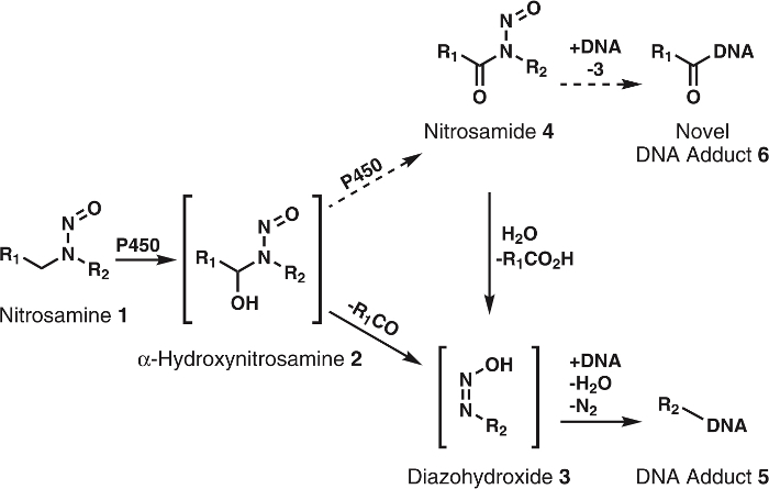
N-nitrosamines are a large class of carcinogens found in the diet, tobacco products, and the general environment; they can also be formed endogenously in the human body1. More than 300 N-nitroso compounds have been tested and >90% were evaluated as carcinogenic in animal models2,3. To exhibit their carcinogenicity, these compounds must first be activated by cytochrome P450s1,2,3. Research shows that cytochrome P450s readily oxidize nitrosamines to α-hydroxynitrosamines (Figure 1), which are highly reactive compounds with half-lives of ~5 s before spontaneously decomposing to alkyldiazohydroxides. The latter can alkylate DNA after the loss of H2O and N2. The resulting DNA adducts, if unrepaired, can cause mutations that, if in critical onco- or tumor suppressor genes, lead to cancer development1. For this reason, much effort has been expended to acquire a full understanding of the metabolic pathways, DNA adducts, and downstream metabolites of cytochrome P450 oxidation of carcinogenic nitrosamines. This knowledge has potential application in individual cancer risk assessment4.

Figure 1: General and proposed metabolism of nitrosamines. Nitrosamines (1) are oxidized by P450s to α-hydroxynitrosamines (2) that spontaneously decompose to alkyldiazohydroxides (3). These compounds can bind to DNA to form DNA adducts. It is hypothesized that 2 are further oxidized by P450s to nitrosamides 4. These can directly bind to DNA to form novel DNA adducts or be hydrolyzed to 3 to form known DNA adducts. R1 and R2 represent any alkyl group. Please click here to view a larger version of this figure.
Although the α-hydroxynitrosamine hypothesis is solidly supported by extensive data, there are a few inconsistencies; a major one is the short half-life of α-hydroxynitrosamines5,6. It is known that these compounds are produced at the endoplasmic reticulum membrane and later alkylate nuclear DNA. Given their lifetime of a few seconds, it is puzzling how these intermediates survive the required travel though the cytosol. One hypothesis is that a portion of the α-hydroxynitrosamines are processively oxidized to nitrosamides7,8, which are quite stable in comparison9. This would presumably occur via retention of α-hydroxynitrosamines in the cytochrome P450 active site. Precedent for this type of oxidation has been seen with nicotine10, alcohols11, and simple alkylnitrosamines12,13. Additionally, nitrosamides are direct-acting carcinogens2,3. Based on their reactivity9, these compounds are believed to produce DNA adducts identical to those resulting from α-hydroxynitrosamines along with new, unexplored DNA adducts (Figure 1). Thus, this hypothesis not only explains the transport through the cytosol, but also the formation of DNA damage products.
In this paper, a general protocol for assessing the in vitro cytochrome P450-mediated conversion of nitrosamines to nitrosamides is described. The previously reported conversion of N′-nitrosonornicotine (NNN) to N′-nitrosonorcotinine (NNC) by cytochrome P450 2A6 is showcased as an example14. Application of this protocol to a wide range of substrate-enzyme systems will help determine the importance of nitrosamides in overall nitrosamine metabolism.
Protocol
1. Materials and general procedures
Synthesize NNN as previously described15. Obtain norcotinine, P450 2A6 Baculosomes, NADPH regeneration system, 0.5x reaction buffer, and all other chemicals or solvents from commercial sources in reagent grade.
Record NMR spectra on a 500 MHz spectrometer. Report chemical shifts as parts per million (ppm). Use residual solvent peaks as internal references for 1H-NMR (7.26 ppm CDCl3) and 13C-NMR (77.2 ppm CDCl3). NOTE: Peak splitting used the following abbreviations: s = singlet, d = doublet, dd = doublet of doublets, dt = doublet of triplets, dq = doublet of quartets, ddd = doublet of doublet of doublets, and m = multiplet.
Perform high resolution mass spectrometry (HRMS) for selected compounds on an LTQ Orbitrap Velos and report data as m/z.
Use polygram pre-coated silica gel TLC plates (40 mm x 80 mm, 0.2 mm thick) with 254 nm fluorescent indicator for thin-layer chromatography (TLC). Visualize TLC plates by UV lamp irradiation.
Perform flash chromatography on a 60 Å, 70-150 mesh silica gel using a 1.5 cm x 15 cm glass column.
2. Preparation of nitrosamide positive control (N′-nitrosonorcotinine, NNC)
Dry, in an oven overnight (16 h, ~140 °C), a 25 mL, round-bottom flask containing a magnetic stir bar. In the morning, cool the flask under a stream of N2 while using an oil bubbler to keep the N2 flow rate under one bubble per second. NOTE: After cooling, no precautions to keep the flask under N2 are required.
In a fume hood, add norcotinine (31.8 mg, 0.196 mmol), acetic anhydride (5 mL), and acetic acid (1 mL) to the cooled flask. Stir this mixture continuously while cooling to 0 °C with a water-ice bath. Caution: Acetic anhydride and acetic acid are corrosive.
Add NaNO2 (33.3 mg, 0.483 mmol) in one portion and continuously stir the mixture for 2.5 h. Monitor reaction progress by TLC (100% EtOAc, Rf = 0.19, see step 1.4). NOTE: Over this time, the mixture will become increasingly yellow with occasional bubbling.
Quench the reaction by pouring the mixture into ice-cold H2O (18 mL). Immediately extract the aqueous solution with 18 mL of CH2Cl2 in a 100 mL separatory funnel. Extract the aqueous layer at least 2 additional times with CH2Cl2 (9 mL each).
Dry the pooled organics over ~100 mg of MgSO4 for 2 min. Filter and concentrate the solution by rotary evaporation to yield a crude, yellow oil. Heat the water bath to 30 °C during evaporation to remove the residual acetic acid. NOTE: The protocol can be paused here if the compound is dissolved in CH2Cl2 and the solution stored in the dark at 2 - 8 °C.
Purify the crude compound by column chromatography (see step 1.5) using silica gel as the stationary phase and 100% EtOAc as the eluent16. Caution: NNC and related nitrosamides should be handled with care as they are assumed to be human carcinogens.
Dissolve the pure compound in CDCl3 and use NMR (see step 1.2) to confirm the structure and determine the molarity of this solution17. Store this solution in the dark at 2 - 8 °C until needed. NOTE: This solution will be used as the positive control in the in vitro assay (step 3.5). NMR spectra and chemical shifts are available in the Supporting Information.
With a pure NNC solution, confirm the accurate parent mass and determine product ion masses by direct infusion on a high-resolution mass spectrometer (HRMS) under parameters described in steps 1.3 and 4.3. NOTE: Product masses will be used for detecting this compound in the in vitro metabolism assay (step 4.3).
3. An example nitrosamine-P450 in vitro incubation
Remove P450 2A6 Baculosomes, Vivid-NADPH-Regeneration System, and 10 mM NADP+ Stock Solution from a -80 °C freezer and let them thaw on ice. Once thawed, dilute the Baculosomes and NADPH-Regeneration System 1:10 and 1:50, respectively, into a single 1 mL tube with 0.5X reaction Buffer. Similarly, add 3 μL of the NADP+ Stock Solution to 97 μL of 0.5X Reaction Buffer. NOTE: NADP+ is light sensitive. Keep this solution protected by wrapping its container in aluminum foil. Enzyme solutions are sensitive to freeze-thaw cycles; limit this to no more than 2 cycles. Prepare aliquots as necessary for future experiments.
For each incubation, add 50 μL of enzyme solution (containing 5 pmol P450) to a 1 mL tube containing 40 μL of 4 μM NNN solution made with Reaction Buffer. Pre-incubate this new mixture for 2 min at 37 °C using a water bath and then add 10 μL of diluted NADP+ solution. Incubate the full system for 1 - 30 min at 37 °C. NOTE: Using 5-min intervals for incubation times (e.g. 5, 10, 15 min, etc.) will provide a sufficient nitrosamide formation time course.
After the desired incubation time, quench by first adding 10 μl of 3.0 N ZnSO4 and then 10 μL of 3.0 N Ba(OH)2. Vortex this solution and a white precipitate will form. Centrifuge the sample at 8000 x g for 4 min to pellet the precipitate. NOTE: The procedure must not pause at this step as the formed nitrosamides have limited stability in aqueous solutions. Long pauses may result in false negatives.
Immediately pipette the supernatant and analyze 2 μL by liquid chromatography positive nanoelectrospray-ionization high-resolution tandem mass spectrometry (LC-NSI+-HRMS/MS, Steps 4.1 - 4.3).
For positive control incubations, evaporate an aliquot containing 100 fmol of synthesized NNC solution into a 1 mL tube under a stream of N2. To this vial, add the full enzyme system from step 3.2 and proceed as described for steps 3.3 - 3.4.
For negative control incubations, perform the experiment as described (steps 3.2 - 3.4) except replace the 50 μL enzyme solution with 0.5X reaction buffer.
4. Example parameters for nitrosamide detection by LC-NSI+-HRMS/MS
For the LC component, apply the following multi-step gradient in step 4.2 to a UPLC system using a commercial, hand-packed18 C18 (5 μm), 100 mm x 75 μm, 15 μm orifice capillary column.
Using 5 mM NH4OAc as solvent A and MeCN as solvent B, load the sample onto the column by running at 5% B at 1 μL/min from 0 - 5 min, and then slowing the flow rate to 0.3 μL/min afterwards. Next, run a gradient from 5 to 20% B over 4 min, followed by a ramp to 55% B over 10 min, and then re-equilibration to 5% B.
Perform the LC-NSI+-HRMS/MS on a LTQ Orbitrap. Monitor for NNC by both full scan and MS2 fragmentation. Perform full scan at a resolution of 60,000 and extract the accurate parent mass (192.07670) at a mass tolerance of 5 ppm. Use MS2 fragmentation to isolate parent ions (2.0 amu) and fragment by collision-induced dissociation (CID) with a collision energy of 25 eV, resolution of 15,000, and scan time of 30 ms. Extract the accurate masses for product ions of NNC (m/z 192 m/z 134.04739 and 162.07874) at a mass tolerance of 5 ppm.
Representative Results
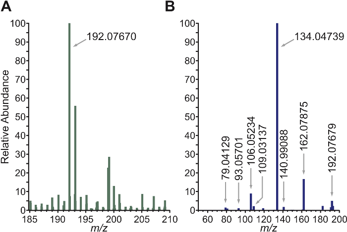
Based on the work of White et al.19, norcotinine was nitrosated to NNC cleanly and in high yield (80 - 92%) to produce a standard for the in vitro experiment. Structural evidence for a successful reaction was obtained from spectroscopic analyses including 1H-NMR, 13C-NMR, COSY, and HSQC (Supporting Information) along with HRMS which confirmed the parent mass [M + H]+ within 5 ppm of the theoretical value (Figure 2). MS2 fragmentation of NNC was used to determine the most abundant product ion masses. The determined accurate parent mass and product ion masses were used in the LC-NSI+-HRMS/MS method of the in vitro assay. If stored in the dark at 2 - 8 °C, the NNC solution is stable for at least 3 months. As this is a very general reaction, it has application to any 2° amide.

Figure 2: HRMS and HRMS2 spectrum of NNC. A) Accurate parent mass of NNC with a mass tolerance of 5 ppm: Calculated: [M+H]+ = 192.07675, Found: 192.07670. B) MS2 fragmentation of m/z 192 with an isolation width of 2.0 amu. The two most abundant product ions are 134.04739 and 162.07875. Reprinted with permission from Chem. Res. Toxicol., 2016, 29, pp 2194-2205. Copyright 2016 American Chemical Society. Please click here to view a larger version of this figure.
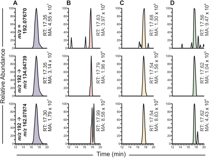
Synthesized NNC was utilized as the standard for the in vitro P450 metabolism assay. Cytochrome P450 2A6 was used because it is known to be an effective catalyst of NNN oxidation20. Likewise, the NNN concentration was set to 2 μM as it approximates the Km for α-hydroxylation21. Under the chromatographic conditions described, synthetic NNC elutes at ~17.5 min with well resolved peaks in both the parent ion and product ion channels. In accordance with the positive control, formation of NNC was noted in the 1, 5, and 10 min incubations.

Figure 3: LC-NSI-HRMS chromatograms resulting from the NNN-cytochrome P450 2A6 incubations. For all sections, the top chromatogram is the accurate parent mass extracted from full scan for NNC. The middle and bottom chromatograms are two accurate product ion masses extracted from MS2 fragmentation for NNC. Sections are as follows: NNC standard (A), NNC-cytochrome P450 2A6 incubations containing all relevant enzymes and cofactors with incubation times of 1 min (B), 5 min (C), and 10 min (D). RT = retention time; MA = Mass Area. Please click here to view a larger version of this figure.
NNC levels were maximal at 5 min and decreased at later time points. Relative quantitation using synthetic NNC as a standard estimated maximum NNC levels to be 10 nM. In the negative control lacking the P450 system, no NNC formation was detected (data not shown), thus indicating enzymatic conversion was required. Together, this protocol shows direct evidence for the conversion of NNN to NNC by P450 2A6, although the extent of formation is minor compared to α-hydroxylation of NNN.
Discussion
Elucidating the metabolism of nitrosamines is a critical component to understanding their carcinogenicity. Since the involved cytochrome P450s and other metabolic enzymes are polymorphic, further application of this knowledge could potentially identify high risk individuals1,4. New data indicates that further oxidation of α-hydroxynitrosamines, the presumed major metabolites of nitrosamines involved in DNA binding, to nitrosamides is possible; however, this has not been robustly tested across a wide range of substrates. We have described a protocol for determining the extent of processive oxidation of nitrosamines to nitrosamides in vitro. Wide application of this method will help determine the overall importance of this route of metabolism.
The method starts by preparing a standard for the nitrosamide of interest. In the example study, it is NNC, a hypothesized metabolite of the tobacco carcinogen NNN, a carcinogen found in tobacco products14,22. As described, nitrosation of norcotinine proceeded smoothly with no discernable side products. The crude product was contaminated by only the starting amide and acetic acid, which were easily removed by column chromatography. This was followed by confirmation of structure by NMR and HRMS. A benefit of this method is that it is suitable for virtually any amide, which allows multiple nitrosamine/nitrosamide systems to be investigated. Additionally, use of NMR for quantitation gives a direct measurement of molarity for the standard solution17. This allows storage of the compound under inert conditions rather than requiring measurement by destructive techniques such as reverse-phase HPLC. Then various dilutions can be made to suit the experiment design.
With a positive control and a well characterized mass fragmentation pattern, the highly sensitive and selective LC-NSI+-HRMS/MS method allows detection of nitrosamides in a relatively complex matrix. As shown, the method estimated an NNC concentration of 1-10 nM or 2 - 20 fmol on-column despite the 1000-fold excess of starting nitrosamine, enzyme, and other cofactors. The sensitivity and selectivity of the method are due to the use of high resolution mass monitoring, which standard LC-MS systems lack. Parent ion selection with a 5-ppm mass tolerance ensures that only compounds with the desired molecular formula are isolated. Likewise, accurate mass detection of the two most abundant product ions along with their retention time gives additional analyte confirmation. Because of this, the mass spectrometry method requires that many parameters are optimized including isolation width, collision energy, and scan time. The LC gradient can also be modified to improve sensitivity as co-elution with other substances will limit filling the ion trap with the target nitrosamide.
In the example study14, NNC formation was minor. Based on mass area, NNC was ~4000-fold less abundant than the hydrolyzed products of α-hydroxyNNN21. Though this indicates NNC is a very minor metabolite of NNN, other systems may differ. Chowdhury et al. found considerable, processive oxidation of dimethylnitrosamine and diethylnitrosamine; however, the nitrosamide intermediate was undetectable under their conditions12. It is possible that reanalyzing their system by the technique described here could reveal nitrosamide formation. Regardless of their formation level, the higher stability of nitrosamides compared to α-hydroxynitrosamines may indicate that these compounds have biological importance as they should more readily transport the cell.
Though this method will be applicable to most nitrosamine-enzyme systems, there are some limitations. The most significant one is that the method is not quantitative. In the example experiment, formation was estimated from the mass spectrometric peak area of a positive control sample. For a more accurate measurement, an isotope-dilution method utilizing an isotopically labelled nitrosamide could be developed. Another approach would be implementing a trapping strategy with compounds such as N-acetyllysine or N-acetylcysteine, but this was unsuccessful to date in our hands14.
Another limitation is the tendency of nitrosamides to hydrolyze easily9; thus samples must be processed individually. This slows the workflow and prevents samples from being sent to collaborators for analysis. Lastly, it should be noted that although cytochrome P450 2A6 and many others are commercially available, some require protein expression and purification to obtain. For example, this assay was applied to cytochrome P450 2A1314, but it is only available after a rigorous isolation process23.
In summary, we have described a protocol for detecting nitrosamides resulting from cytochrome P450-catalyzed oxidation of nitrosamines in vitro. The protocol includes a general synthesis for a nitrosamide standard as a positive control and an in vitro cytochrome P450 metabolism assay using LC-NSI+-HRMS/MS for detection. In the example study, NNC was detected at all incubation times ranging from 1 - 10 min, but only as a minor metabolite of NNN. Application of this protocol to other nitrosamines will assess its generality and the potential role of nitrosamides in nitrosamine carcinogenesis.
Disclosures
The authors have nothing to disclose.
Acknowledgments
This study was supported by grant no. CA-81301 from the National Cancer Institute. We thank Bob Carlson for editorial assistance, Dr. Peter Villalta and Xun Ming for mass spectrometry assistance in the Analytical Biochemistry Shared Resource of the Masonic Cancer Center, and Dr. Adam T. Zarth and Dr. Anna K. Michel for their valuable discussions and input. The Analytical Biochemistry Shared Resource is partially supported by National Cancer Institute Cancer Center Support Grant CA-77598
References
- Rom WN, Markowitz S. Environmental and Occupational Medicine. 4th ed. Wolters Kluwer/Lippincott Williams & Wilkins; 2007. pp. 1226–1239. [Google Scholar]
- Preussmann R, Stewart BW. In: Chemical Carcinogens, ACS Monograph 182. 2nd ed. Searle CE, editor. Vol. 2. American Chemical Society; 1984. pp. 643–828. [Google Scholar]
- Magee PN, Montesano R, Preussmann R. In: Chemical Carcinogens. ACS monograph 173. Searle CE, editor. American Chemical Society; 1976. pp. 491–625. [Google Scholar]
- Zhu AZ, et al. Alaska Native smokers and smokeless tobacco users with slower CYP2A6 activity have lower tobacco consumption, lower tobacco-specific nitrosamine exposure and lower tobacco-specific nitrosamine bioactivation. Carcinogenesis. 2013;34(1):93–101. doi: 10.1093/carcin/bgs306. [DOI] [PMC free article] [PubMed] [Google Scholar]
- Mesić M, Revis C, Fishbein JC. Effects of structure on the reactivity of alpha-hydroxydialkynitrosamines in aqueous solutions. J. Am. Chem. Soc. 1996;118:7412–7413. [Google Scholar]
- Mochizuki M, Anjo T, Okada M. Isolation and characterization of N-alkyl-N- (hydroxymethyl)nitrosamines from N-alkyl-N- (hydroperoxymethyl)nitrosamines by deoxygenation. Tetrahedron Lett. 1980;21:3693–3696. [Google Scholar]
- Guttenplan JB. Effects of cytosol on mutagenesis induced by N-nitrosodimethylamine, N-nitrosomethylurea and à-acetoxy-N-nitrosodimethylamine in different strains of Salmonella:evidence for different ultimate mutagens from N-nitrosodimethylmine. Carcinogenesis. 1993;14:1013–1019. doi: 10.1093/carcin/14.5.1013. [DOI] [PubMed] [Google Scholar]
- Elespuru RK, Saavedra JE, Kovatch RM, Lijinsky W. Examination of a-carbonyl derivatives of nitrosodimethylamine in ethylnitrosomethyamine as putative proximate carcinogens. Carcinogenesis. 1993;14:1189–1193. doi: 10.1093/carcin/14.6.1189. [DOI] [PubMed] [Google Scholar]
- Chow YL. ACS Symposium Series. Vol. 101. American Chemical Society; 1979. pp. 13–37. [Google Scholar]
- von Weymarn LB, Retzlaff C, Murphy SE. CYP2A6- and CYP2A13-catalyzed metabolism of the nicotine delta5'(1')iminium ion. J. Pharmacol. Exp. Ther. 2012;343(2):307–315. doi: 10.1124/jpet.112.195255. [DOI] [PMC free article] [PubMed] [Google Scholar]
- Bell-Parikh LC, Guengerich FP. Kinetics of cytochrome P450 2E1-catalyzed oxidation of ethanol to acetic acid via acetaldehyde. J Biol Chem. 1999;274(34):23833–23840. doi: 10.1074/jbc.274.34.23833. [DOI] [PubMed] [Google Scholar]
- Chowdhury G, Calcutt MW, Nagy LD, Guengerich FP. Oxidation of methyl and ethyl nitrosamines by cytochrome P450 2E1 and 2B1. Biochemistry. 2012;51(50):9995–10007. doi: 10.1021/bi301092c. [DOI] [PMC free article] [PubMed] [Google Scholar]
- Chowdhury G, Calcutt MW, Guengerich FP. Oxidation of N-nitrosoalkylamines by human cytochrome P450 2A6: sequential oxidation to aldehydes and carboxylic acids and analysis of reaction steps. J Biol Chem. 2010;285(11):8031–8044. doi: 10.1074/jbc.M109.088039. [DOI] [PMC free article] [PubMed] [Google Scholar]
- Carlson ES, Upadhyaya P, Hecht SS. Evaluation of nitrosamide formation in the cytochrome P450-mediated metabolism of tobacco-specific nitrosamines. Chem Res Toxicol. 2016;29(12):2194–2205. doi: 10.1021/acs.chemrestox.6b00384. [DOI] [PMC free article] [PubMed] [Google Scholar]
- Amin S, Desai D, Hecht SS, Hoffmann D. Synthesis of tobacco-specific N-nitrosamines and their metabolites and results of related bioassays. Crit. Rev. Toxicol. 1996;26:139–147. doi: 10.3109/10408449609017927. [DOI] [PubMed] [Google Scholar]
- Clark AG, Wong ST. A rapid chromatographic technique for the detection of dye-binding. Anal Biochem. 1978;89(2):317–323. doi: 10.1016/0003-2697(78)90357-3. [DOI] [PubMed] [Google Scholar]
- Pauli GF, et al. Importance of purity evaluation and the potential of quantitative (1)H NMR as a purity assay. J Med Chem. 2014;57(22):9220–9231. doi: 10.1021/jm500734a. [DOI] [PMC free article] [PubMed] [Google Scholar]
- van der Heeft E, et al. A microcapillary column switching HPLC-electrospray ionization MS system for the direct identification of peptides presented by major histocompatibility complex class I molecules. Anal Chem. 1998;70(18):3742–3751. doi: 10.1021/ac9801014. [DOI] [PubMed] [Google Scholar]
- White EH. The Chemistry of the N-Alkyl-N-nitrosoamides. I. Methods of Preparation. J. Am. Chem. Soc. 1955;77:6008–6010. [Google Scholar]
- Patten C, et al. Evidence for cytochrome P450 2A6 and 3A4 as major catalysts for N'-nitrosonornicotine alpha-hydroxylation by human liver microsomes. Carcinogenesis. 1997;18:1623–1630. doi: 10.1093/carcin/18.8.1623. [DOI] [PubMed] [Google Scholar]
- Wong HL, Murphy SE, Hecht SS. Cytochrome P450 2A-catalyzed metabolic activation of structurally similar carcinogenic nitrosamines: N'-nitrosonornicotine enantiomers, N-nitrosopiperidine, and N-nitrosopyrrolidine. Chem. Res. Toxicol. 2004;18:61–69. doi: 10.1021/tx0497696. [DOI] [PubMed] [Google Scholar]
- Hecht SS. Biochemistry, biology, and carcinogenicity of tobacco-specific N-nitrosamines. Chem. Res. Toxicol. 1998;11:559–603. doi: 10.1021/tx980005y. [DOI] [PubMed] [Google Scholar]
- von Weymarn LB, Zhang QY, Ding X, Hollenberg PF. Effects of 8-methoxypsoralen on cytochrome P450 2A13. Carcinogenesis. 2005;26(3):621–629. doi: 10.1093/carcin/bgh348. [DOI] [PubMed] [Google Scholar]


