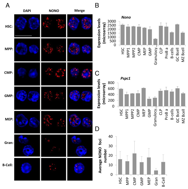
Figure 3. Paraspeckle protein NONO presents aggregated clusters in hematopoietic cells’ nuclei and reduced expression in granulocytes.
(A) Confocal microscopy analysis of NONO, a paraspeckle protein, using an immunofluorescent staining technique, in representative primary cell types through hematopoiesis, HSC, MPP, CMP, GMP, MEP, granulocyte, B cell. NONO (red) is superimposed over nuclei stained with DAPI (blue). Scale bar denotes 10 μ.m. (B and C) Expression levels of mRNA of the paraspeckles proteins NONO and PSPC1, respectively. Data is from ImmGen’s microarray database, showing averages of at least triplicates per cell type except for MPP and MEP that are duplicates; and SD per cell type. (D) Quantification of the foci number per cell type from immunofluorescent stained cells for NONO. Histograms show averages ±SD.

