Endoscopic ultrasonography-guided fine-needle aspiration (EUS-FNA) is a safe and powerful modality for diagnosis of gallbladder lesions.[1,2,3] We report an extremely rare case in which EUS-FNA was useful for the definitive diagnosis of metastatic melanoma of the gallbladder.
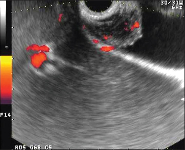
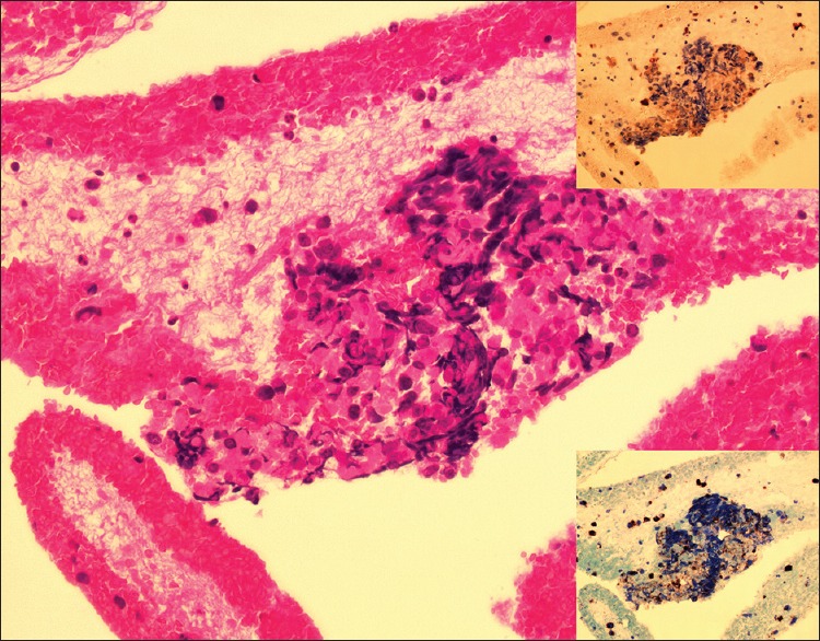
A 53-year-old woman was treated of a vaginal malignant melanoma. Enhanced computed tomography (CT) showed a heterogeneously enhanced mass of 3.5 cm × 3.0 cm in the vagina. No other lesions were detected in the liver, gallbladder, or other organs. Three months of radiation therapy resulted in regression of the tumor revealed by image modalities. Six months after the radiation, follow-up enhanced CT revealed a heterogeneously well-enhanced mass in the neck of the gallbladder. EUS demonstrated a 12 mm × 7 mm well-defined hypoechoic heterogeneous mass with a hyperechoic-layered structure arising from the gallbladder wall. Power Doppler EUS revealed spotty increased flow on the tumor [Figure 1]. Transduodenal EUS-FNA of the gallbladder mass using a 25-gauge FNA needle was performed without any complications. Histopathological examination demonstrated a cluster of small- to medium-sized cells without melanin pigment. The nuclei were round or oval and they were hyperchromatic with inconspicuous nucleoli [Figure 2]. Immunohistochemistry was positive for S-100 protein and HMB-45 [Figure 2], consistent with malignant melanoma. The patient received chemotherapy; however, she deceased 4 months after EUS-FNA.
Figure 1.

Endoscopic ultrasonography image showing a well-defined hypoechoic heterogeneous mass with a hyperechoic-layered structure arising from the gallbladder wall. Power Doppler endoscopic ultrasonography revealed spotty increased flow on the tumor
Figure 2.

Endoscopic ultrasonography-guided fine-needle aspiration specimen from the neck of the gallbladder showing a cluster of small- to medium-sized cells without melanin pigment. Some tumor cells have intranuclear inclusion. The nuclei are round or oval and they were hyperchromatic with inconspicuous nucleoli (inset; the tumor cells are positive for S-100 protein [above] and HMB-45 [below])
Since the first case of malignant melanoma from gallbladder,[4] the interest in gallbladder melanoma has been increasing. Although more cases have been reported since then, both primary and metastatic melanomas of the gallbladder are extremely rare. Because of the rarity of gallbladder melanoma, definitive diagnosis is still difficult even if clinically suspicious. To the best of our knowledge, this is the second report of metastatic malignant melanoma of the gallbladder,[3] which was successfully diagnosed by EUS-FNA.
Declaration of patient consent
The authors certify that they have obtained all appropriate patient consent forms. In the form the patient has given her consent for her images and other clinical information to be reported in the journal. The patients understand that her name and initial will not be published and due efforts will be made to conceal her identity, but anonymity cannot be guaranteed.
Financial support and sponsorship
Nil.
Conflicts of interest
There are no conflicts of interest.
REFERENCES
- 1.Varadarajulu S, Eloubeidi MA. Endoscopic ultrasound-guided fine-needle aspiration in the evaluation of gallbladder masses. Endoscopy. 2005;37:751–4. doi: 10.1055/s-2005-870161. [DOI] [PubMed] [Google Scholar]
- 2.Meara RS, Jhala D, Eloubeidi MA, et al. Endoscopic ultrasound-guided FNA biopsy of bile duct and gallbladder: Analysis of 53 cases. Cytopathology. 2006;17:42–9. doi: 10.1111/j.1365-2303.2006.00319.x. [DOI] [PubMed] [Google Scholar]
- 3.Antonini F, Acito L, Sisti S, et al. Metastatic melanoma of the gallbladder diagnosed by EUS-guided FNA. Gastrointest Endosc. 2016;84:1072–3. doi: 10.1016/j.gie.2016.02.015. [DOI] [PubMed] [Google Scholar]
- 4.Wieting H, Hamdi A. Über die physiologische und pathologische melanin pigmentierung und den epithelialen ursprung der melanoblastome. Ein primäres melanoblastum der Gallenblase. Beitr Pathol Anat. 1907;42:23–84. [Google Scholar]


