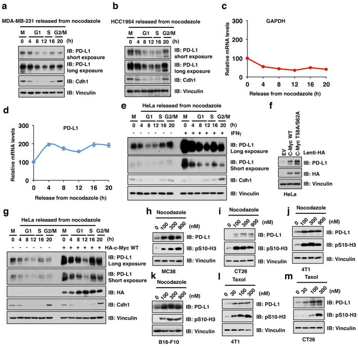
Extended Data Figure 1. PD-L1 fluctuates during cell cycle progression.
a, b, Immunoblot (IB) of whole cell lysates (WCL) derived from MDA-MB-231 or HCC1954 cells synchronized in M phase by nocodazole treatment prior to releasing back into the cell cycle for the indicated times.
c, d, Quantitative real-time PCR (qRT-PCR) analyses of relative mRNA levels of PD-L1 and GAPDH from samples derived from HeLa cells synchronized in M phase by nocodazole treatment prior to releasing back to the cell cycle for the indicated time points.
e, IB of WCL derived from HeLa cells pre-treated with/without IFNγ (10 ng/ml) for 12 hours and then synchronized in M phase by nocodazole treatment prior to releasing back into the cell cycle for the indicated times.
f, IB of WCL derived from HeLa cells stably expressing HA-c-Myc WT, or HA-T58A/S62A-c-Myc as well as empty vector (EV) as a negative control.
g, IB of WCL derived from HeLa cells with/without stably expressing HA-c-Myc WT synchronized in M phase by nocodazole treatment prior to releasing back into the cell cycle for the indicated times.
h–j, IB of WCL derived from MC38, CT26, 4T1, or B16-F10 mouse tumor cells treated with the indicated concentration of nocodazole for 20 hours before harvesting. (k–m) IB of WCL derived from B16-F10, 4T1, or CT26 mouse tumor cells treated with the indicated concentration of taxol for 20 hours before harvesting.

