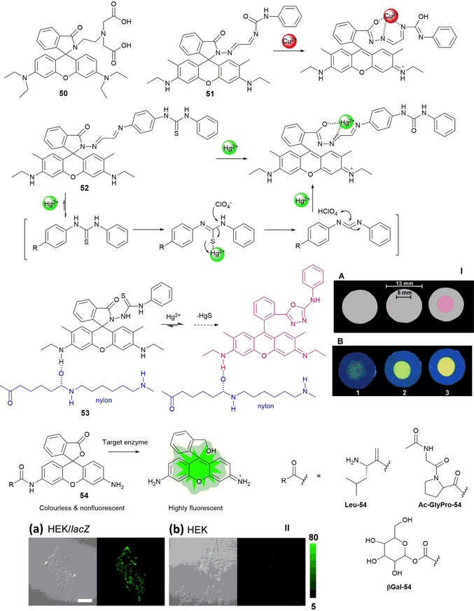
Figure 10.

Chemical structures and reaction mechanisms of probes 50–54. Panel I) Photographs of 53‐doped nylon membranes irradiated with a) visible and b) UV lamps, after treatment with 10 mL of 1) HgII‐free water, 2) 5 ng mL−1 HgII solution, and 3) 12 ng mL−1 HgII solution. The bars indicate the diameters of the nylon membrane and the area exposed to the solution flow. Reproduced from Ref. 105 with permission from The Royal Society of Chemistry. Panel II) Fluorescence confocal imaging of a) HEK 293/lacZ cells and b) HEK 293 cells loaded with βGal‐HMRG. Scale bar represents 50 μm. Reprinted (adapted) with permission from Ref. 106. Copyright 2013 American Chemical Society.
