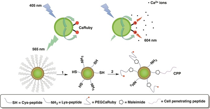
Figure 32.

FRET‐based Ca2+ biosensor 186. Step 1) The QD TOP/TOPO passivating layer was replaced by a peptide coating made by mixing cysteine (SH function) and lysine (NH2 function) terminated peptides [pC, Ac‐CGSESGGSESG(FCC)3F‐amide; and pK, NH2‐KGSESGGSESG(FCC)3F‐amide, respectively]. Both components (hydrophobic QDs and peptides) were first dissolved in their respective solvents, pyridine and DMSO. After mixing, surfactant exchange and peptide binding were initiated by raising the pH. Step 2) Nanoparticles were further functionalized by adding CaRuby (red dots) and cell‐penetrating peptides (CPP, purple wiggles) onto peptide‐coated QDs by using a SH/maleimide linking reaction. Reprinted (adapted) with permission from Ref. 258. Copyright (2014) American Chemical Society.
