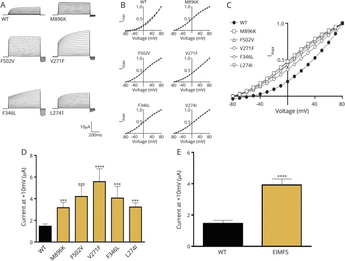
Figure 2. Functional investigation of KCNT1 mutations in a xenopus oocyte model.
(A) Representative current traces obtained from oocytes expressing WT and EIMFS mutants (M896K, F502V, V271F, F346L, and L274I). Oocytes were held at −90 mV and stepped from −80 to 80 mV for 600 milliseconds every 5 seconds. Scale bars apply to all traces. (B) Current-voltage relationships for WT (n = 32), M896K (n = 15), F502V (n = 13), V271F (n = 9), F346L (n = 11), and L274I (n = 12). Currents were averaged and then normalized to the value at a test potential of 80 mV (Imax). (C) Comparison of current-voltage relationships between WT (solid circles, n = 32) and EIMFS mutations (M896K [squares, n = 15], F502V [triangles, n = 13], V271F [hexagons, n = 9], F346L [diamonds, n = 11], and L274I [inverted triangles, n = 12]). Currents were averaged and then normalized to the value at a test potential of 80 mV (Imax). (D) Average peak currents at 10 mV for WT (n = 44), M896K (n = 19), F502V (n = 16), V271F (n = 10), F346L (n = 11), and L274I (n = 12) channels. Peak currents for each mutant channel at 10 mV were compared to the peak currents for the WT channel at 10 mV. ***p < 0.001, ****p < 0.0001. (E) Comparison of pooled WT (n = 44) and EIMFS (n = 68) currents at 10 mV. ****p < 0.0001. EIMFS = epilepsy of infancy with migrating focal seizures; WT = wild-type.

