Abstract
Previous studies have demonstrated strong anti-tumor effects of ent-11α-hydroxy-15-oxo-kaur-16-en-19-oic-acid (5F), an extract from Pteris semipinnata, in liver, lung, stomach and anaplastic thyroid cancer cells. However, whether 5F inhibits the growth of breast cancer cells remains unclear. The present study assessed the effect of 5F on breast cancer cells. The breast cancer cell lines MCF-7, MDA-MB-231 and SK-BR-3 were each treated with 0, 5, 10, 20 and 40 µg/ml 5F. Morphological changes in the breast cancer cells were assessed using fluorescence microscopy. The proliferation and apoptosis of the breast cancer cells were also examined using Cell Counting Kit-8 and flow cytometry. The levels of B-cell lymphoma 2 (Bcl-2), Bcl-2-associated X apoptosis regulator (Bax), Bcl-2 antagonist/killer (Bak) 1 and caspase-3 in the breast cancer cells were assessed. The results of the present study demonstrated that 5F inhibited the proliferation of MCF-7, MDA-MB-231 and SK-BR-3 breast cancer cells in a concentration- and time-dependent manner. Treatment with 5F also induced the apoptosis of breast cancer cells. MDA-MB-231, MCF-7, and SK-BR-3 cells exhibited apoptotic rates of 40.13, 60.44, and 70.49%, respectively, following incubation with 5F for 24 h. Furthermore, 5F significantly decreased the expression of Bcl-2 and increased the expression of Bax, Bak, and caspase-3 in a concentration-dependent manner. The results of the present study revealed that the P. semipinnata extract 5F inhibited the growth of human breast cancer cells in a time- and concentration-dependent manner, and that 5F induced apoptosis of human breast cancer cells.
Keywords: Pteris semipinnata, ent-11α-hydroxy-15-oxo-kaur-16-en-19-oic-acid, breast cancer cells, apoptosis
Introduction
Breast cancer is the most common malignant tumor in females (1). The prognosis of breast cancer depends on the age of the patient, tumor grade and treatment received. Individualizing treatment plans based on histopathological types of tumor is recommended by tumor experts for the treatment of breast cancer (2). Based on the phenotypes of breast cancer, at least four molecular subtypes of breast cancer have been identified, including luminal A, luminal B, triple negative/basal-like and human epidermal growth factor receptor 2 (HER2) (3). The multiple molecular subtypes of breast cancer exhibit distinct sensitivities to chemotherapy, and different prognoses. Patients with luminal breast cancer typically have good prognoses and benefit less from chemotherapy compared with patients with other subtypes of breast cancer (4). Currently, no effective chemotherapy drugs are available for triple negative/basal-like breast cancer. Multi-drug resistance is common in patients with advanced breast cancer. Furthermore, since the majority of chemotherapy drugs possess side effects, selecting effective and safe chemotherapy drugs for patients with advanced breast cancer poses a substantial challenge. The development of chemotherapy drugs that are independent of HER2, estrogen and progesterone receptors is crucial for the future successful treatment of breast cancer, particularly advanced breast cancer.
Multiple Chinese herbs have been hypothesized to facilitate the treatment of tumors. However, the molecular mechanisms underlying the anti-tumor effects associated with certain Chinese herbs remain to be fully understood. Pteris semipinnata is a traditional Chinese herb of the Pteridaceae family. P. semipinnata has been used to treat snake bites, hepatitis and enteritis due to its detoxifying, swelling-reducing and pain-relieving effects (5). Ent-11α-hydroxy-15-oxo-aur-16-en-19-olic acid (5F), a compound isolated from the leaves of P. semipinnata, crystallizes in the monoclinic system. The structure consists of three six-membered rings. Additionally, 5F possesses seven chiral atoms, the configuration of which, as obtained by anomalous dispersion, is R-C4, S-C5, R-C8, S-C9, R-C12 and R-C15 (6). Previous studies have reported that 5F induces apoptosis in and inhibits the proliferation of lung, liver, anaplastic thyroid, stomach, colon and throat cancer cells (7–11).
However, whether 5F exerts anti-tumor effects during the progression of breast cancer remains unclear. The present study examined the functions of 5F in the growth and apoptosis of three breast cancer cell lines, including MDA-MB-231, MCF-7 and SK-BR-3.
Materials and methods
Materials
The human breast cancer cell lines MDA-MB-231, MCF-7, and SK-BR-3 were obtained from the Central Laboratory of the Third Affiliated Hospital of Sun Yat-sen University (Guangzhou, China). Dulbecco's modified Eagle's medium (DMEM), fetal bovine serum, trypsin, and dimethyl sulfoxide were purchased from Gibco; Thermo Fisher Scientific, Inc. (Waltham, MA, USA). Cell Counting Kit-8 (CCK-8) was purchased from Shanghai Dongren Chemical Technology Co., Ltd. (Shanghai, China). The Annexin V-fluorescein isothiocyanate (FITC)/propidium iodide (PI) apoptosis kit and ethanol were purchased from Nanjing KeyGen Biotech Co., Ltd. (Nanjing, China). PI and ribonuclease (RNase) were purchased from Sigma-Aldrich; Merck KGaA (Darmstadt, Germany). The BCA Protein Assay Kit for protein quantification was purchased from Applygen Technologies, Inc. (Beijing, China). The Cell Total Protein Extraction Kit used for protein extraction was purchased from Beyotime Institute of Biotechnology (Haimen, China). B-cell lymphoma 2 (Bcl-2; cat. no. sc-509), Bcl-2-associated X, apoptosis regulator (Bax; cat. no. sc-6236), Bcl-2 antagonist/killer (Bak; cat. no. sc-832), β-actin (cat. no. sc-130300) and caspase-3 (cat. no. sc-271759) antibodies were purchased from Santa Cruz Biotechnology, Inc. (Dallas, TX, USA). The reactive oxygen species detection kit was purchased from Shanghai GenePharma Co., Ltd. (Shanghai, China). The Key Laboratory of Natural Drug Research and Development (Guangdong Medical College, Zhanjiang, China) provided the 5F. 5F was extracted and diluted from P. semipinnata as described in a previous study (10). The 5F was dissolved in propylene glycol, volume fraction 0.15, and diluted with distilled water to prepare 5F stock solutions (1,000 mg/l). The 5F solutions were sterilized using filtration and stored in a −20°C freezer.
Analysis of cell proliferation
MDA-MB-231, MCF-7 and SK-BR-3 cells were digested using trypsin, concentration 0.25%, during the logarithmic growth phase. Using DMEM, breast cancer cell suspensions ranging from 2.0–10.0×10⁴/ml in concentration were prepared. Cancer cells were inoculated onto a 96-well culture plate (5.0×10³ cells/100 µl/well). Following culture for 24 h, DMEM was removed and 100 µl 5F solution (0, 5, 10, 20 or 40 µg/ml) was added to the culture. Eight parallel wells were used for a combination of one drug and one type of breast cancer cell. Following incubation for 24, 48 and 72 h at 37°C, the medium containing 5F at different concentrations was removed, and 100 µl DMEM and 10 µl CCK-8 were added and incubated for 2 h at 37°C. The absorbance value (A) at 450 nm was determined using a microplate reader (BioTek Instruments, Inc., Winooski, VT, USA). The proliferation-inhibiting effect of 5F on cancer cells was evaluated according to the following cancer cell proliferation inhibition rate equation, as obtained from triplicate experiments: Cancer cell proliferation rate (%) = [1 - (A value of the positive group - A value of the blank group)/A value of the negative group - A value of the blank group)] × 100%.
Morphological changes of breast cancer cells
The concentration of MDA-MB-231, MCF-7 and SK-BR-3 cells at the logarithmic growth phase was set to 2.0×10⁵/ml. Cancer cells (2 ml) were added to a 6-well plate with coverslips and incubated for 24 h at 37°C until cancer cells were 100% confluent. Following this, 100 µl 5F (0 or 40 µg/ml) was added and the cells were cultured in DMEM for 48 h at 37°C. Cells were subsequently rinsed in cold PBS twice and fixed in 4% paraformaldehyde at 4°C for 30 min followed by paraffin embedding at room temperature for 30 min in accordance with the standard methodology (12). Slices were cut automatically and manually at thickness of 5 µm by an automatic microtome. Morphological changes of cells were identified under transmission electron microscopy (TEM) (Hitachi Model H-7000; Hitachi, Ltd., Tokyo, Japan), magnification, ×200. Subsequent to treatment with 0 or 40 µg/ml 5F for 24 h, cells were rinsed with PBS twice and stained in 10 mg/ml Hoechst 33342 at 37°C for 15 min. Cells were subsequently rinsed with PBS twice and observed using fluorescence microscopy.
Flow cytometry
The concentration of MDA-MB-231, MCF-7 and SK-BR-3 cells at the logarithmic growth phase was set to 2.0×10⁵/ml. Cells (2 ml) were inoculated onto a 6-well plate. Subsequent to incubation for 24 h, when the cells were 100% confluent, 5F was added to the culture medium at concentrations of 0 and 40 µg/ml. Following incubation in DMEM for 48 h at 37°C, the morphology of the cells was observed using an inverted microscope. Subsequently, cells were rinsed with cold PBS twice and digested with trypsin, concentration 0.25%. Cells were harvested via centrifugation at 1,000 × g for 5 min at 4°C, rinsed with PBS and suspended in Annexin V-FITC (5 µl) for 30 min for flow cytometry analysis on a FACS Calibur flow cytometer (BD Biosciences, Franklin Lakes, NJ, USA). Data were collected and analyzed using FlowJo software version 7.6.1 (Tree Star, Inc., Ashland, OR, USA).
Western blotting
The expression of multiple apoptosis-associated genes, including Bcl-2, Bax and caspase-3, was evaluated using western blotting. The concentration of MDA-MB-231, MCF-7 and SK-BR-3 cells at the logarithmic growth phase were set to 2.0×10⁵/ml. Cells (2 ml) were inoculated onto a 6-well plate. Following 24 h of incubation, during which cells attached to the wells, 5F was added to the culture to reach concentrations of 0, 20, and 40 µg/ml. Following incubation in DMEM for 24 h at 37°C, cells were harvested via centrifugation at 1,000 × g for 5 min at 4°C and rinsed with cold PBS twice at 4°C. The harvested cells were subsequently digested with 200 µl lysate buffer (Gibco; Thermo Fisher Scientific, Inc.) for 30 min at 4°C. Supernatant was collected via centrifugation at 1,000 × g for 5 min at 4°C and proteins were quantified using the Bradford protein quantification method. Proteins (50 µg) were separated using 12.5% SDS-PAGE and transferred onto a nitrocellulose membrane. Following blocking in 5% nonfat milk at room temperature for 1 h, the nitrocellulose membrane was rinsed with Tris buffered saline with Tween [TBST, 20 mM Tris-Hcl (pH=7.6), 137 mM Nacl and 0.01% Tween-20] and incubated with primary antibodies against Bak (1:200), Bcl-2 (1:200), or caspase-3 (1:300) at room temperature overnight. β-actin (1:500) was used as an internal control. Following three rinses with TBST for 15 min, the nitrocellulose membrane was incubated with horseradish peroxidase (HRP)-labeled goat-anti rabbit immunoglobulin G (IgG; 1:5,000; cat. no. sc-2004; Santa Cruz Biotechnology, Inc. TX, USA) at room temperature for 1 h. Subsequently, the nitrocellulose membrane was rinsed with TBST for 15 min three times. Following rinsing with PBS for 1 min, the nitrocellulose membrane was stained with 10 ng/ml enhanced chemiluminescence reagents (Tiangen, Beijing, China) at 4°C for 1 h and exposed three times. The western blot analysis was repeated three times. Then, the blots were analyzed by Image J software version 1.41 (National Institutes of Health, Bethesda, MD, USA).
Statistical analyses
Experimental data were presented as the mean ± standard deviation. Statistical analyses were performed using SPSS 16.0 statistical software (SPSS, Inc., Chicago, IL, USA). Comparison between two groups was performed by means of independent samples t test. Comparison among multiple groups was performed by one-way analysis of variance followed by Tukey's and Tamhane's T2 post-hoc tests. P<0.05 was considered to indicate a statistically significant difference.
Results
5F inhibited the proliferation of human breast cancer cells
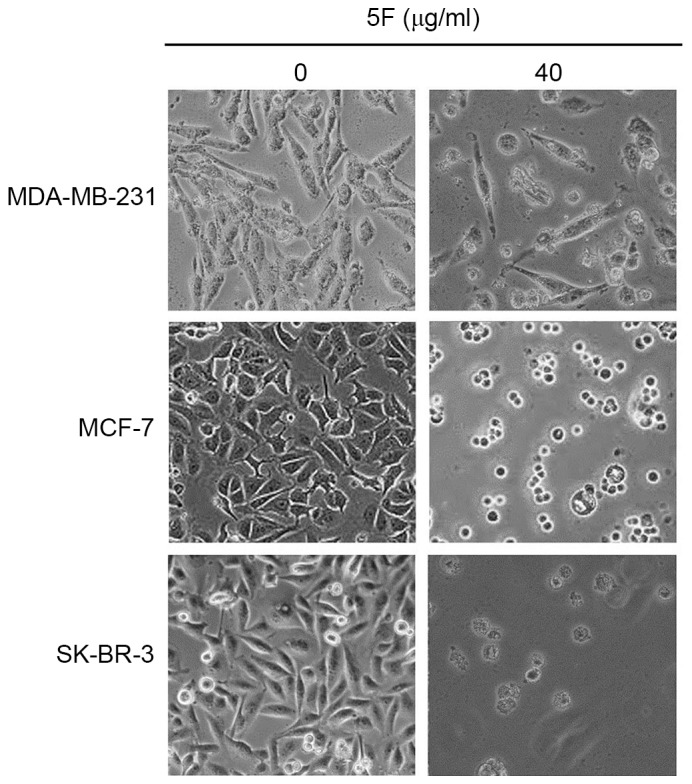
The CCK-8 staining experiment demonstrated that 5F inhibited the proliferation of the three types of human breast cancer cell assessed in the present study, MDA-MB-231, MCF-7 and SK-BR-3 cells, in a time- and concentration-dependent manner (Fig. 1). SK-BR-3 cells were the most sensitive to 5F of the three human breast cancer cells; <6% of SK-BR-3 cells survived following culturing in DMEM containing 5F at a concentration of 40 µg/ml for 72 h. Following culture in DMEM containing 5F at a concentration of 40 µg/ml for 72 h, ~50.00 and 44.13% of MCF-7 and MDA-MB-231 cells survived. Morphological changes, including membrane rupture and atrophy, disconnection from wells, and granular bodies in the cytoplasm were observed in all cancer cells following incubation with DMEM containing 5F at a concentration of 40 µg/ml for 24 h (Fig. 2).
Figure 1.
The survival rate of the human breast cancer cells MDA-MB-231, MCF-7 and SK-BR-3 following incubation in Dulbecco's modified Eagle's medium containing ent-11α-hydroxy-15-oxo-kaur-16-en-19-oic-acid of different concentrations (0, 5, 10, 20, and 40 µg/ml) for 24, 48, and 72 h. (A) MDA-MB-231 cells, (B) MCF-7 cells, (C) SK-BR-3 cells. *P<0.05 vs. control.
Figure 2.

Morphological changes of the human breast cancer cells MDA-MB-231, MCF-7 and SK-BR-3 following incubation in Dulbecco's modified Eagle's medium containing 5F (40 µg/ml) for 48 h Magnification, ×200. 5F, ent-11α-hydroxy-15-oxo-kaur-16-en-19-oic-acid.
5F induced the apoptosis of human breast cancer cells
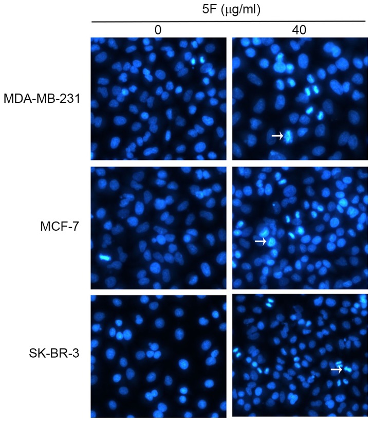
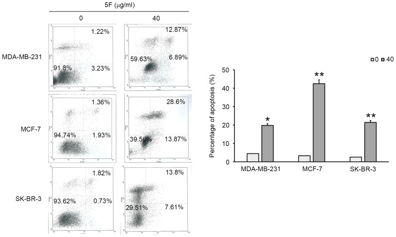
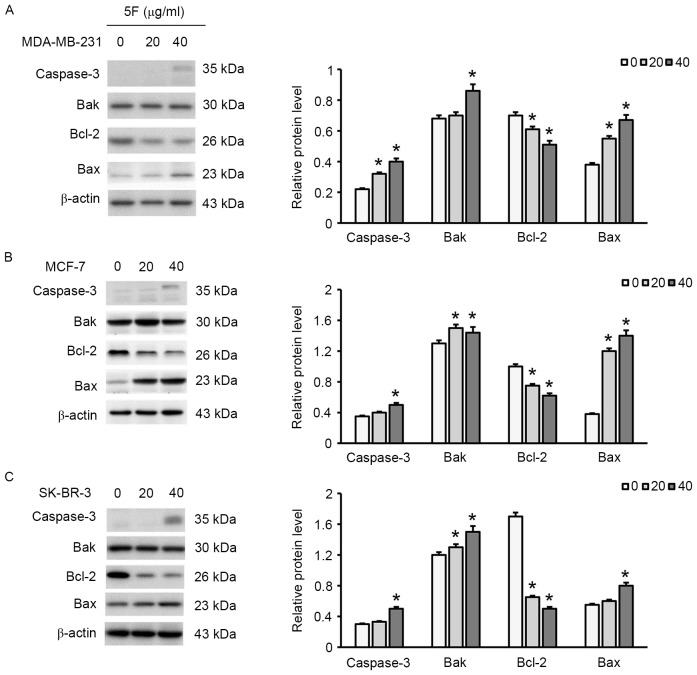
To assess whether 5F had any effect on the apoptosis of MDA-MB-231, MCF-7 and SK-BR-3 cells the morphological changes and the expression of multiple apoptosis-associated proteins in these human breast cancer cells were evaluated. The cells were incubated in 5F for 48 h and the morphological changes of the cells were subsequently observed using TEM. Since few MDA-MB-231 (50%), MCF-7 (44.13%) and SK-BR-3 (6%) cells survived the 48 h incubation (Fig. 2), fluorescence microscopy was used to evaluate the morphological changes of cancer cells incubated in DMEM containing 5F (40 µg/ml) for 24 h. Cells were stained with Annexin V/FITC. Condensation and degradation of cancer cell nuclei were induced by 5F under fluorescence microscopy (Fig. 3). The results of flow cytometry demonstrated that 6.89, 13.87 and 7.61% of MDA-MB-231, MCF-7, and SK-BR-3 cells, respectively, exhibited in early apoptosis. The results of flow cytometry also demonstrated that 12.87, 28.6 and 13.8% of MDA-MB-231, MCF-7, and SK-BR-3 cells, respectively, exhibited late apoptosis (Fig. 4). Furthermore, a decrease in the expression of Bcl-2 and an increase in the expression of Bax, Bak and caspase-3 were observed (Fig. 5), suggesting that 5F induced breast cancer cell apoptosis by regulating the expression of these apoptosis-associated proteins. In addition, 40 µg/ml 5F exhibited greater effects on the expression of Bcl-2, Bax, Bak and caspase-3 than 20 µg/ml 5F, suggesting that 5F affected the expression of these apoptosis-associated proteins in a concentration-dependent manner.
Figure 3.

The human breast cancer cells MDA-MB-231, MCF-7 and SK-BR-3 following incubation in Dulbecco's modified Eagle's medium containing 5F (40 µg/ml) for 24 h. Cells were stained with Hoechst 33342 and examined under fluorescence microscopy. Magnification, ×200. Arrows indicate 5F-induced apoptosis of the human breast cancer cells MDA-MB-231, MCF-7 and SK-BR-3. 5F, ent-11α-hydroxy-15-oxo-kaur-16-en-19-oic-acid.
Figure 4.
Flow cytometry analysis. The apoptotic rates of the human breast cancer cells MDA-MB-231, MCF-7 and SK-BR-3 following incubation with 5F for 48 h, assessed by flow cytometry. *P<0.05 and **P<0.01 vs. control. 5F, ent-11α-hydroxy-15-oxo-kaur-16-en-19-oic-acid.
Figure 5.
The expression of Bcl-2, Bax, Bak and caspase-3 in the human breast cancer cells MDA-MB-231, MCF-7 and SK-BR-3 following treatment with 5F for 24 h. (A) MDA-MB-231 cells, (B) MCF-7 cells and (C) SK-BR-3 cells. *P<0.05 vs. control. Bcl-2, B-cell lymphoma 2; Bax, Bcl-2-associated X, apoptosis regulator; Bak, Bcl-2 antagonist/killer; 5F, ent-11α-hydroxy-15-oxo-kaur-16-en-19-oic-acid.
Discussion
5F exerts anti-tumor effects against liver (8), lung (7), stomach (13), and anaplastic thyroid (5) cancers, but whether 5F exhibits anti-tumor effects against breast cancer remains unclear. The present study assessed the potential function of 5F in the growth of breast cancer in three different types of breast cancer cell line: MCF-7, MDA-MB-231 and SK-BR-3. Each line possesses different hormone receptors: Thyroid hormone receptors, steroid hormone receptors and estrogen receptor in MCF-7, MDA-MB-231 and SK-BR-3 cell lines, respectively (14,15), and different HER2 and p53 expression levels. The results of the present study demonstrated that 5F inhibited the proliferation of each of these three types of breast cancer cell in a concentration- and time-dependent manner, suggesting that the anti-tumor effects of 5F are independent of the hormone receptors, HER2 expression, and p53 expression in breast cancer cells.
The results of the present study also suggested that 5F promoted the apoptosis of the three breast cancer cell lines in a concentration- and time-dependent manner. However, the rate of apoptosis induced by 5F differed among the three breast cancer cell lines, suggesting that the three lines differed in 5F sensitivity. This result may be explained by the different characteristics of the three breast cancer cell lines. However, certain key molecules may serve crucial functions in the proliferation and apoptosis of breast cancer cells, though the underlying molecular mechanisms remain to be fully understood. Apoptosis is an important step in cell development. The dysregulation of apoptosis leads to numerous diseases, including the development of cancer (16). Intrinsic and extrinsic pathways are involved in apoptosis. Extrinsic apoptosis signals are transduced into cells from cell surface death receptors, a pathway known as the death receptor pathway (17). Cell surface death receptors include the tumor necrosis factor and nerve growth factor superfamilies. Extracellular death signals are transduced into cells through the binding of death receptors and ligands. Intrinsic apoptosis pathways include the mitochondrial pathway and the endoplasmic reticulum pathway (18). In the mitochondrial pathway, the Bax subfamily protein inserts into the mitochondrial membrane from the outer mitochondrial membrane or cytoplasm, causing changes in the permeability of the mitochondrial membrane. Subsequently, the mitochondrial membrane potential decreases and cytochrome C, multiple other proteins and apoptosis-inducing factors are released from the mitochondrion into the cytoplasm (19). The release of cytochrome C is key in the mitochondrial apoptosis pathway; activated cytochrome C activates caspase-9 and downstream caspase-3, and induces apoptosis (20).
The activation, expression and regulation of numerous genes is crucial in apoptosis (21). Of these genes, the Bcl-2 family genes serve key functions in the regulation of apoptosis. Bcl-2 family proteins are divided into two groups: Anti-apoptotic and pro-apoptotic. Anti-apoptotic members include Bcl-2, Bcl-W and Bcl-2 family apoptosis regulator; pro-apoptotic members include Bax, Bak, Bcl-extra small, Bcl-2 associated agonist of cell death, harakiri, and Bcl-2 homology region 3 interacting domain death agonist (22). Apoptosis occurs when the activities of anti-apoptotic proteins are lower than those of pro-apoptotic proteins (23). The western blotting results of the present study demonstrated that 5F increased the expression of Bax and decreased the expression of Bcl-2 in breast cancer cells. Furthermore, increasing the concentration of 5F increased the expression of caspase-3. A previous study reported that 5F was involved in the regulation of apoptosis and of the cell cycle by affecting the expression of extracellular regulated kinase 1/2, c-jun N-terminal kinase, and p38 (24). Therefore, the present study speculated that 5F arrests the cell cycle and promotes apoptosis by downregulating Bcl-2 and Bcl-extra large, upregulates Bax and Bak and inhibits the expression of proliferation-associated proteins (10). The results of the present study suggested that 5F induced the apoptosis of breast cancer cells by affecting the balance of activity of apoptosis-promoting and -inhibiting proteins of the Bcl-2 family.
To conclude, 5F inhibited the proliferation of breast cancer cells in a time- and concentration-dependent manner. Furthermore, 5F induced the apoptosis of breast cancer cells.
Acknowledgements
The present study was supported by the Guangdong Medical Scientific Research Fund (grant no. A2016004) and the Medical Scientific Research Foundation of Guangdong Province [grant no. (2016) 568].
References
- 1.Benson JR, Jatoi I. The global breast cancer burden. Future Oncol. 2012;8:697–702. doi: 10.2217/fon.12.61. [DOI] [PubMed] [Google Scholar]
- 2.Nandy A, Gangopadhyay S, Mukhopadhyay A. Individualizing breast cancer treatment-The dawn of personalized medicine. Exp Cell Res. 2014;320:1–11. doi: 10.1016/j.yexcr.2013.09.002. [DOI] [PubMed] [Google Scholar]
- 3.Slamon DJ, Leyland-Jones B, Shak S, Fuchs H, Paton V, Bajamonde A, Fleming T, Eiermann W, Wolter J, Pegram M, et al. Use of chemotherapy plus a monoclonal antibody against HER2 for metastatic breast cancer that overexpresses HER2. N Engl J Med. 2001;344:783–792. doi: 10.1056/NEJM200103153441101. [DOI] [PubMed] [Google Scholar]
- 4.Lim E, Winer EP. Adjuvant chemotherapy in luminal breast cancers. Breast. 2011;20(Suppl 3):S128–131. doi: 10.1016/S0960-9776(11)70309-5. [DOI] [PubMed] [Google Scholar]
- 5.Liu ZM, Chen GG, Vlantis AC, Liang NC, Deng YF, van Hasselt CA. Cell death induced by ent-11alpha-hydroxy-15-oxo-kaur-16-en-19-oic-acid in anaplastic thyroid carcinoma cells is via a mitochondrial-mediated pathway. Apoptosis. 2005;10:1345–1356. doi: 10.1007/s10495-005-1730-5. [DOI] [PubMed] [Google Scholar]
- 6.Brunocolmenarez J, Peña A, Alarcón L, Usubillaga A, Delgadoméndez P. Structure of ent-15a-hydroxy-kaur-16-en-19-oic acid. Avances En Química. 2011;6:16–20. [Google Scholar]
- 7.Li L, Chen GG, Lu YN, Liu Y, Wu KF, Gong XL, Gou ZP, Li MY, Liang NC. Ent-11α-Hydroxy-15-oxo-kaur-16-en-19-oic-acid inhibits growth of human lung cancer A549 cells by arresting cell cycle and triggering apoptosis. Chin J Cancer Res. 2012;24:109–115. doi: 10.1007/s11670-012-0109-8. [DOI] [PMC free article] [PubMed] [Google Scholar]
- 8.Chen GG, Leung J, Liang NC, Li L, Wu K, Chan UP, Leung BC, Li M, Du J, Deng YF, et al. Ent-11α-hydroxy-15-oxo-kaur-16-en-19-oic-acid inhibits hepatocellular carcinoma in vitro and in vivo via stabilizing IkBα. Invest New Drugs. 2012;30:2210–2218. doi: 10.1007/s10637-011-9791-5. [DOI] [PubMed] [Google Scholar]
- 9.Liu Z, Ng EK, Liang NC, Deng YF, Leung BC, Chen GG. Cell death induced by Pteris semipinnata L. Is associated with p53 and oxidant stress in gastric cancer cells. FEBS Lett. 2005;579:1477–1487. doi: 10.1016/j.febslet.2005.01.050. [DOI] [PubMed] [Google Scholar]
- 10.Chen GG, Liang NC, Lee JF, Chan UP, Wang SH, Leung BC, Leung KL. Over-expression of Bcl-2 against Pteris semipinnata L-induced apoptosis of human colon cancer cells via a NF-kappa B-related pathway. Apoptosis. 2004;9:619–627. doi: 10.1023/B:APPT.0000038041.57782.84. [DOI] [PubMed] [Google Scholar]
- 11.Vlantis AC, Lo CS, Chen GG, Liang Ci N, Lui VW, Wu K, Deng YF, Gong X, Lu Y, Tong MC, van Hasselt CA. Induction of laryngeal cancer cell death by Ent-11-hydroxy-15-oxo-kaur-16-en-19-oic acid. Head Neck. 2010;32:1506–1518. doi: 10.1002/hed.21357. [DOI] [PubMed] [Google Scholar]
- 12.Mgbonyebi OP, Russo J, Russo IH. Roscovitine induces cell death and morphological changes indicative of apoptosis in MDA-MB-231 breast cancer cells. Cancer Res. 1999;59:1903–1910. [PubMed] [Google Scholar]
- 13.Chen JF, Chen YX, Li P, Fu M, Lv YN, Li L. Effect of Ent-11α-hydroxy-15-oxo-kaur-16-en-19-oic-acid on human gastric cancer cells and its mechanism. Nan Fang Yi Ke Da Xue Xue Bao. 2011;31:1345–1348. (In Chinese) [PubMed] [Google Scholar]
- 14.Crépin M, Salle V, Raux H, Berger R, Hamelin R, Brouty-Boyé D, Israel L. Steroid hormone receptors and tumorigenicity of sublines from breast tumor metastatic MDA-MB 231 cell line. Anticancer Res. 1990;10:1661–1666. [PubMed] [Google Scholar]
- 15.Duncan RE, Archer MC. Farnesol induces thyroid hormone receptor (THR) beta1 but inhibits THR-mediated signaling in MCF-7 human breast cancer cells. Biochem Biophys Res Commun. 2006;343:239–243. doi: 10.1016/j.bbrc.2006.02.145. [DOI] [PubMed] [Google Scholar]
- 16.Reed JC. Dysregulation of apoptosis in cancer. J Clin Oncol. 1999;17:2941–2953. doi: 10.1200/JCO.1999.17.9.2941. [DOI] [PubMed] [Google Scholar]
- 17.Gupta S. Molecular steps of death receptor and mitochondrial pathways of apoptosis. Life Sci. 2001;69:2957–2964. doi: 10.1016/S0024-3205(01)01404-7. [DOI] [PubMed] [Google Scholar]
- 18.Pinton P, Ferrari D, Rapizzi E, Di Virgilio F, Pozzan T, Rizzuto R. The Ca2+ concentration of the endoplasmic reticulum is a key determinant of ceramide-induced apoptosis: Significance for the molecular mechanism of Bcl-2 action. EMBO J. 2001;20:2690–2701. doi: 10.1093/emboj/20.11.2690. [DOI] [PMC free article] [PubMed] [Google Scholar]
- 19.Gross A, McDonnell JM, Korsmeyer SJ. BCL-2 family members and the mitochondria in apoptosis. Genes Dev. 1999;13:1899–1911. doi: 10.1101/gad.13.15.1899. [DOI] [PubMed] [Google Scholar]
- 20.Eldering E, Mackus WJ, Derks IA, Evers LM, Beuling E, Teeling P, Lens SM, van Oers MH, van Lier RA. Apoptosis via the B cell antigen receptor requires Bax translocation and involves mitochondrial depolarization, cytochrome C release, and caspase-9 activation. Eur J Immunol. 2004;34:1950–1960. doi: 10.1002/eji.200324817. [DOI] [PubMed] [Google Scholar]
- 21.Chen F, Jiang X, Chen X, Liu G, Ding J. Effects of downregulation of inhibin alpha gene expression on apoptosis and proliferation of goose granulosa cells. J Genet Genomics. 2007;34:1106–1113. doi: 10.1016/S1673-8527(07)60126-X. [DOI] [PubMed] [Google Scholar]
- 22.Pilchova I, Klacanova K, Chomova M, Tatarkova Z, Dobrota D, Racay P. Possible contribution of proteins of Bcl-2 family in neuronal death following transient global brain ischemia. Cell Mol Neurobiol. 2015;35:23–31. doi: 10.1007/s10571-014-0104-3. [DOI] [PMC free article] [PubMed] [Google Scholar]
- 23.Guo B, Zhai D, Cabezas E, Welsh K, Nouraini S, Satterthwait AC, Reed JC. Humanin peptide suppresses apoptosis by interfering with Bax activation. Nature. 2003;423:456–461. doi: 10.1038/nature01627. [DOI] [PubMed] [Google Scholar]
- 24.Li MY, Liang NC, Chen GG. Ent-11α-hydroxy-15-oxo-kaur-16-en-19-oic-acid induces apoptosis of human malignant cancer cells. Curr Drug Targets. 2012;13:1730–1737. doi: 10.2174/138945012804545623. [DOI] [PubMed] [Google Scholar]


