Abstract
Lingual hematoma is a severe situation, which is rare and endangers the airway. It can develop due to trauma, vascular abnormalities, and coagulopathy. Due to its sudden development, it can be clinically confused with angioedema. In patients who applied to the doctor with complaints of a swollen tongue, lingual hematoma can be confused with angioedema, in particular, at the beginning if the symptoms occurred after drug use. It should especially be considered that dystonia in the jaw can present as drug-induced hyperkinetic movement disorder. Early recognition of this rare clinical condition and taking precautions for providing airway patency are essential. In this case report, we will discuss mimicking angioedema and caused by a bite due to dystonia and separation of the tongue from the base of the mouth developing concurrently with lingual hematoma.
Key words: Airway obstruction, antipsychotic, dystonia, hyperkinesis
Introduction
Lingual hematoma is a severe situation, which is rare and endangers the airway. It can develop due to trauma, vascular abnormalities, and coagulopathy.[1] Due to its sudden development, it can be clinically confused with angioedema. Early recognition of this rare clinical condition and taking precautions for providing airway patency are essential. In this case report, we will discuss lingual hematoma mimicking angioedema and caused by a bite due to dystonia and separation of the tongue from the base of the mouth developing concurrently with lingual hematoma.
Case Report
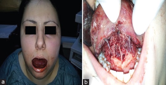
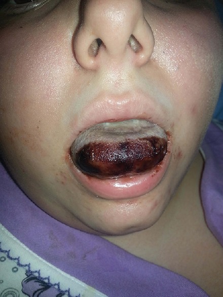
A 20-year-old female was admitted to the emergency department (ED) with swelling of the tongue [Figure 1a]. According to the medical history, she was taking quetiapine 300 mg due to bipolar disorder. She had no history of smoking, alcohol consumption, chronic drug use, or disease. According to the information received from a relative, a medication was applied to her in another medical center. They said that they did not know the name of the drug. Angioedema was considered due to rising of swelling symptoms soon after the drug application. Pheniramine maleate 45.5 mg intravenous (IV) and methylprednisolone 100 mg IV were immediately administered to the patient in our ED. Later, the patient's relative informed that intramuscular haloperidol had been administered due to acute agitation at another medical center 11 h ago and biperiden was administered due to developing a lisp 1 h after haloperidol administration in the same medical center. It was also informed that shrinkage and contraction developed in her face 1–2 h after the application of biperiden and meanwhile she bit her tongue involuntarily. Her general state was good, and she was conscious with full orientation and cooperation. Vital signs were as follows: blood pressure 110/80 mmHg, body temperature 36.2°C, and O2 saturation 98%. In the physical examination, the patient's tongue was edematous and protruding from the mouth. Respiratory sounds were normal. The uvula could not be evaluated, as the tongue filled the inside of the mouth. Neurological examination was normal. It was observed that hematoma occurred due to biting; there were lacerations in the sublingual region and tongue dorsum, and the tongue was separated from the base of the mouth [Figure 1b]. It was considered as an acute dystonic reaction, which is a type of hyperkinetic movement disorder due to drug use. Accordingly, it was considered that the lingual hematoma occurred due to biting, and the tongue was separated from the base of the mouth. A fiberoptic laryngoscopic examination was conducted by the doctor on duty at the ENT clinic, and it was determined that the tongue base was normal and the airway was open. She was followed up with methylprednisolone 1 g and antibiotics. Edema of the tongue diminished, and it was decided to continue monitoring without obstructing airway. A demarcation line was observed on the front part of tongue on the 3rd day [Figure 2]. On the 5th day of follow-up, granulation tissue was observed in the sublingual region. After 10 days of follow-up, the patient was discharged as no complications were present. The case was followed-up for 6 months, and no problems were observed.
Figure 1.

(a) The view of swelling of the tongue. (b) Separation of the tongue from the base of the mouth
Figure 2.

Demarcation line on the 3rd day
Discussion
Lingual or sublingual hematomas can develop secondary to trauma anticoagulant use, or tonic–clonic seizures.[2] Saah et al.[1] reported a case in which a hematoma occurred due to biting the tongue caused by eclamptic seizures. In the current case, lingual hematoma occurred due to a tongue bite, which was a result of hyperkinetic movement disorders related to the use of antipsychotics. Haloperidol was a probable causing agent according to the WHO-UMC causality assessment criteria and Naranjo probability scale (score 6). The severity of reaction was moderate according to Modified Hartwig and Siegel scale. We observed the separation of the tongue from the base of the mouth with a hematoma. We could not found any case in the literature with separation of the tongue from the base of the mouth and accompanying hematoma due to trauma. As an adverse effect of antipsychotic drugs, hyperkinetic movement disorders may be observed. Furthermore, many drug types such as dopamine-receptor blockers, antidepressants can cause drug-induced hyperkinetic movement disorder. The main symptoms of hyperkinetic movement disorder are tremor, akathisia, dyskinesia, myoclonus, and dystonia.[3] In atypical antipsychotic use, the prevalence of acute dystonic reactions is approximately 2%–3%. Acute dystonic reactions are generally seen before the age of 20 with an incidence of 60% and more.[4] Acute dystonic reactions may be seen clinically, such as torticollis, limb cramps, opisthotonus, oculogyric crisis, and jaw trismus.[4] In our case, dystonia in the jaw was seen as a movement disorder. In the jaw dystonia, lingual and sublingual hematomas can occur due to tongue biting. In the differential diagnosis of lingual hematoma, angioedema is present. Especially, if comprehensive medical history cannot be obtained, it can be confused with tongue swelling and angioedema after the drug use. The most important complication of lingual hematoma and separation of the tongue from the base of the mouth is acute upper airway obstruction. In these cases, different methods can be applied such as close monitoring of the airway or ensuring surgical airway and waiting for the decline of the hematoma. Good results are achieved in treatment with corticosteroids and antibiotics.[5] Hematoma evacuation is not possible because of bleeding is into the intrinsic muscles instead of potential spaces. To achieve a secured airway, methods such as orotracheal or nasotracheal intubation, tracheotomy can be used.[5] By evaluating the structures such as the posterior of the tongue, uvula, and epiglottis with flexible fiber-optic laryngoscopy, unnecessary intubation can be avoided. Furthermore, in our case, the base of the tongue and airway was evaluated by flexible fiber-optic laryngoscopy. It was considered that there was no need for emergency intubation or tracheotomy and waiting for the decline of the hematoma under close monitoring was decided.
Declaration of patient consent
The authors certify that they have obtained all appropriate patient consent forms. In the form the patient(s) has/have given his/her/their consent for his/her/their images and other clinical information to be reported in the journal. The patients understand that their names and initials will not be published and due efforts will be made to conceal their identity, but anonymity cannot be guaranteed.
Financial support and sponsorship
Nil.
Conflicts of interest
There are no conflicts of interest.
References
- 1.Saah D, Braverman I, Elidan J, Nageris B. Traumatic macroglossia. Ann Otol Rhinol Laryngol. 1993;102:729–30. doi: 10.1177/000348949310200915. [DOI] [PubMed] [Google Scholar]
- 2.Dhaliwal HS, Dhaliwal SS, Heckel RD, Quereshy FA, Baur DA. Diagnosis and management of upper airway obstruction due to lingual hematoma: Report of a case. J Oral Maxillofac Surg. 2011;69:558–63. doi: 10.1016/j.joms.2009.11.007. [DOI] [PubMed] [Google Scholar]
- 3.Burkhard PR. Acute and subacute drug-induced movement disorders. Parkinsonism Relat Disord. 2014;20(Suppl 1):S108–12. doi: 10.1016/S1353-8020(13)70027-0. [DOI] [PubMed] [Google Scholar]
- 4.Loochtan MJ, Shafiq Q, Baugh RF. Flexible laryngoscopy in post-seizure lingual hematoma. Clin Neurol Neurosurg. 2013;115:1530–1. doi: 10.1016/j.clineuro.2012.12.028. [DOI] [PubMed] [Google Scholar]
- 5.Song Z, Laggan B, Parulis A. Lingual hematoma treatment rationales: A case report. J Oral Maxillofac Surg. 2008;66:535–9. doi: 10.1016/j.joms.2006.09.023. [DOI] [PubMed] [Google Scholar]


