Abstract
db/db mice is one of most widely used animal models in studying the cellular and molecular mechanisms of metabolic disorders, such as diabetes, hyperlipidemia, and obesity. The mice carry spontaneous point mutations in the gene encoding the leptin receptor, leading to leptin receptor inactivation. Since homozygous db/db mice are sterile, the maintenance of db/db mice requires breeding between heterozygous pairs, which makes genotyping essential for the identification of offspring. The aim of this study was to develop a quick and highly repeatable method for genotyping db/db mice, which comprised only three simple steps: genomic DNA is extracted from either tail tips or ear notches via alkaline lysis (∼20 min); samples are then subjected to tetra-primer amplification refractory mutation system-polymerase chain reaction (ARMS-PCR) using specially designed and validated primer sets (∼1.5 h); finally, genotypes are be determined by resolving PCR products on regular DNA electrophoresis (∼10 min). The entire db/db mice genotyping procedure can be performed using regular Taq polymerase and PCR amplification within 2 h. Other advantages of this method include high sensitivity and reproducibility. Minimal amounts of tissue from mice are required, and genomic DNA samples can be stably stored at room temperature for up to one month. In conclusion, the method is simple, cost effective, sensitive and reliable, which will greatly facilitate studies using db/db mice.
Keywords: db/db, genotyping, ARMS-PCR, leptin receptor, gene mutation
Introduction
db/db mice serve as a valuable model for studying obesity, diabetes, and dyslipidemia, wherein leptin receptor activity is deficient due to the mice being homozygous for a point mutation (G/T) in the gene for the leptin receptor1,2. The leptin receptor, also known as LEP-R or OB-R, is a protein that is encoded by the lepr gene in humans. LEP-R functions as a single-transmembrane-domain receptor of the cytokine receptor family for the fat cell-specific hormone leptin3. The severity of disease in this genetic background leads to an uncontrolled rise in blood sugar, severe depletion of insulin-producing beta-cells in pancreatic islets, and death by 10 months of age4,5. Although homozygous db/db mice (leprdb) become identifiably obese at approximately three to four weeks of age, the maintenance of db/db mice requires breeding between heterozygous pairs, which makes genotyping essential for the identification of offspring.
A few methods have been developed for identifying single-point mutations, including PCR-RFLP, real-time PCR, high-resolution melting analysis, restriction enzymatic digestion, and the minisequencing-ligation method6,7,8,9,10; however, these methods are time consuming (multiple complicated steps are involved), technique and resource demanding (requires precise analysis of the results and real-time PCR) and unstable (the restriction enzyme is expensive and its activity is not stable). Therefore, genotyping has become one of the primary rate-limiting steps in db/db-related research.
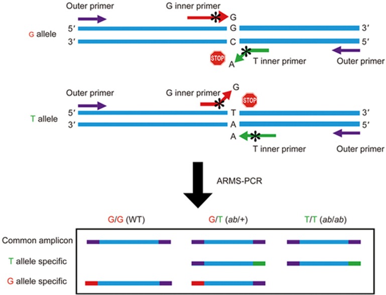
Here, we adapted a novel, simple and time-saving genotyping method from other studies for the identification of db/db mice11,12,13, called tetra-primer amplification refractory mutation system PCR (ARMS-PCR)11,12,13,14. The ARMS assay is based on the principle that PCR amplification is inefficient or completely refractory if there is a mismatch between the 3' terminal nucleotide of a primer and its template sequence. The reason is that Taq polymerase lacks 3′-exonucleolytic proofreading activity and therefore cannot correct the 3' terminal mismatch (please see “stop sign” in Figure 1). This technique employs two primer pairs to amplify two alleles (one wild-type allele (G allele in Figure 1) and one mutant allele (T allele in Figure 1) in a single PCR reaction. One pair of primers are a forward outer primer and a reverse outer primer, and they are completely complimentary to the corresponding genomic DNA sequence. The region flanking the mutation is amplified by these two outer primers, producing non-allele-specific positive-control bands. The other pair of primers are a forward inner primer (G allele-specific in Figure 1) and a reverse inner primer (T allele-specific in Figure 1). While designing the outer primers is straightforward, the design of the inner primers must follow certain rules: There must be not only a 3′ terminus mismatch (A C mismatch or G T mismatch in Figure 1) but also a second deliberate mismatch at position -2 from 3′ terminal of the same allele-specific primer to increase the specificity of the reaction (labeled as “*” in Figure 1 and highlighted in red in Table 1). Two allele-specific inner primers are designed in opposite orientations and, in combination with the outer primers, can simultaneously amplify both the wild-type and the mutant amplicons. Finally, the two allele-specific amplicons have significantly different lengths, allowing for easy separation by EB-stained agarose gel electrophoresis (as shown in Figure 1 for a demonstration and in the center panel of Figure 2 for a real DNA gel image).
Figure 1.
Schematic summary of ARMS-PCR primer design and DNA gel patterns of the different genotypes. Different colors indicate different primers participating in the PCR reaction. Purple: outer primers; red: inner G-allele-specific primer; green: inner T-allele-specific primer; asterisk: the second mismatch of the inner primer.
Table 1. PCR primers for identification of lepr gene G/T point mutation.
| Name | Primers (5′–3′)a | Genotyping pattern (bp) |
|---|---|---|
| Primers from referenceb | FO: AGGGGTTAGAGATCTTTCATCTTTAGCT | 407 bp (outer) |
| RO: ATGCAGAGTCCATGAATATCAACTTTAA | 268 bp (T allele) | |
| FI: ATTAGAAGATGTTTACATTTTGATGGAGGG | 199 bp (G allele) | |
| RI: GTCATTCAAACCATAGTTTAGGTTTGTTTA | ||
| Primer set-1 | FO-1: TTGTTCCCTTGTTCTTATACCTATTCTGA | 610 bp (outer) |
| RO-1: CTGTAACAAAATAGGTTCTGACAGCAAC | 406 bp (T allele) | |
| FI: ATTAGAAGATGTTTACATTTTGATGGAAGG | 264 bp (G allele) | |
| RI: GTCATTCAAACCATAGTTTAGGTTTGTCTA | ||
| Primer set-2 | FO-2: AGGATACAATACAAGAACAAAAAGCCTG | 387 bp (outer) |
| RO-2: AACTTTAAATTTTTGATGCCCTGAAA | 268 bp (T allele) | |
| FI: ATTAGAAGATGTTTACATTTTGATGGAAGG | 179 bp (G allele) | |
| RI: GTCATTCAAACCATAGTTTAGGTTTGTCTA |
aSpecificity is increased by introducing a deliberate second mismatch at position -2 from the same allele-specific primer, which were indicated in bold. Meantime, the original point mutation was indicated by underlined letters.
bThe primers were copied from the reference17. Annotations: FO-forward outside primer; RO-reverse outside primer; FI-forward inner primer; RI-reverse inner primer.
Figure 2.
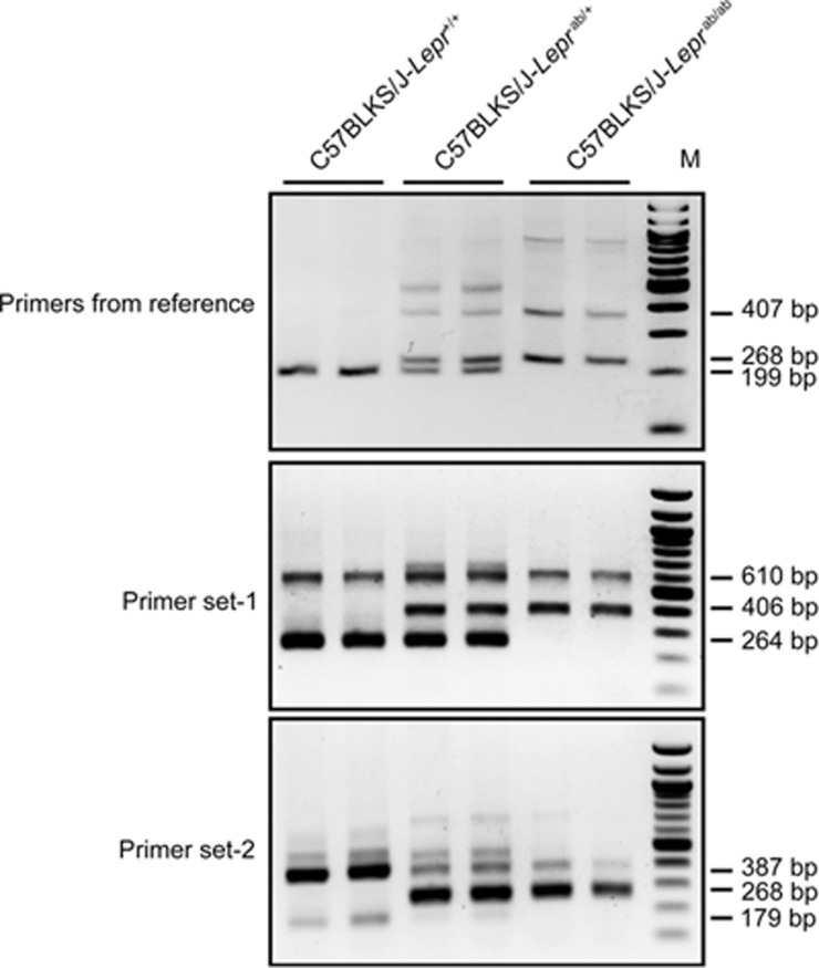
Comparison of genotyping results by using primers from reference17 (upper panel) and our primer set-1 and primer set-2 (middle and bottom panels). Since the primers from reference produced two bands similar in size (268 bp for T-allele band and 199 bp for G-allele band), we have to use 3% agarose gel and longer running time for a clear separation. While both of our primer sets produce well separated PCR products, which can be easily resolved by 1.5% agarose DNA gel with regular running time. The DNA templates are from wild type mice, db/+ heterozygous mice and db/db homozygous mice. Each lane represents one independent sample. M: 100 bp DNA ladder.
Here, we have established a sensitive, quick and low-cost ARMS-PCR method to reliably detect lepr point mutations. In summary, our method will not only greatly facilitate db/db-related research but also potentially be useful for genotyping other animal models that harbor single-nucleotide point mutation.
Materials and methods
Mice
db/db heterozygote mice (BKS.Cg-Dock7m +/+ Leprdb/J) were obtained from Jackson Laboratories (https://www.jax.org/mouse-search) and maintained in our facilities in accordance with NIH guidelines and IACUC approval by the Ohio State University.
Crude extraction of genomic DNA
One-millimeter mouse tail or ear notches were placed into 1.5 mL microcentrifuge tubes containing 180 μL of alkaline lysis reagent (50 mmol/L NaOH). The samples were then heated to 95 °C for 10 min to release genomic DNA. The tubes are cooled to 4 °C, and then 20 μL of neutralization buffer (1 mol/L Tris-HCl, pH 8.0) was added to the alkaline lysis buffer. The sample was then centrifuged to pellet the tissue debris, and the supernatant was immediately used for PCR. Two microliters of supernatant were used in each 20 μL PCR reaction. The samples can be stored at 4 °C for at least 3 months or at -20 °C for longer without degrading the PCR signal15. For genotyping of pups, toes can be used for genomic DNA extraction following the same protocol.
Primer design
The primers for ARMS-PCR were designed using a web-based program, available at: http://primer1.soton.ac.uk/primer1.html13. Briefly, the target sequence is copied and pasted to the website, after which required parameters are input, including the position of the point mutation in the sequence, the length of the PCR products, melting temperature (TM), primer size, and the percentage of GC content. The software will generate a few primer sets in the output window. Each set of primers includes four primers: one pair of primers comprises the forward and reverse outer primers, which are completely complimentary to the corresponding nucleotide. The other pair of primers comprises the forward (G allele-specific, wild-type allele-specific) and reverse inner primer (T allele-specific, mutant allele-specific). Importantly, in tetra-primer ARMS-PCR, there is not only a 3′ terminus mismatch but also a second deliberate mismatch at position -2 from the 3′ terminal of the same allele-specific primer to increase the specificity of the reaction. For more detailed information, please see screenshots of this website-based software shown in Figure S1.
ARMS PCR assay
PCR was carried out as described previously16. Briefly, the PCR samples comprised 1–2 μL of genomic DNA, 10 μL of Taq 2×PCR master mix (NEB, catalogue number: M0270L), 1 μL of dNTPs (2.5 mmol/L) and 2 μL of mixed primers (2 μmol/L of primer each were mixed at 1:1:1:1 volume ratio, or outer primers: inner primers at a 2:1 ratio) at a final concentration of 0.2 μmol/L. Target DNA was amplified in a thermocycler machine (Mastercycler, Pro S, Eppendorf), with initial denaturation at 94 °C for 5 min, followed by 40 cycles of denaturation at 95 °C for 30 s, annealing at 55 °C for 30 s, extension at 68 °C for 1 min and a final elongation step at 68 °C for 5 min. The PCR products were electrophoresed on a 2% EB-stained agarose gel.
Results
The mouse leptin receptor sequence (C57BL/6J) was retrieved from NCBI (gene ID: 16847). The leprdb mice have a single-point mutation (G→T) within intron 18 of the leptin receptor gene. Mutations in this gene have been associated with obesity and pituitary dysfunction. We download the partial sequence of the lepr gene from NCBI and then designed 2 sets of primer groups (all primer information is listed in Table 1). The primers used for amplification from normal and mutant leptin receptor alleles were designed using web-based ARMS-PCR software. Partial sequences of the normal and mutant alleles of lepr gene are shown in Figure 3.
Figure 3.
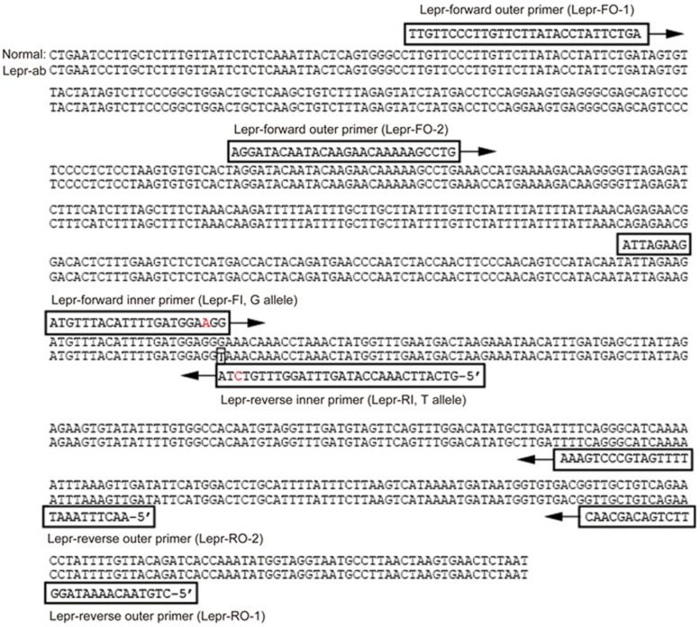
Primer sets sequence alignment to normal and mutant leptin receptor allele sequences (NCBI, Gene ID: 16847). The primers for ARMS-PCR were designed using the primer design web service for tetra-primer ARMS-PCR. Mouse leptin receptor (Lepr), on chromosome 4, range of 161510-162209 bp of complete sequence. Box indicates transverse point mutation (G→T) in intron 18 of the leptin receptor. The second mismatch of the 3′ terminus of the inner primers is indicated in red.
Primers used in the PCR reaction are boxed, and arrows indicate the PCR extension orientation. Lepr-forward outer primer (ie, Lepr-FO-1 or Lepr-FO-2) and lepr-reverse outer primer (ie, Lepr-RO-1 or Lepr-RO-2) were combined to amplify 610-bp or 387-bp PCR products from either allele. The PCR products generated from the outside primers were used as positive controls in the ARMS-PCR system to ensure the normality of PCR reaction. Lepr-forward inner primer (ie, Lepr-FI) and lepr-reverse outer primer (ie, Lepr-RO-1 or Lepr-RO-2) were combined to amplify G-allele specific (wild-type allele specific) PCR products (264 bp or 179 bp). Lepr-reverse inner primer (ie, Lepr-RI) and Lepr-forward outer primer (ie, Lepr-FO-1 or Lepr-FO-2) were combined to amplify T-allele specific (mutant allele specific) PCR products (406 bp or 268 bp). The PCR products were separated on a 2% EB-stained agarose gel. Since wild-type mice have two normal G-alleles of the lepr gene, only the positive-control and G-allele specific amplicons will be observed. The db/db heterozygous mice, which have one normal lepr gene and one mutant T-allele of lepr gene, will yield the control amplicon, a G-allele specific amplicon and a T-allele specific amplicon. Finally, db/db homozygous mice, which have two T-alleles of the lepr point mutation gene, will yield the common and T-allele specific amplicons. Because of the different sizes of the PCR products, the genotypes can be easily determined from the band patterns on DNA gels. The principle of primer design and the genotype DNA gel patterns are schematically summarized in Figure 1.
Interestingly, a previous study also introduced a genotyping method for db/db mice using tetra-primer ARMS-PCR method17. After studying this article, we identified a few key differences with our method17. First and most importantly, the primers from the previous study sets fail to meet the requirements for ARMS primer design, with only one mismatch in the end of 3′ terminus of inner reverse primers, whereas in our primer design, we not only retain the original point mutation but also introduce a second mismatch at positon of -2 in the same allele-specific primer, which is critical to increase the specificity of the PCR reaction (primer information detailed in Table 1).
Second, our PCR products are more easily identified as their size differences are larger than the PCR products generated in the previous article. To confirm the advantages of our primer design, tetra-primer PCR was performed using the same DNA templates from three different mouse genotypes by using primers from17, as well as primer set-1 and primer set-2 from our study. As shown in Figure 2, although all three primer sets produced similar results, our primers (set-1 and set-2) clearly produced brighter and more distinguishable T-allele-specific bands than those from17 on a 2% agarose gel, whereas we had to run a 3% agarose gel for a longer time to resolve the PCR products derived from primer sets from reference. Notably, although primer set-1 and primer set-2 were both generated through ARMS primer design, the PCR results are more specific and easier to read from primer set-1 than from primer set-2, as evidenced by the better-separated amplicons and more specific PCR bands from primer set-1. In conclusion, our primer set-1 is best for genotyping db/db mice. We also found that primer-set-1 has a broader melting temperature range, from 50 °C to 60 °C (pleases see temperature-gradient PCR results in Figure S2), which makes the genotyping protocol less sensitive to PCR reaction conditions. Furthermore, we found that adding slightly more dNTPs (0.1 mmol/L final concentration) to the reaction and adding more PCR cycles increased the intensity of the T-allele-specific PCR bands. Finally, the PCR products were sequenced to determine whether they corresponded to the expected sequences. As shown in Figure S3, the 406-bp and 264-bp bands indeed corresponded to the mutant and wild-type alleles, respectively. Taken together, we used primer set-1 in our subsequent tests for the sensitivity and stability of our genotyping method.
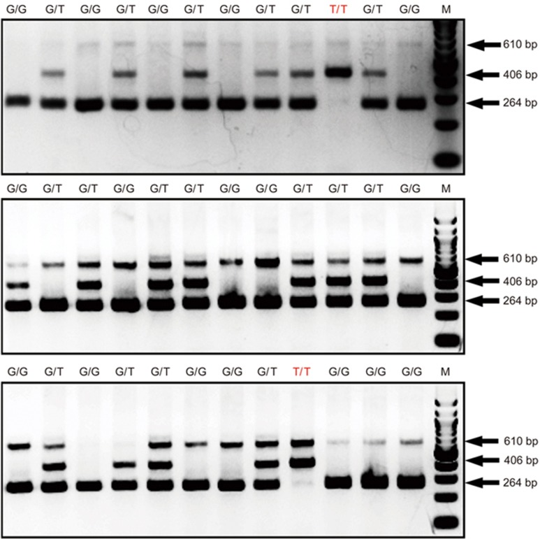
To validate the sensitivity and stability of our genotyping method, 36 long-term stored genomic DNA samples were used. These samples have been stored at -20 °C for more than 5 years, and all of the samples have been used to perform enzymatic digestion genotyping assays, as recommended by the Jackson laboratory, which provided these mice. As shown in Figure 4, our method successfully identified the genotypes of all 36 samples, consistent with the previous restriction enzymatic digestion results (data not shown).
Figure 4.
Sensitivity and stability test of our db/db genotyping method. Thirty-six genomic DNA samples were used to test our established genotyping method. These genomic DNA samples were extracted 5 years ago and kept at -20 °C after extraction. The results showed that our genotyping method can work perfectly on very old samples. M: 100 bp DNA ladder. G/G: 610+264 bp, wild type mice; G/T: 610+264+406 bp, db/db heterozygous mice; T/T: 610+406 bp, db/db mutant homozygous mice. Each lane represents one independent sample.
However, in some samples, we did observed that the 610-bp common amplicon was not clearly present (lane 1 of upper panel and lanes 3 and 4 of lower panel in Figure 4). Although this 610-bp band is not critical for determining the genotype of the mice, its absence might cause confusion. We think there might be three possible reasons for this observation: First, the genomic DNA samples used in Figure 4 were stored at -20 °C for more than five years, which might cause less efficient amplification of the larger common amplicon (610 bp vs 406 bp and 264 bp); second, our PCR system contains four primers, which compete with one another in the system, and DNA polymerase typically prefers to amplify shorter amplicons. To test this hypothesis, we adjusted the ratio of the outer and inner primer sets and used different ratios of outer and inner primers (outer primer: inner primer=1:4; 1:2; 1:1; 2:1; 4:1). The common amplicon appears brighter with increasing amounts of the outer primers (Figure S4). Thus, researchers can increase the outer primer content to obtain a stronger 610-bp band. Finally, the concentrations of the DNA template and the primer sets might require adjustment for optimal outcomes. For beginners trying this genotyping method, we suggest performing three separate PCR reactions (one each for the 610-bp, 406-bp or 264-bp amplicons) with each genomic DNA sample (Figure S5). This approach should be good for beginners to quickly gain experience before starting to use tetra primers. In summary, the common amplicon might not always be present in the final genotyping results; however, this will not affect the interpretation of the genotyping results.
In summary, we found that our method is a quick, easy, sensitive and reliable technique for db/db genotyping. As summarized in Figure 5, it takes only 2 h from initial sample collection to final results. Thus, we are confident that our method can greatly facilitate db/db-related research and potentially be applied to genotyping other animal strains with single-nucleotide mutations.
Figure 5.
Work flow chart of ARMS-PCR procedure for our db/db genotyping protocol.
Author contribution
Bao-yu PENG participated in manuscript writing and experimental design and performed the experiments. Qiang WANG participated in performing part of the experiments. Yan-hong LUO, Jian-feng HE, and Tao TAN participated in manuscript writing, editing and data analysis. Hua ZHU participated in manuscript writing, experimental design and data analysis. All authors have read and approved the manuscript.
Acknowledgments
This study was supported by grants from the National Institutes of Health (R01 #HL124122 and #AR067766), the American Heart Association (No 12SDG12070174) and the National Natural Science Foundation of China (No 81401155).
Footnotes
Supplementary information is available on the website of Acta Pharmacologica Sinica.
Supplementary Information
The screen shot of the online software showing the design and parameters of ARMS-PCR protocol.
Gradient PCR was used to test Tm temperature in ARMS-PCR reaction.
To determine the 406 bp and 264 bp bands are corresponding to db/db allele and wild type allele, respectively.
The ratio between outer primer and inner primer was adjusted from 1:4 to 4:1.
Due to the fact that the common band (610 bp band) could potentially be weak.
References
- Arounleut P, Bowser M, Upadhyay S, Shi XM, Fulzele S, Johnson MH, et al. Absence of functional leptin receptor isoforms in the POUND (Lepr(db/lb)) mouse is associated with muscle atrophy and altered myoblast proliferation and differentiation. PLoS One 2013; 8: e72330. [DOI] [PMC free article] [PubMed] [Google Scholar]
- Tartaglia LA, Dembski M, Weng X, Deng N, Culpepper J, Devos R, et al. Identification and expression cloning of a leptin receptor, OB-R. Cell 1995; 83: 1263–71. [DOI] [PubMed] [Google Scholar]
- Nordstrom V, Willershauser M, Herzer S, Rozman J, von Bohlen Und Halbach O, Meldner S, et al. Neuronal expression of glucosylceramide synthase in central nervous system regulates body weight and energy homeostasis. PLoS Biol 2013; 11: e1001506. [DOI] [PMC free article] [PubMed] [Google Scholar]
- Kondo Y, Hasegawa G, Okada H, Senmaru T, Fukui M, Nakamura N, et al. Lepr(db/db) Mice with senescence marker protein-30 knockout (Lepr(db/db)Smp30(Y/-)) exhibit increases in small dense-LDL and severe fatty liver despite being fed a standard diet. PLoS One 2013; 8: e65698. [DOI] [PMC free article] [PubMed] [Google Scholar]
- Tonra JR, Ono M, Liu X, Garcia K, Jackson C, Yancopoulos GD, et al. Brain-derived neurotrophic factor improves blood glucose control and alleviates fasting hyperglycemia in C57BLKS-Lepr(db)/lepr(db) mice. Diabetes 1999; 48: 588–94. [DOI] [PubMed] [Google Scholar]
- Syvanen AC. From gels to chips: “minisequencing” primer extension for analysis of point mutations and single nucleotide polymorphisms. Hum Mutat 1999; 13: 1–10. [DOI] [PubMed] [Google Scholar]
- Ota M, Fukushima H, Kulski JK, Inoko H. Single nucleotide polymorphism detection by polymerase chain reaction-restriction fragment length polymorphism. Nat Protoc 2007; 2: 2857–64. [DOI] [PubMed] [Google Scholar]
- Han Y, Khu DM, Monteros MJ. High-resolution melting analysis for SNP genotyping and mapping in tetraploid alfalfa (Medicago sativa L.). Mol Breed 2012; 29: 489–501. [DOI] [PMC free article] [PubMed] [Google Scholar]
- Fu YB, Peterson GW, Dong Y. Increasing genome sampling and improving SNP genotyping for genotyping-by-sequencing with new combinations of restriction enzymes. G3 (Bethesda) 2016; 6: 845–56. [DOI] [PMC free article] [PubMed] [Google Scholar]
- Horvat S, Bunger L. Polymerase chain reaction-restriction fragment length polymorphism (PCR-RFLP) assay for the mouse leptin receptor (Lepr(db)) mutation. Lab Anim 1999; 33: 380–4. [DOI] [PubMed] [Google Scholar]
- Newton CR, Graham A, Heptinstall LE, Powell SJ, Summers C, Kalsheker N, et al. Analysis of any point mutation in DNA. The amplification refractory mutation system (ARMS). Nucleic Acids Res 1989; 17: 2503–16. [DOI] [PMC free article] [PubMed] [Google Scholar]
- Newton CR, Heptinstall LE, Summers C, Super M, Schwarz M, Anwar R, et al. Amplification refractory mutation system for prenatal diagnosis and carrier assessment in cystic fibrosis. Lancet 1989; 2: 1481–3. [DOI] [PubMed] [Google Scholar]
- Ye S, Dhillon S, Ke X, Collins AR, Day IN. An efficient procedure for genotyping single nucleotide polymorphisms. Nucleic Acids Res 2001; 29: E88–8. [DOI] [PMC free article] [PubMed] [Google Scholar]
- Chiapparino E, Lee D, Donini P. Genotyping single nucleotide polymorphisms in barley by tetra-primer ARMS-PCR. Genome 2004; 47: 414–20. [DOI] [PubMed] [Google Scholar]
- Meeker ND, Hutchinson SA, Ho L, Trede NS. Method for isolation of PCR-ready genomic DNA from zebrafish tissues. BioTechniques 2007; 43: 610, 12,, 14. [DOI] [PubMed] [Google Scholar]
- Zhang H, Kong F, Wang X, Liang L, Schoen CD, Feng J, et al. Tetra-Primer ARMS PCR for rapid detection and characterization of Plasmopara viticola phenotypes resistant to carboxylic acid amide (CAA) fungicides. Pest Manage Sci 2016 2016; 2: 557–63. [DOI] [PubMed] [Google Scholar]
- Jung H, Nam H, Suh JG. Rapid and efficient identification of the mouse leptin receptor mutation (C57BL/KsJ-db/db) by tetra-primer amplification refractory mutation system-polymerase chain reaction (ARMS-PCR) analysis. Lab Anim Res 2016; 32: 70–3. [DOI] [PMC free article] [PubMed] [Google Scholar]
Associated Data
This section collects any data citations, data availability statements, or supplementary materials included in this article.
Supplementary Materials
The screen shot of the online software showing the design and parameters of ARMS-PCR protocol.
Gradient PCR was used to test Tm temperature in ARMS-PCR reaction.
To determine the 406 bp and 264 bp bands are corresponding to db/db allele and wild type allele, respectively.
The ratio between outer primer and inner primer was adjusted from 1:4 to 4:1.
Due to the fact that the common band (610 bp band) could potentially be weak.


