Abstract
The differential diagnosis of epithelioid mesothelioma from lung adenocarcinoma and squamous cell carcinoma requires the positive and negative immunohistochemical markers of mesothelioma. The IMIG guideline has suggested the use of Calretinin, D2–40, WT1, and CK5/6 as mesothelial markers, TTF-1, Napsin-A, Claudin 4, CEA as lung adenocarcinoma markers p40, p63, CK5/6, MOC-31 as squamous cell markers. However, use of other immunohistochemical markers is still necessary. We evaluated 65 epithelioid mesotheliomas, 60 adenocarcinomas, and 57 squamous cell carcinomas of the lung for MUC4 expression by immunohistochemistry and compared with the previously known immunohistochemical markers. MUC4 expression was not found in any of 65 cases of epithelioid mesothelioma. In contrast, MUC4 expression was observed in 50/60(83.3%) cases of lung adenocarcinoma and 50/56(89.3%) cases of lung squamous cell carcinoma. The negative MUC4 expression showed 100% sensitivity, 86.2% specificity and accuracy rate of 91.2% to differentiate epithelioid mesothelioma from lung carcinoma. The sensitivity, specificity, and accuracy of MUC4 are comparable to that of previously known markers of lung adenocarcinoma and squamous cell carcinoma, namely CEA, Claudin 4 and better than that of MOC-31. In conclusion, MUC4 immunohistochemistry is useful for differentiation of epithelioid mesothelioma from lung carcinoma, either adenocarcinoma or squamous cell carcinoma.
Introduction
Malignant mesothelioma is a highly aggressive malignant neoplasm with unfavorable prognosis. The mortality from mesothelioma is increasing in Japan, and other developing countries1. Some of the non-mesotheliomatous peripheral lung cancer, adenocarcinoma or squamous cell carcinoma, may present as the pleurotropic growth resembling that of mesothelioma2. As the prognosis and treatment protocols of epithelioid mesothelioma are different from that of the lung cancer, accurate diagnosis of mesothelioma is crucial.
Mesothelioma is classified into three major histological subtypes: epithelioid, biphasic, and sarcomatoid mesothelioma. Epithelioid mesothelioma shows numerous histological growth pattern3. Due to this resemblance of clinical growth pattern and histology of epithelioid mesothelioma with lung cancers, immunohistochemical markers are essential for accurate diagnosis of epithelioid mesothelioma.
Although IMIG (International Mesothelioma Interest Group) recommended Calretinin, D2–40 (podoplanin), WT1 as mesothelioma markers, CEA, TTF-1, Napsin-A, as lung adenocarcinoma markers, and p63, p40, MOC-31 as for lung squamous carcinoma markers4, the additional immunohistochemical markers will surely benefit in some unusual cases.
We recently proposed the addition of two immunohistochemical markers, Intelectin-1 and DAB2, as the positive immunohistochemical markers of epithelioid mesothelioma5. In this study, we also found the down regulation of many other genes including MUC4 in epithelioid mesothelioma as mentioned in its supplementary data.
Therefore, we evaluated the diagnostic applicability of MUC4 immunohistochemistry for differentiation of epithelioid mesothelioma to lung adenocarcinoma and squamous cell carcinoma.
Materials and Methods
Patients and Histologic Samples
Pathological specimens (formalin-fixed paraffin-embedded tissue blocks) of 65 epithelioid mesotheliomas and 60 lung adenocarcinoma and 57 squamous cell carcinoma were obtained from the tissue archives of the Department of Pathology, Hiroshima University. We also reviewed the patient’s clinical details and chest computed tomography findings for confirmation of the tumor localization. The mean age of lung adenocarcinoma patients was 69 with range from 43 to 84 (male 38, female 22), that of lung squamous cell carcinoma was 69 with range from 39 to 86 (male 49, female 7), and that of epithelioid mesothelioma 69 with range from 33–92 (male 61, female 4).
All histological sections were examined and reclassified by three pathologists (VJA, KK, and YT) according to recent WHO classification6. Pathologic diagnosis was confirmed by histologic findings and the immunohistochemical marker panel recommended by 2012 International Mesothelioma Interest Group (IMIG) guideline4. The collection of tissue specimens for this study was carried out in accordance with the “Ethics Guidelines for Human Genome/Gene Research” enacted by the Japanese Government. Ethical approval was obtained from the institutional ethics review committee (Hiroshima University E-974). All experimental procedures were in accordance with the with ethical guidelines. Samples used were linked-anonymised archival specimens, and individual consent was not required for this research.
Immunohistochemical Procedures and Evaluation of MUC4 Expression
Immunohistochemistry was performed using 3 μm tissue sections prepared from the best representative formalin-fixed paraffin-embedded blocks of epithelioid mesothelioma, lung adenocarcinoma, and squamous cell carcinoma cases. Immunohistochemical staining was performed using the Ventana Benchmark GX automated immunohistochemical station (Roche Diagnostics, Tokyo, Japan). The antigen retrieval methods and antibodies used in this study are summarized in Table 1. Incubation with the secondary antibody and detection was performed with Ventana ultraView Universal DAB Detection Kit (Roche Diagnostics). Immunoreactivity was evaluated as either negative or positive. Nuclear staining of calretinin, WT1, p40, p63, and TTF-1, cytoplasmic staining of MUC4 and Napsin-A, CK5/6, CEA, membranous staining of podoplanin (clone: D2–40), Epithelial Related Antigen (clone: MOC-31) and claudin 4 were considered as ‘positive’.
Table 1.
List of antibodies with their clone, commercial source, and reaction conditions.
| Antibody to | Clone | Source | Dilution | Antigen Retrieval |
|---|---|---|---|---|
| MUC4 | 8G7 | Santa Cruz Biotech | 1:25 | 60 min, CC1 |
| Calretinin | SP65 | Ventana-Roche | prediluted | 30 min, CC1 |
| Podoplanin | D2–40 | Nichirei Bioscience | prediluted | 60 min, CC1 |
| WT1 | 6F-H2 | Ventana-Roche | 1:25 | 60 min, CC1 |
| CEA | COL-1 | Nichirei Bioscience | Prediluted | 8 min, CC1 |
| Claudin 4 | 3E2C1 | Life Technologies | 1:100 | 60 min, CC1 |
| TTF-1 | 8G7G3/q | Dako-Agilent | 1:25 | 30 min, CC1 |
| Napsin-A | MRQ-60 | Ventana-Roche | prediluted | 60 min, CC1 |
| Epithelial Related Antigen | MOC-31 | Dako-Agilent | 1:25 | 10 min, Protease |
| p63 | DAK-p63 | Dako-Agilent | 1:25 | 60 min, CC1 |
| CK5/6 | D5/16 B4 | Dako-Agilent | 1:25 | 60 min, CC1 |
| p40 | BC28 | Biocare Medical | 1:100 | 60 min, CC1 |
WT1: Wilm’s tumor 1; CEA: carcinoembryonic antigen; TTF-1: thyroid transcription factor-1; CK: cytokeratin.
Abbreviation: CC1, cell conditioning buffer 1 (Tris-based buffer, pH 8.5 from Ventana-Roche).
Positive Immunoreactivity was semi quantitatively scored as 0 for none to trace, 1+ for up to 10%, 2+ for 10–50%, and 3+ for >50% tumor cells showing positive expression.
Statistical analyses were performed using the Fisher’s exact test for calculation of p-value of positivity of individual markers, Mann Whitney U test for calculation Z-score and the p-value of immunohistochemical score of individual markers. Sensitivity, specificity, and accuracy rate are calculated using a 2 × 2 contingency table model.
Results
Immunohistochemical Result
The percentages and immunohistochemical scores of MUC4 expression in epithelioid mesothelioma, lung adenocarcinoma, and squamous cell carcinoma and other immunohistochemical markers are shown in Table 2.
Table 2.
Immunohistochemical Result of Epithelioid Mesothelioma, Lung Adenocarcinoma, and Squamous Cell Carcinoma.
| Antibody | Epithelioid Mesothelioma (65 cases) | Lung Adenocarcinoma (60 cases) | Lung Squamous Cell Carcinoma (56 cases) | Z-score! | |||||||||||||
|---|---|---|---|---|---|---|---|---|---|---|---|---|---|---|---|---|---|
| Positive cases (%) | Immunohistochemical Score | Positive cases (%) | Immunohistochemical Score | Positive cases (%) | Immunohistochemical Score | EM vs LAC | EM vs LSC | ||||||||||
| 0 | 1+ | 2+ | 3+ | 0 | 1+ | 2+ | 3+ | 0 | 1+ | 2+ | 3+ | ||||||
| MUC4 | 0 (0) | 65 | 0 | 0 | 0 | 50 (83.3) | 10 | 20 | 9 | 21 | 50 (89.3) | 6 | 19 | 10 | 21 | 8.0* | 8.4* |
| Calretinin | 64 (98.5) | 1 | 5 | 2 | 57 | 17 (28.3) | 43 | 11 | 6 | 0 | 28 (50) | 28 | 13 | 10 | 5 | 9.1* | 7.8* |
| D2–40 | 63 (96.9) | 2 | 4 | 5 | 54 | 7 (11.7) | 53 | 4 | 3 | 0 | 34 (60.7) | 22 | 7 | 18 | 9 | 9.2* | 6.7* |
| WT1 | 56 (86.2) | 9 | 17 | 7 | 32 | 0 (0) | 60 | 0 | 0 | 0 | 2 (3.6) | 54 | 2 | 0 | 0 | 9.3* | 9.1* |
| CEA | 0 (0) | 65 | 0 | 0 | 0 | 58 (96.7) | 2 | 7 | 12 | 39 | 56 (100) | 0 | 20 | 21 | 15 | 9.3* | 9.5* |
| Claudin 4 | 0 (0) | 65 | 0 | 0 | 0 | 57 (95) | 3 | 2 | 10 | 45 | 55 (98.2) | 1 | 3 | 34 | 18 | 9.5* | 9.2* |
| TTF-1 | 0 (0) | 65 | 0 | 0 | 0 | 54 (90) | 6 | 2 | 6 | 46 | 5 (8.9) | 51 | 5 | 0 | 0 | 8.7* | 1.01 |
| Napsin-A | 0 (0) | 65 | 0 | 0 | 0 | 48 (80) | 12 | 9 | 3 | 36 | 2 (3.6) | 54 | 2 | 0 | 0 | 7.7* | 0.34 |
| MOC-31 | 8 (12.3) | 57 | 5 | 2 | 1 | 55 (91.7) | 5 | 9 | 8 | 38 | 51 (91.1) | 5 | 8 | 18 | 25 | 8.3* | 7.8* |
| P63 | 15 (23.1) | 50 | 11 | 3 | 1 | 32 (53.3) | 28 | 16 | 10 | 6 | 56 (100) | 0 | 1 | 2 | 53 | 3.2 | 9.0* |
| CK5/6 | 45 (69.2) | 20 | 8 | 11 | 26 | 13 (21.7) | 47 | 6 | 5 | 2 | 55 (98.2) | 1 | 1 | 5 | 49 | 5.2* | 4.6* |
| P40 | 3 (4.6) | 62 | 3 | 0 | 0 | 6 (10) | 54 | 5 | 1 | 0 | 55 (98.2) | 1 | 0 | 3 | 52 | 0.52 | 9.1* |
WT1: Wilm’s tumor 1; CEA: carcinoembryonic antigen; TTF-1: thyroid transcription factor-1; CK: cytokeratin.
Immunohistochemical score 0: 0% positive cells or trace staining; 1+: 1–10% positive cells; 2+: 11–50% positive cells; 3+: >51% positive cells! The Z-score is calculated by the MannWhitney ‘U’ test between immunohistochemical scores of epithelioid mesothelioma and lung cancer (adenocarcinoma or squamous cell carcinoma).
EM: epithelioid mesothelioma; LAC: lung adenocarcinoma; LSqCC: lung squamous cell carcinoma *significant p-value (<0.0001).
MUC4 expression
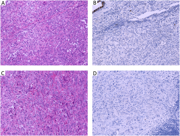
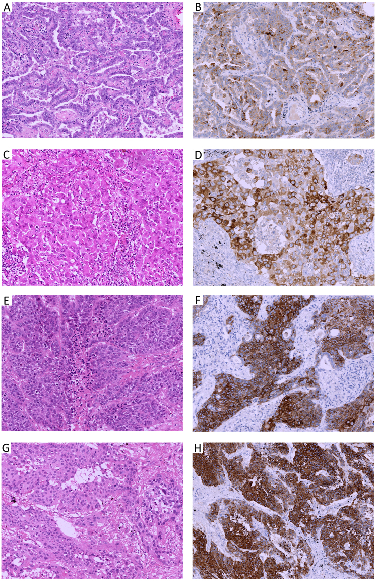
MUC4 expression was not present in any of 65 epithelioid mesotheliomas (0%, Fig. 1B,D). MUC4 expression was present in the cytoplasm of the tumor cell of lung adenocarcinoma and squamous cell carcinoma. It was also evident in the normal bronchial epithelium, considered as an internal positive control. It was present in 50 of 60 cases of adenocarcinoma (83.3%, Fig. 2B,D) and 50/56 cases of squamous cell carcinoma (89.3%, Fig. 2F,H). Among lung adenocarcinoma cases, 21 cases showed immunohistochemical score 3+, 9 cases in score 2+, and 20 cases in score 1+. In lung squamous cell carcinoma, 21 cases showed score 3+, 10 cases score 2+, and 19 cases score 1+.
Figure 1.
Representative cases of epithelioid mesothelioma with papillo-tubular (A) and solid (C) growth showing no MUC4 expression (B,D). Note: normal bronchial epithelium as internal control showing MUC4 expression. (A,C) H & E stain × 40 high power field; (B,D) MUC4 immunohistochemistry).
Figure 2.
Representative cases of lung adenocarcinomas, papillary (2 A) or solid (2 C) growth and non-keratinizing squamous cell carcinoma (E,G) showing MUC4 expression. (A,C,E,G): H & E stain × 40 high power field, (B,D,F,H): MUC4 immunohistochemistry).
In addition, we found no MUC4 expression in mesothelial layer of visceral pleura present in 39 lung cancer cases.
Calretinin, D2–40, WT1, and CK5/6
Calretinin expression was present in 64 cases (98.5%) of epithelioid mesothelioma with the immunohistochemical score of 3+ in 57. Calretinin expression was also present in 17 of 60 (28.3%) cases of lung adenocarcinoma, and 28 of 56 (50%) cases of squamous cell carcinoma. The immunohistochemical of 3+ was not present in adenocarcinoma but was present in 5 cases of squamous cell carcinoma. D2–40 expression was present in 63 cases (96.9%) of epithelioid mesothelioma with the immunohistochemical score of 3+ in 54 cases. D2–40 expression was also present in 7 of 60 (11.7%) cases of lung adenocarcinoma and 34 of 56 (60.7%) cases of squamous cell carcinoma. The immunohistochemical score of 3+ was not present in adenocarcinoma but was present in 9 cases of squamous cell carcinoma. WT1 expression was present in 56 (86.2%) epithelioid mesothelioma, with the immunohistochemical score of 3+ in 32 cases. WT expression was not present in any of the 60 adenocarcinomas but was present in 2 of 56 (3.6%) squamous cell carcinoma. CK5/6 expression was present in 45 of 65 (69.2%) epithelioid mesothelioma, and 55 of 56 (98.2%) squamous cell carcinoma, and 13 of 60 (21.7%) adenocarcinoma. The majority of epithelioid mesothelioma and squamous cell carcinoma showed the immunohistochemical score of 3+ while in adenocarcinoma showed the immunohistochemical score of 1+ to 3+.
CEA, Claudin 4, MOC-31
CEA and Claudin 4 expressions were not present in any of the epithelioid mesotheliomas. In contrast, CEA Expression was present in both 58 (96.7%) cases of lung adenocarcinoma and 56 (100%) cases of squamous cell carcinoma. The majority of lung adenocarcinoma showed the immunohistochemical score of 3+, and only one-fourth of CEA-positive lung squamous cell carcinoma showed the immunohistochemical score of 3+.
Membranous Claudin 4 expression was present in 57 (95%) cases of adenocarcinoma with 39 cases showing the immunohistochemical score of 3+ and 55 (98.2%) cases of squamous cell carcinoma with 34 cases showing the immunohistochemical score of 2+.
MOC-31 expression was present in 55 (91.7%) and 51 (91.1%) cases of adenocarcinoma and squamous cell carcinoma. However, MOC-31 was also expressed in 8 (12.3%) epithelioid mesothelioma, of which, 5 cases showed the immunohistochemical score of 1+, 2 cases showed 2+, and 1 case showed 3+.
TTF-1, Napsin-A, p63, p40
Nuclear expression of TTF-1 and cytoplasmic expression of Napsin-A are the positive immunohistochemical markers of lung adenocarcinoma. TTF-1 and Napsin-A expression were not present in any of 65 (0%) cases of epithelioid mesothelioma and only 54 (90%) cases and 48 (80%) cases of lung adenocarcinoma. The immunohistochemical score of 3+ for TTF-1 and Napsin-A was observed in 46 and 36 cases respectively. TTF-1 expression was also present in 5 (8.9%) cases, and Napsin-A expression in 2 (3.6%) cases of lung squamous cell carcinoma but with the immunohistochemical score of 1+.
Nuclear p63 expression and recently nuclear p40 expression are the positive immunohistochemical markers of lung squamous cell carcinoma. However, we found p63 and p40 expression in 15 (23%) and 3 (4.6%) cases of epithelioid mesothelioma although the immunohistochemical score was 1+. The p63 and p40 expression were present in 56 (100%), 55(98.2%) cases of squamous cell carcinoma and most of them with the immunohistochemical score of 3+. However, 32 (53.3%) and 6 (10%) cases of adenocarcinoma also showed immunoreactivity for p63 and p40 respectively but with the immunohistochemical score of 1+.
Sensitivity, Specificity and accuracy rate of immunohistochemical markers
Table 3 shows the sensitivities, specificities, and accuracy rates of the immunohistochemical markers to differentiate epithelioid Mesothelioma from lung adenocarcinoma and squamous cell carcinoma. Among the negative mesothelioma markers, MUC4, CEA, Claudin 4, TTF-1, and Napsin-A showed 100% sensitivity, p40 showed 95.4%, P63 showed 76.9%, and CK5/6 showed 69.2%. When the histological type was known as lung adenocarcinoma or squamous cell carcinoma, the specificity of TTF-1, Napsin-A and p40 were high, but with combined lung adenocarcinoma and squamous cell carcinoma, it remains around or less than 50%. Among the positive mesothelioma markers, WT1 showed the highest specificity of 98.6%, but sensitivity was limited to 86.2%. The sensitivities of calretinin and D2–40 were high with 98.5% and 96.9% respectively, but the specificities were low with 61.2%and 64.7% specificity. Among the negative mesothelial markers, CEA has the highest accuracy rate of 98.9% followed by Claudin 4 of 97.8%. The accuracy of MUC4 is 91.2% which is better than that of MOC-31.
Table 3.
Sensitivity, Specificity, and Accuracy Rate of Immunohistochemical Markers.
| Antibody | Sensitivity, % | Specificity, % | Accuracy Rate, % | p-value* |
|---|---|---|---|---|
| A. Differentiation of Epithelioid Mesothelioma from Lung Cancer including Adenocarcinoma and Squamous Cell Carcinoma | ||||
| MUC4(−) | 100 | 86.2 | 91.2 | <0.0001 |
| Calretinin(+) | 98.5 | 61.2 | 74.6 | <0.0001 |
| D2–40(+) | 96.92 | 64.7 | 76.2 | <0.0001 |
| WT1(+) | 86.15 | 98.6 | 94.8 | <0.0001 |
| CK5/6(+) | 69.2 | 41.4 | 51.4 | 0.2 |
| CEA(−) | 100 | 98.3 | 98.9 | <0.0001 |
| Claudin 4(−) | 100 | 96.6 | 97.8 | <0.0001 |
| MOC31(−) | 87.7 | 91.4 | 90 | <0.0001 |
| TTF-1(−) | 100 | 50.9 | 68.5 | <0.0001 |
| NapsinA(−) | 100 | 43.1 | 63.5 | <0.0001 |
| P63(−) | 76.9 | 75.2 | 76.2 | <0.0001 |
| P40(−) | 95.4 | 52.6 | 68 | <0.0001 |
| B. Differentiation of Epithelioid Mesothelioma from Lung Adenocarcinoma | ||||
| MUC4(−) | 100 | 83.3 | 92 | <0.0001 |
| Calretinin(+) | 71.7 | 98.5 | 85.6 | <0.0001 |
| D2–40(+) | 88.3 | 96.9 | 92.8 | <0.0001 |
| WT1(+) | 100 | 86.2 | 92.8 | <0.0001 |
| CK5/6(+) | 69.2 | 78.3 | 73.6 | <0.0001 |
| CEA(−) | 100 | 96.7 | 98.4 | <0.0001 |
| Claudin 4(−) | 100 | 95 | 97.6 | <0.0001 |
| MOC31(−) | 87.7 | 91.7 | 89.6 | <0.0001 |
| TTF-1(−) | 100 | 90 | 95.2 | 0.02 |
| NapsinA(−) | 100 | 80 | 90.4 | 0.21 |
| P63(−) | 76.9 | 53.3 | 65.6 | <0.0001 |
| P40(−) | 95.4 | 10 | 54.4 | <0.0001 |
| C. Differentiation of Epithelioid Mesothelioma from Lung Squamous Cell Carcinoma | ||||
| MUC4(−) | 100 | 89.3 | 95 | <0.0001 |
| Calretinin(+) | 98.5 | 50 | 76 | <0.0001 |
| D2–40(+) | 97 | 39.3 | 70.2 | <0.0001 |
| WT1(+) | 86.2 | 96.4 | 91 | <0.0001 |
| CK5/6(+) | 69.2 | 1.8 | 38 | <0.0001 |
| CEA(−) | 100 | 100 | 100 | <0.0001 |
| Claudin 4(−) | 100 | 98.2 | 99.2 | <0.0001 |
| MOC31(−) | 87.7 | 91.1 | 89.3 | <0.0001 |
| TTF-1(−) | 100 | 8.9 | 57.9 | <0.0001 |
| NapsinA(−) | 100 | 3.6 | 55.4 | <0.0001 |
| P63(−) | 76.9 | 100 | 88.5 | 0.008 |
| P40(−) | 95.4 | 98.2 | 96.7 | 0.31 |
WT1: Wilm’s tumor 1; CEA: carcinoembryonic antigen; TTF-1: thyroid transcription factor-1; CK: cytokeratin.
*p-value is calculated by Fisher’s exact test.
Discussion
MUC4 is a transmembrane mucin expressed in normal epithelial cells including epithelial mucosa of the digestive tract, ductal epithelium of salivary gland and lacrimal gland, larynx and trachea, lung, stomach, intestine, uterus, cervix, mammary gland, ovary, and kidney7. Many human malignancies like carcinomas of the pancreas8, ovary9, salivary gland10, lung11, stomach12, breast13 show MUC4 expression. MUC4 expression has been reported in the non-small cell lung cancer more in adenocarcinoma than do squamous cell carcinomas and large cell carcinomas14. MUC4 expression was also present significantly in solid adenocarcinoma of the lung15. In addition to poor prognosis correlated with MUC4 expression in the biliary tract, pancreas, ovary, and colorectal junction16, the MUC4 mRNA expression is related to the tumor histological type and its differentiation17. The tubular and papillary patterns of adenocarcinomas and the nests and in squamous pearls of squamous cell carcinomas show strong membranous and less cytoplasmic expression of MUC418. MUC4 plays a role in cell differentiation rather than cell proliferation of the normal goblet cells, the stratified squamous epithelial cells, and malignant epithelial tumor cells19. We previously reported the MUC4 expression in sarcomatoid carcinoma of the lung and its utility to differentiate from sarcomatoid mesothelioma20.
In this study, We found MUC4 expression in 50/60 (83.3%) cases of lung adenocarcinoma, 50/56 (89.3%) cases of squamous cell carcinoma, and none (0%) of epithelioid mesothelioma. Kwon et al.14 and Llinares et al.21 previously reported the diagnostic value of MUC4 expression in distinguishing epithelioid mesothelioma and lung adenocarcinoma. They reported the diagnostic value of MUC4 immunostaining in distinguishing mesothelioma and lung adenocarcinoma. They raised the polyclonal antibody against a KLH conjugate of a synthetic peptide corresponding to the tandem repeat sequence of MUC4 for their study. They found that MUC4 was expressed in 0 of the 41 epithelioid mesotheliomas and 32 of the 35 (91%) lung adenocarcinoma. Our study utilized the commercially available antibody and is the validation of the previous study. Other things different from this study is the inclusion of squamous cell carcinoma which also showed similar frequencies of positive cases like that of lung adenocarcinoma.
Our data of MUC4 expression in adenocarcinoma and squamous cell carcinoma of lung is also similar to another study by Kwon et al. who studied MUC4 expression in lung adenocarcinoma and squamous cell carcinoma of the lung but without mesothelioma.
In this study, we analyzed the utility of MUC4 to distinguish epithelioid mesothelioma from lung adenocarcinoma and squamous cell carcinoma. We have previously reported the MUC4 expression in sarcomatoid carcinoma of the lung and its utility to differentiate from sarcomatoid mesothelioma20. In our previous report, we emphasized the MUC4 expression in spindled cells of sarcomatoid carcinoma of lung. None of sarcomatoid mesothelioma had the expression of MUC4 expression. MUC4 was expressed in 72% of the sarcomatoid carcinoma of lung.
Ordonez et al.22 reported that there is no absolute specific and sensitive marker of epithelioid mesothelioma, although the various immunohistochemical markers are currently available for the diagnosis of epithelioid mesotheliomas. He also added the location and histologic features of the tumor, the sex of the patient, and the clinical findings need to be considered when selecting the markers for accurate diagnosis of epithelioid mesothelioma from the various other carcinomas22.
The sensitivity and specificity of MUC4 to differentiate epithelioid mesothelioma from lung adenocarcinoma were 100% and 83.3% respectively with the accuracy rate of 92% and those of MUC4 to differentiate epithelioid mesothelioma from lung squamous cell carcinoma 100% and 89.3% respectively with the accuracy rate of 95%. We found CEA is the best marker with the sensitivity of 100%, specificity of 98.3%, and accuracy rate of 98.9% followed by Claudin 4 which had the sensitivity of 100%, specificity of 96.6%, and accuracy rate of 97.8%. MUC4 expression showed a similar sensitivity of 100%, but the lower specificity of 86.2% and accuracy rate of 91.2%. However, it showed better sensitivity, specificity and accuracy rate than that of MOC-31. Also, we found positive immunoreactivity for MUC4 in some cases of adenocarcinoma and squamous cell carcinoma with no CEA and/or Claudin 4 expression. Two cases of CEA-negative adenocarcinoma, one of which was negative for Claudin 4 too, showed MUC4 expression (immunohistochemical score of 2+ or 3+). Moreover, 3 cases of Claudin 4-negative adenocarcinoma, two of which were negative for CEA too, showed MUC4 expression (immunohistochemical score of 1+ or 2+). There was one adenocarcinoma case which was negative for both CEA and Claudin-4 but positive MUC4 expression. So, MUC4 has the potential for the use as an additional negative marker of epithelioid mesothelioma for differentiation from lung cancer including adenocarcinoma or squamous cell carcinoma.
International Mesothelioma Interest Group Guideline for pathologic diagnosis of malignant mesothelioma has recommended various immunohistochemical negative markers, CEA, MOC-31, TTF-1, Napsin-A to differentiate it from adenocarcinoma and p40, p63, MOC-31 to differentiate it from squamous cell carcinoma4. Ordonez et al. later added Claudin 4 as one of the best broad-spectrum carcinoma markers to discriminate epithelioid mesotheliomas from both lung adenocarcinomas and squamous cell carcinomas23. In our previous publication too, we found CEA and Claudin 4 were the best positive carcinoma markers for discriminating epithelioid mesotheliomas from squamous cell carcinomas24. Although p63 is the marker of squamous cell carcinoma, it was at least focally positive in many of adenocarcinoma (32/60, 53.3%) and epithelioid mesothelioma (15/65, 23.1%) in the present study. So, limiting the value of the p63 expression for differentiation of epithelioid mesothelioma from squamous cell carcinoma.
In conclusion, MUC4 can be added as additional positive marker of non-small cell carcinoma (both lung adenocarcinoma and squamous cell carcinoma) and negative immunohistochemical marker to differentiate epithelioid mesothelioma from lung adenocarcinoma and squamous cell carcinoma. This study includes the limited cases (65 epithelioid mesothelioma and 116 cases of lung adenocarcinoma and needs follow up study consisting of large numbers of cases.
Acknowledgements
The authors thank Ms. Yukari Go of the Technical Center in Hiroshima University for excellent technical assistance and Ms. Naomi Fukuhara for administrative assistance. This work was supported by JSPS KAKENHI Grant Number 17K08742.
Author Contributions
Amany Sayed Mawas and Vishwa Jeet Amatya performed the immunohistochemical expression and statistical interpretation of the data and prepared Figs 1 and 2 and preparation of the manuscript. Yuichiro Kai, Yoshihiro Miyata, and Morihito Okada have contributed in the collection of the samples. All the authors reviewed the paper. Vishwa Jeet Amatya, Kei Kushitani and Yukio Takeshima contributed with pathological diagnosis of the cases. Vishwa Jeet Amatya and Yukio Takeshima has designed the research idea and ensured that all the authors involved in this study were included in the author list, and the list order has been agreed upon by all authors.
Competing Interests
The authors declare that they have no competing interests.
Footnotes
Amany Sayed Mawas and Vishwa Jeet Amatya contributed equally to this work.
Publisher's note: Springer Nature remains neutral with regard to jurisdictional claims in published maps and institutional affiliations.
References
- 1.Delgermaa, V. et al. Global mesothelioma deaths reported to the World Health Organization between 1994 and 2008. Bull World Health Organ89, 716–724, 724A-724C, 10.2471/BLT.11.086678 (2011). [DOI] [PMC free article] [PubMed]
- 2.Attanoos RL, Gibbs AR. ‘Pseudomesotheliomatous’ carcinomas of the pleura: a 10-year analysis of cases from the Environmental Lung Disease Research Group, Cardiff. Histopathology. 2003;43:444–452. doi: 10.1046/j.1365-2559.2003.01674.x. [DOI] [PubMed] [Google Scholar]
- 3.Galateau-Salle F, Churg A, Roggli V, Travis WD, World Health Organization Committee for Tumors of the, P The 2015 World Health Organization Classification of Tumors of the Pleura: Advances since the 2004 Classification. J Thorac Oncol. 2016;11:142–154. doi: 10.1016/j.jtho.2015.11.005. [DOI] [PubMed] [Google Scholar]
- 4.Husain AN, et al. Guidelines for pathologic diagnosis of malignant mesothelioma: 2012 update of the consensus statement from the International Mesothelioma Interest Group. Arch Pathol Lab Med. 2013;137:647–667. doi: 10.5858/arpa.2012-0214-OA. [DOI] [PubMed] [Google Scholar]
- 5.Kuraoka, M. et al. Identification of DAB2 and Intelectin-1 as Novel Positive Immunohistochemical Markers of Epithelioid Mesothelioma by Transcriptome Microarray Analysis for its Differentiation From Pulmonary Adenocarcinoma. Am J Surg Pathol, 10.1097/PAS.0000000000000852 (2017). [DOI] [PubMed]
- 6.Travis WD, et al. The2015 World Health Organization Classification of Lung Tumors: Impact of Genetic, Clinical and Radiologic Advances Since the 2004 Classification. J Thorac Oncol. 2015;10:1243–1260. doi: 10.1097/JTO.0000000000000630. [DOI] [PubMed] [Google Scholar]
- 7.Carraway KL, et al. Multiple facets of sialomucin complex/MUC4, a membrane mucin and erbb2 ligand, in tumors and tissues (Y2K update) Front Biosci. 2000;5:D95–D107. doi: 10.2741/carraway. [DOI] [PubMed] [Google Scholar]
- 8.Choudhury A, et al. Human MUC4 mucin cDNA and its variants in pancreatic carcinoma. J Biochem. 2000;128:233–243. doi: 10.1093/oxfordjournals.jbchem.a022746. [DOI] [PubMed] [Google Scholar]
- 9.Giuntoli RL, II, Rodriguez GC, Whitaker RS, Dodge R, Voynow JA. Mucin gene expression in ovarian cancers. Cancer Res. 1998;58:5546–5550. [PubMed] [Google Scholar]
- 10.Weed DT, et al. MUC4 and ERBB2 expression in major and minor salivary gland mucoepidermoid carcinoma. Head Neck. 2004;26:353–364. doi: 10.1002/hed.10387. [DOI] [PubMed] [Google Scholar]
- 11.Lopez-Ferrer A, et al. Mucins as differentiation markers in bronchial epithelium. Squamous cell carcinoma and adenocarcinoma display similar expression patterns. Am J Respir Cell Mol Biol. 2001;24:22–29. doi: 10.1165/ajrcmb.24.1.4294. [DOI] [PubMed] [Google Scholar]
- 12.Lopez-Ferrer A, et al. Role of fucosyltransferases in the association between apomucin and Lewis antigen expression in normal and malignant gastric epithelium. Gut. 2000;47:349–356. doi: 10.1136/gut.47.3.349. [DOI] [PMC free article] [PubMed] [Google Scholar]
- 13.Carraway KL, et al. Muc4/sialomucin complex in the mammary gland and breast cancer. J Mammary Gland Biol Neoplasia. 2001;6:323–337. doi: 10.1023/A:1011327708973. [DOI] [PubMed] [Google Scholar]
- 14.Kwon KY, et al. MUC4 expression in non-small cell lung carcinomas: relationship to tumor histology and patient survival. Arch Pathol Lab Med. 2007;131:593–598. doi: 10.5858/2007-131-593-MEINCL. [DOI] [PubMed] [Google Scholar]
- 15.Rokutan-Kurata, M. et al. Lung Adenocarcinoma With MUC4 Expression Is Associated With Smoking Status, HER2 Protein Expression, and Poor Prognosis: Clinicopathologic Analysis of 338 Cases. Clin Lung Cancer, 10.1016/j.cllc.2016.11.013 (2016). [DOI] [PubMed]
- 16.Huang X, et al. Clinicopathological and prognostic significance of MUC4 expression in cancers: evidence from meta-analysis. Int J Clin Exp Med. 2015;8:10274–10283. [PMC free article] [PubMed] [Google Scholar]
- 17.Yu CJ, et al. Mucin mRNA expression in lung adenocarcinoma cell lines and tissues. Oncology. 1996;53:118–126. doi: 10.1159/000227547. [DOI] [PubMed] [Google Scholar]
- 18.Moniaux N, et al. Complete sequence of the human mucin MUC4: a putative cell membrane-associated mucin. Biochem J. 1999;338(Pt 2):325–333. doi: 10.1042/bj3380325. [DOI] [PMC free article] [PubMed] [Google Scholar]
- 19.Weed DT, et al. MUC4 and ErbB2 expression in squamous cell carcinoma of the upper aerodigestive tract: correlation with clinical outcomes. Laryngoscope. 2004;114:1–32. doi: 10.1097/00005537-200408001-00001. [DOI] [PubMed] [Google Scholar]
- 20.Amatya VJ, et al. MUC4, a novel immunohistochemical marker identified by gene expression profiling, differentiates pleural sarcomatoid mesothelioma from lung sarcomatoid carcinoma. Mod Pathol. 2017;30:672–681. doi: 10.1038/modpathol.2016.181. [DOI] [PubMed] [Google Scholar]
- 21.Llinares K, et al. Diagnostic value of MUC4 immunostaining in distinguishing epithelial mesothelioma and lung adenocarcinoma. Mod Pathol. 2004;17:150–157. doi: 10.1038/modpathol.3800027. [DOI] [PubMed] [Google Scholar]
- 22.Ordonez NG. Application of immunohistochemistry in the diagnosis of epithelioid mesothelioma: a review and update. Hum Pathol. 2013;44:1–19. doi: 10.1016/j.humpath.2012.05.014. [DOI] [PubMed] [Google Scholar]
- 23.Ordonez NG. Value of claudin-4 immunostaining in the diagnosis of mesothelioma. Am J Clin Pathol. 2013;139:611–619. doi: 10.1309/AJCP0B3YJBXWXJII. [DOI] [PubMed] [Google Scholar]
- 24.Ordonez NG. The diagnostic utility of immunohistochemistry in distinguishing between epithelioid mesotheliomas and squamous carcinomas of the lung: a comparative study. Mod Pathol. 2006;19:417–428. doi: 10.1038/modpathol.3800544. [DOI] [PubMed] [Google Scholar]


