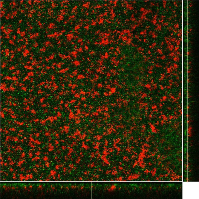
FIG 2 .

CLSM images of dual-species biofilms formed by S. pneumoniae EF3030-GFP (green) and S. aureus UAMS1-dsRED (red) following 48 h of coculture. The image shows the average intensity projections through the confocal image stack, with the maximum-intensity x-z and y-z projections shown along the bottom and side of the image; a representative image of a single slice from the central region of the biofilm is shown. Images were produced using ImageJ.
