ABSTRACT
It is widely known that visuospatial neglect and hemianopia maybe superimposed. We considered the differences in implicit information processing which is effective in patients with neglect but not with hemianopia. We then hypothesize that a prime-word in the neglected field should determine a semantic activation effect but not in a blind hemifield. Moreover eye movements could provide further details. In this work we considered a patient with a bilateral with the presence of either a left visual neglect and a right homonymous hemianopia. Our results supported implicit information processing in the space affected by neglect but not by hemianopia.
KEYWORDS: Eye movements, hemianopia, neglect, semantic priming
Introduction
The difficulty in exploring the surrounding space after a brain damage could be caused by an attentional deficit, such as visuospatial neglect, or by reduction of visual-field width, such as hemianopia. Anatomical sites involved in brain lesion may be fundamental to orient the clinician towards an effective diagnostic assessment. On one hand, unilateral spatial neglect (USN) as visuospatial attentional disorder determines tendency to ignore contralesional space, omit stimuli, and reduce accuracy in exploring surrounding space and, more in general, to perceive part of the space. It frequently arises after lesions involving right parietal lobe, inferior parietal lobule, temporo-parietal junction, and commonly areas and connections on the posterior dorsal pathway.1,2 On the other hand, homonymous hemianopia (HH) consists of a reduction of visual field as consequence of a lesion involving the optic tract or the geniculo-striate pathway or the occipital cortex: in particular, patients with HH show an inability to see part of the contralateral space. Even though scientific literature can clearly describe the underlying neuroanatomical substrates of these two different disturbances, clinical evaluation could be affected by their possible interaction effects; as also, the performances of the patients may provide similar and confounding results at the specific examinations. In particular, it is not yet clear if the observation at the neuropsychological tests is a unique expression of neglect nor if the result at the visual-field examination is exclusively dependent by hemianopia.
In the last 20 years, some contributions focused on the presence of either HH or USN (e.g., Walker et al.3 and Müller-Oehring et al.4,5). Unfortunately, most of them investigated aspects concerning the interactions between HH and USN, namely, the severity of USN in relation to the presence of HH, or the recovery of USN when patients presented HH in addition. Only few of them tried to differentiate between these two different disorders: Walker et al.3 analysed performance of a single patient, whilst other works observed different performance between HH patients and USN patients in paper-and-pencil tasks.6–9
In particular, these authors found some interesting results concerning the variation of perceptual bias in line bisection task as resulting from the presence of neglect or hemianopia.
Daini and coworkers,9 for example, found differences between patients with neglect and patients with hemianopia at the line bisection task with the Muller-Leyer illusion; namely, they found an effect generated by the illusion in neglect patients and no effects in hemianopic patients even though both of them did not explicitly perceive the illusion. This last work is in line with the wide research field concerning implicit perception in patients with USN.
Moreover, notwithstanding this amount of results concerning patients with visual neglect, very few observations were collected about the implicit perception of patients with homonymous hemianopia and no test is at present available with this specific aim. That is, even when neglect patients say they do not see something, they show a sort of unaware information processing of the unseen stimuli. Actually, a great number of studies provide evidence concerning implicit information processing in patients with neglect. In 1962, Kinsbourne and Warrington10 claimed that “in some rudimentary manner the total word length was perceived” in relation to their observations on reading errors committed by patients with neglect in word reading tasks. Some years later, Marshall and Halligan11 provided the description of a neglect patient, now widely known, who was unable to find the only difference between two drawings representing a house, precisely a fire from the left window; nonetheless, she was totally sure to select the house without fire if the examiner asked her in which of the two she would have lived. Implicit information processing could be seen in a number of tasks and at different stage of visual scan: for example, Peru et al.12 described how patients with neglect were strongly affected in copying chimeric shapes by the left side of the figure, or Van Fleet and Robertson13 found that neglect patients appeared aware of feature conjunctions on the neglect side while they showed an effect caused by implicit priming from single features. In more recent years, Della Sala and colleagues14 examined three patients with neglect by means of experiments in which participants were required to find the proverb best fitting with the picture presented. The tasks required a visual analysis of the figure that could be presented as complex scenes in which the main character was holding some objects either with left or right hand; the authors found an above-average level of correct response even when there was congruency between the left part of picture (neglected side) and the proverb; moreover, they found an effect in judging the familiarity of proverbs also in case of subliminal presentation of the figures. These results were strongly in line with an effect of an unaware perceptual processing. This implicit activation may in turn determine a direct access to the semantic memory without a specific activation of working memory.14
Not only USN is characterised by implicit processing. There is a large amount of studies concerning the presence of blindsight, which is the ability of the patient to respond to stimuli occurring in a blind field even if consciously aware of being blind.15 Blindsight phenomena in effect were studied either on monkeys or humans; researchers found that in many cases of lesions involving post-chiasmatic pathways or visual cortex, animals and patients were able to detect and process stimuli presented in the blind field with an accuracy significantly above chance.16 Nonetheless, studies mainly report abilities in processing features concerning shapes, colours, or spatial location17 and facial expressions or emotional stimuli.18,19 However, no contributions seem to be available concerning blindsight and implicit processing for simple words.
One of the most used experimental paradigm for implicit information processing is the so-called priming effect, that a prime-word to be ignored by the subjects, presented before a target-word, can accelerate target-word processing, therefore reducing response times (RTs). This reduction effect is due to unaware processing of prime-word, which determines activation of semantic knowledge and consequently prepares the subject to provide response.20,21 Evidence of priming effect is explained as a hierarchical information processing within a theoretical construct of spreading activation model in which prime processing, even if implicit, determines activation of related semantic nodes and a consequent facilitation in explicit target processing.22–24 From the number of studies concerning priming effect in neglect patients, Viggiano and coworkers25 found significant priming effects in picture implicit processing presented in neglect field, confirming the possibility to activate semantic knowledge by means of pictures. Semantic activation after priming was found by Kanne26 in his work concerning the different level of implicit processing. This author studied priming effect at a semantic, orthographic, or phonological level, finding that processing of a neglected word was possible mainly at a semantic level but not at a phonological or orthographic level. Moreover, Schweinberger and Stief27 provided strong results in favour of an implicit information processing in patients with neglect and hemianopia by means of a repetition priming paradigm. In their study, patients were required to express a lexical judgement, i.e., indicate if the target was a word or a non-word and target could be preceded by the same word (repetition priming) or by an unrelated word. In this work, we will describe the case of a patient with a bilateral brain lesion that determines right homonymous hemianopia and left unilateral neglect. Actually, the clinical profile of this patient allows us to observe different modalities in spatial information processing in the same individual, depending on where the stimulus is presented (i.e., neglected space vs. hemianopic field). Patient’s behavioural responses were integrated with eye-movement measure in order to monitor the gaze orientation during a visual search task and, in particular, to explore how the visual space is scanned during task execution.
Prime-word could occur either on the left or on the right on the computer screen. Their results showed that no priming effects were present in patients with hemianopia, whilst a facilitation effect arose in patients with neglect for words but not for pseudowords. Basing on those observations, the authors claimed that word implicit processing is present either in neglect and non-neglect space until a lexical access level. Thus, it is globally assumed that prime words could be processed in neglect patients but no evidences are provided on implicit processing in hemianopia. Moreover, studies so far limited the observations to left and right hemispaces without exploring variations on a gradient perception of space (28).
Based on previous findings, in the present study we aim to explore the distinct effect determined by neglect and hemianopia in the patient by using priming effect. More specifically, we hypothesise that a prime-word in the neglected field should determine a facilitation effect (the expected priming effect), since the prime is perceived even if it is not consciously processed by patients with neglect; in contrast, if the priming word occurs in a blind hemifield (as for the hemianopic field), it should not determine any facilitation effect, since for this part of the field the patient is unable to perceive the stimulus at all.
Secondly, we aim to demonstrate if the presence/absence of implicit processing in the case of neglected field could be observed along a spatial continuum (based on a sort of gradient effect), instead of a simple left/right hemispace difference. Indeed, we suppose that neglect can vary and progressively reduces through rightward direction28 (from the neglected to the preserved hemispace). For this reason, we may expect different effects of priming along this spatial continuum (from less to more significant effect) whilst no gradient variation should be observed in hemianopic visual field, where the prime effect is supposed to be absent.
Material and methods
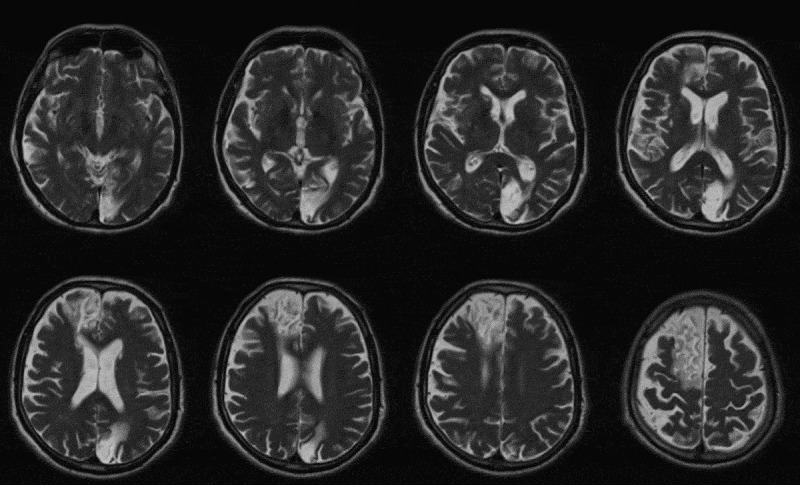
AAP is a 73-year-old man admitted to the neurorehabilitation unit of Casa Cura Policlinico following a right hemispheric stroke conditioning slight left hemiparesis. Brain magnetic resonance imaging (MRI) showed a recent wide subacute ischaemic lesion involving cortico-subcortical right frontal-temporal area and a chronic lesion localised in left occipital lobe caused by a previous ischaemic stroke occurred in 2004 (see Figure 1).
Figure 1.

Brain MRI, T2/FLAIR weighted. Neuroimaging shows bilateral lesion involving both left and right hemispheres, in particular fronto-temporal areas on the right and occipital lobe on the left.
Neuro-ophthalmological examination
AAP underwent an examination including visual-field assessment using standard automated perimetry. Visual-field analysis was carried out using 30-2 and 10-2 Swedish interactive threshold algorithm (SITA) Standard strategy (Humphrey Field Analyser; Carl Zeiss Meditec) with Goldman size III target; near refraction was used.
Best-corrected visual acuity was 20/20 in both eyes; colour vision was normally at Ishihara test. Slit-lamp examination of anterior segment and intraocular pressure were normal; on funduscopy, no retinal nor optic disc changes were found bilaterally. Ocular motility was normal.
Visual field showed right complete homonymous right hemianopia on 30-2 SITA Standard strategy and a partial sparing in superior central 10° on 10-2 SITA.
Neuropsychological evaluation
We examined AAP 1 month after the last stroke with psychometric tests aimed at evaluating the presence of visuospatial neglect, attention, mental reasoning, and memory.
In task involving selective attention, such as star cancellation and bell cancellation, the patient omitted some of the item on the left, highlighting an asymmetry in exploration on a A3 paper sheet. When we asked the patient to explore the room, he was able to explore the right part of the space, finding objects and providing an accurate description of that part of the room. This spontaneous exploration stopped at the median body line and dramatically failed in exploring and finding objects on the left. These observations were compatible with the presence of slight-to-moderate unilateral left visuospatial neglect. In addition, we administered a series of neuropsychological tests in order to evaluate cognitive functioning in the following domains: executive functions, memory, and language. AAP did not show any difficulty in problem solving, mental reasoning, and lexical retrieval: he was able to find a high number of correct responses for the Coloured Progressive Matrices; the execution of Clock Drawing Test provided a near-ceiling score (9 on 10); and verbal fluency was normal. Memory tests did not show any impairment in learning abilities neither in explicitly retrieving the acquired information after some minute interference.
Language subtest highlighted spared abilities in naming objects: the performance was characterised by the presence of a unique visual error: naming “bag” instead of “pocket;” all the other 29 items were correct. This performance provided two-fold information: there was a direct index of lack of impairment in language abilities, name retrieval, and semantic knowledge, with the indirect information of spared visual abilities and absence of visual agnosia.
To test our hypothesis, we realised a categorisation task using a repetition priming paradigm, modified from one described by Schweinberger and Stief.27 This paradigm is usually based on simple go-no-go task, in order to test the priming effect by observing the response time (RTs) and accuracy in responding or not to the prime.
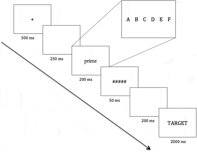
The task consisted of the following series of event (see Figure 2): a fixation point (+), which lasted in the centre of the screen for 500 ms, after that a blank for 250 ms and subsequently a prime-word occurred in six possible positions on the central horizontal line of the screen, corresponding to three positions on the left and three on the right. The extreme position was at 9.6° of visual field to the left or right from the midpoint of the monitor; medial position was at 5.8° from the midpoint and central position at 1.9°. The prime lasted for 200 ms and was immediately followed by a 50-ms mask (string of dashes). After a blank of 200 ms, the target-word appeared and lasted for 2000 ms. Patient sat in front of the monitor at a stable distance of 1 m; monitor dimensions were 41.0 cm width and 26.5 cm height.
Figure 2.

The arrow in bold indicates the sequence of events for any experimental trial. Fixation point lasting for 500 ms; a blank followed by prime-word, which lasted on the screen for 200 ms. The square projection indicates the six possible positions in which the prime-word could occur (A = extreme left to F = extreme right). After the 250-ms blank, the target-word appeared in the centre of the screen.
Target-words were 90 Italian, bi-syllabic, high-frequency words belonging either to living category (e.g., CANE, PAPA [DOG, DAD]) or to non-living category (NIDO, LIBRO [NEST, BOOK]). Word perceptual characteristic were Arial font and a 26 point size; the dimension corresponded to an average value of 3.97° of visual field (standard deviation, 0.62°). All item were combined in order to obtain three different conditions between prime and target: related (e.g., prime: DOG; target: DOG); unrelated (prime: BOOK; target: DOG), and neutral (prime: XXX; target: DOG). We add 54 more bi-syllabic words in order to produce the unrelated conditions (both for living and non-living targets) and avoid the repetition of the prime-words used in related conditions. Any combination was repeated twice in order to have 36 observations for related condition, target “living;” 36 unrelated conditions, target “non-living;” 36 related conditions, target “non-living;” 36 unrelated conditions, target “non-living;” 18 neutral conditions, target “living;” and 18 neutral conditions, target “non-living.” We had finally a total of 180 observations (and the corresponding number of 178 accuracy and RT measures) The experiment was subdivided in five blocks in order to avoid a reduction of attention levels.
The patient sat in front of the monitor at a distance of 1 m; monitor dimensions were 41.0 cm width and 26.5 cm height.
The task consisted of pressing the space bar only when the target-word, appearing in the middle of the monitor, could be deemed as a “living entity” and to not press any key if the target belongs to the category of “non-living entities.” We chose such categorisation task because this is one of the most used experimental paradigm to evaluate semantic activation.25 Moreover, the experimental procedure involves a go-no go paradigm (press if “living,” do not press if it is a “non-living”) in order to let the task be the most feasible for brain-damaged patients. This last reason justify the permanence of the target for an equal time of 2000 ms, even when the patient gave his response, in order to avoid a displacement in target exposure.
After that the patient underwent to an explicit categorisation task; in other words, we used a modified version of the first task in which no prime will be presented whilst target-words will randomly appear in correspondence of the six different positions previously used for prime stimuli.
During this task, we recorded eye movements by means of an infrared-based video tracking (Tobii X120). This system device registers data at sampling rate of 120 Hz with a spatial resolution of less than 0.3°. It provides an accuracy of gaze position relative to stimulus coordinates of 0.5°.
Eye movements were monitored for the time to first fixation and first fixation duration. Fixation was defined as the stable horizontal and vertical eye positions between the end of one saccade and the start of the following saccade, minimal fixation duration was set to 100 ms.
Results
We matched patient results with those obtained from a control group of healthy participants. Seven aged-matched participants took part in the experiment (mean age: 70.9; SD = 10.5). The overall accuracy was more than 95% of a total number of observations counted in 90 items for each experiment, and we run a 3 × 6 repeated-measures analysis of variance (ANOVA) with the following independent factors: target/prime relation (related, unrelated, and neutral) and position (A, B, C, D, E, F). For all of the ANOVAs, the degrees of freedom were corrected using Greenhouse-Geisser epsilon where appropriate. Post hoc comparisons (contrast analyses) were applied to the data. Bonferroni test was applied for multiple comparisons. Data showed a significant main effect on relation (F(1, 2) = 29.75; p < 0.001; η2 = 0.832), whereas no effect arose for position (F(1, 5) = 1.57; p = 0.197; η2 = 0.208). Moreover, the interaction effect between these two variables did not reach significance (F(1, 10) = 1.41; p = 0.199; η2 = 0.190). These results show faster reaction times in case of relation between prime and target (M = 786; SD = 52) with respect to the unrelated pairwise (M = 929; SD = 59) and neutral (M = 888; SD = 44) conditions independently from the position in which the prime occurred.
Patient AAP
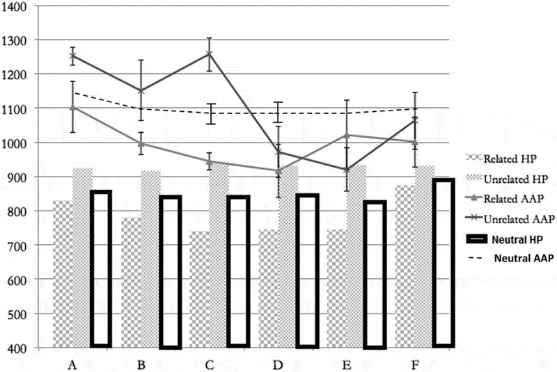
Figure 3 shows AAP’s reaction times compared with those obtained from healthy subjects (patient results are plotted as line). First of all, RTs are on average higher than those obtained from healthy participants. Secondly, AAP showed an effect with respect to the prime position. Actually, we observed a difference between related and unrelated conditions only when the prime appeared in the left side position, namely, AAP had lower RTs in categorising target-word when a related prime appeared in positions A, B, and C. The overall accuracy was inferior to that for normal subjects and was calculated to about 40%, basically due to the number of omissions. We did not find any difference between the two experimental conditions when word-prime was presented on the right side.
Figure 3.

Comparison between patient AAP and healthy participants (HPs) at experimental task. In columns, HP results for both related and unrelated conditions: as expected, HPs have low reaction times for related condition when prime-word occurs in any of the six positions. In lines, AAP results: only when prime-word occurs on the left (A, B, C positions), reaction times are lower in related condition.
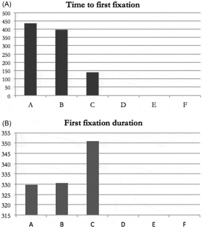
Figure 4A shows the times between prime-word onset and first fixation. AAP took on average 400 ms to orient his gaze for a first fixation towards left extreme positions (columns A and B) and a very short time to move gaze in central position. No movements towards right columns were recorded. In Figure 4B, results concerning the duration of the first fixation are plotted. They seem to be complementary in duration, showing shorter times of fixations in the left columns (A and B) and a longer fixation on the central column.
Figure 4.

(A) Average of AAP’s reaction times to the first fixation. The more extreme was the prime, the longer was time to orient gaze. No right-side movements were recorded. (B) Average of AAP’s durations for first fixation. Length of fixation reduced with a leftward gradient with shorter fixations at the left extremities.
Discussion
The disentanglement between neglect and hemianopia may sometimes present difficulties: the scientific debate points on the different modalities in visual information processing depending on whether there are visual-attentive deficits or a visual-field reduction. The patient here described can thoroughly provide some additional information in this debate mainly because of his clinical characteristics. Indeed, anatomical lesion sites lead to a clear and undoubted clinical phenomenon: a right fronto-temporal lesion determines a left visual neglect, with no visual-field deficits, and from a left occipital lesion originates a right homonymous hemianopia, without neglect. Experimental findings on this patient are in line with our hypothesis: although our patient was unable to find objects or stimuli on the left side, he showed an implicit processing of meaning for the prime-word. On the other hand, in the hemianopic space, none of the prime-words were seen implicitly processed, and this fact determined a total absence of any facilitatory effect on RTs for target-word response.
The choice of this experimental paradigm was based on previous research evidence: the relevance and the type of the stimuli are two variables that dramatically influenced visual processing even without conscious awareness.29 First of all, the use of words instead of pictures or more general stimuli with emotional contents could permit avoiding the possibility of a blindsight effect in hemianopic field. So far, no evidence was found on processing of words presenting in hemianopic fields, and these data seem still to confirm. Secondly, as previously described, words seem to be processed at either lexical or semantic level when presented in neglect space; the use of a repetition priming paradigm was aimed at strongly magnifying the priming effect in order to provide a task that could be used as a sensitive instrument of assessment for neglect and hemianopia possibly superimposed. Actually, although concerning a single patient, our findings show a wide dissociation between related and unrelated conditions only in left space. These relevant effects were observed for both related (with facilitation effect and lower RTs) and unrelated (with interference effect and higher RTs) conditions. Specifically, comparing the left-right spatial gradient, we may suggest that a clear facilitation effect is observable when patient moves from left to right positions. Indeed, a significant reduction of RTs was gradually present from left to right, as well as a significant increased interference effect was revealed with the same direction. It should be noted that a relevant higher interference was revealed in the case of the C position (the more medial position). This is an interesting result, since we can underline the presence of the maximum interference precisely in that position where the gradient effect (reduction of hemispatial neglect) reaches its higher value. In other words, we may suggest a sort of maximum gain for the related primes and maximum interference for unrelated primes exactly as attended in control group.
In this context, our patient represents a rare clinical situation whose peculiarity certainly helped us in studying different types of perception. Thus, on one hand, what we observed confirms the issue about different visual processing; on the other hand, it supports the hypothesis that the presence or absence of priming effect could be a key point to differentiate between visuospatial deficit and visual-field reduction.
Besides, eye-movement analysis leads us to further observations. Firstly, confirming what previously described by some authors, automatic orientation of attention may be spared in patients with neglect. In few words, even though patients are not able to voluntarily orient their attention towards contralesional space, some of them may be captured by occasional and unexpected stimuli occurring in the neglected space, this automatic orientation being accompanied by eye movements.30 Actually, AAP presented this specific pattern showing very rapid gaze orientation at prime-word onset towards the left space. We can briefly describe his explorative behaviour as rapid eye orientations towards the sites of the prime onset, but since the time to first fixation on the left sites were in average 350 ms whereas the prime onset lasted 200 ms, we can reasonably assume that the patient did not overtly see the word but had just an implicit processing of it. Secondly, AAP’s visual exploration was limited to the gaze orientation and followed as expected a gradient of attention, such as a progressive and gradual increase of visual exploration from the extreme left to the right. In our previous works,28,31 we observed behavioural and eye-movement variations along a left-to-right horizontal continuum in patients with neglect. We modified the line bisection test in a space bisection task and asked patients to find the exact half-point between two extremities. According to Bisiach et al.,32 we then modify distance between extremities and dislocation of segments on the screen in order to have an interaction between length (shorter vs. longer) and position (extreme left vs. extreme right). We found a strong variation of rightward bias along the space continuum, providing evidence of an extreme-left gradient of severity of neglect. In the present work, we demonstrate that AAP showed different variations of gaze orientation towards left but priming effect dramatically increased as more prime was presented towards the right side until the position was close to the centre of the screen. As third point concerning eye movements, no explorative eye direction or gaze captures were found on the right side. These data strongly support RT observations: AAP was unable to see prime-word on the right space and he did not process it in any way, and this can thoroughly explain the absence of any facilitation effect on target-word. One may be concerned about the absence of any directional movements toward the right side; in particular, we wondered why AAP, whose cognitive profile was lack of any executive impairments, never had the doubt that if some word “seldom and quickly” appeared on the left “never” appeared on the right. In fact, AAP never tried to orient his gaze towards hemianopic field. We think that the answer could be found in a weakness of anosognosia for hemianopia.33 As a matter of fact, during neuropsychological interviews, AAP never expressed spontaneously the presence of a visual-field defect, but he reported some difficulties in seeing objects on the right only after specific questions (e.g., “how about your sight, can you see everything in the same way both on left and right?”).
In conclusion, the case here described presents some peculiar characteristics that make him exemplificative for a test aimed at disentangling between neglect and hemianopia. Indeed, since our patient showed a double condition (both neglect and hemianopia deficit), he can furnish important evidence to distinguish between these two syndromes. More specifically, the priming paradigm offers relevant indications on how to use the differential diagnosis and how to test the clinical profile (whether neglect or hemianopia) in order to support successive clinical treatments. In fact, our main aim is about the realisation of a tool that can be useful for clinicians and could integrate with available and most used instruments for neuropsychological and neuro-ophthalmological examinations in order to assess the presence of either neglect or hemianopia. In this framework, despite the fact that AAP did not present these two deficits superimposing each other, as in most of cases, he represented a case of control for himself in studying visual information processing in neglect and in hemianopic fields and definitely provided first evidence on the functionality of this test. Finally, as a limit of the present research, we are aware that applicability should be deeply investigated with a wider range of homogeneous groups of brain-damaged patients, in order to obtain stronger and reliable results about if and how half of the world is perceived.
Declaration of interest
The authors report no conflicts of interest. The authors alone are responsible for the content and writing of the article.
References
- [1].Vallar G. Extrapersonal visual unilateral spatial neglect and its neuroanatomy. Neuroimage 2001;14:S52–S58. [DOI] [PubMed] [Google Scholar]
- [2].Karnath H-O. Spatial attention systems in spatial neglect. Neuropsychologia 2015;75:61–73. [DOI] [PubMed] [Google Scholar]
- [3].Walker R, Findlay JM, Young AW, Welch J.. Disentangling neglect and hemianopia. Neuropsychologia 1991;29:1019–1027. [DOI] [PubMed] [Google Scholar]
- [4].Müller-Oehring EM, Kasten E, Poggel DA, Schulte T, Strasburger H, Sabel BA.. Neglect and hemianopia superimposed. J Clin Exp Neuropsychol 2003;25:1154–1168. [DOI] [PubMed] [Google Scholar]
- [5].Müller-Oehring EM, Schulte T, Kasten E, Poggel DA, Müller I. Parallel interhemispheric processing in hemineglect: relation to visual field defects. Neuropsychologia 2009;47:2397–2408. [DOI] [PubMed] [Google Scholar]
- [6].Ota H, Fujii T, Suzuki K, Fukatsu R, Yamadori A.. Dissociation of body-centered and stimulus-centered representations in unilateral neglect. Neurology 2001;57:2064–2069. [DOI] [PubMed] [Google Scholar]
- [7].Kerkhoff G, Schenk T.. Line bisection in homonymous visual field defects—recent findings and future directions. Cortex 2011;47:53–58. [DOI] [PubMed] [Google Scholar]
- [8].Kuhn C, Rosenthal A, Bublak P, Grotemeyer KJ, Reinhart S, Kerkhoff G.. Does spatial cueing affect line bisection in chronic hemianopia? Neuropsychologia 2012;50:1656–1662. [DOI] [PubMed] [Google Scholar]
- [9].Daini R, Angelelli P, Antonucci G, Cappa SF, Vallar G.. Exploring the syndrome of spatial unilateral neglect through an illusion of length. Exp Brain Res 2002;144:224–237. [DOI] [PubMed] [Google Scholar]
- [10].Kinsbourne M, Warrington EK.. A disorder of simultaneous form perception. Brain 1962;85:461–468. [DOI] [PubMed] [Google Scholar]
- [11].Marshall JC, Halligan PW.. Blindsight and Insight in visuo-spatial neglect. Nature 1988;336:766–767 [DOI] [PubMed] [Google Scholar]
- [12].Peru A, Moro V, Avesani R, Aglioti S.. Overt and covert processing of left-side information in unilateral neglect investigated with chimeric drawings. J Clin Exp Neuropsychology 1996;18:621–630. [DOI] [PubMed] [Google Scholar]
- [13].Van Fleet TM, Robertson LC.. Implicit representation and explicit detection of features in patients with hemispatial neglect. Brain 2009;132:1889–1897. [DOI] [PMC free article] [PubMed] [Google Scholar]
- [14].Della Sala S, Van der Meulen M, Bestelmeyer P, Logie RH.. Evidence for a workspace model of working memory from semantic implicit processing in neglect. J Neuropsychol 2010;4:147–166. [DOI] [PubMed] [Google Scholar]
- [15].Weiskrantz L, Warrington EK, Sanders MD, Marshall J.. Visual capacity in the hemianopic field following a restricted occipital ablation. Brain 1974;97:709–728. [DOI] [PubMed] [Google Scholar]
- [16].Peretz C, Chokron S.. Rehabilitation of homonymous hemianopia: insight into blindsight. Front Integr Neurosci 2014;8:82. [DOI] [PMC free article] [PubMed] [Google Scholar]
- [17].Van den Stock J, Vandenbulcke M, Sinke C, Goebel R, de Gelder B.. How affective information from faces and scenes interacts in the brain. Social cognitive and affective neuroscience. Soc Cogn Affect Neurosci 2014;9:1481–1488. [DOI] [PMC free article] [PubMed] [Google Scholar]
- [18].Cecere R, Bertini C, Maier ME, Làdavas E.. Unseen fearful faces influence face encoding: evidence from ERPs in hemianopia patients. J Cogn Neurosci 2014;26:2564–2577. [DOI] [PubMed] [Google Scholar]
- [19].Tamietto M, Castelli L, Vighetti S, Perozzo P, Geminiani G, Weiskrantz L, de Gelder B.. Unseen facial and bodily expressions trigger fast emotional reactions. Proc Natl Acad Sci U S A 2009;106:17661–17666. [DOI] [PMC free article] [PubMed] [Google Scholar]
- [20].McClelland J, Rumelhart D.. An interactive activation model of context effects in letter perception: part 1. An account of basic findings. Psychol Rev 1981;88:375–407. [PubMed] [Google Scholar]
- [21].Tulving E, Schacter DL.. Priming and human memory systems. Science 1990;247:301–306. [DOI] [PubMed] [Google Scholar]
- [22].Arend I, Aisenberg D, Henik A.. Social priming of hemispatial neglect affects spatial coding: evidence from the Simon task. Conscious Cogn 2016;45:1–8. [DOI] [PubMed] [Google Scholar]
- [23].Collins AM, Loftus EF.. A spreading activation of semantic processing. Psychol Rev 1975;82:407–428. [Google Scholar]
- [24].Meyer DE, Schvaneveldt RW.. Facilitation in recognizing pairs of words: evidence of a dependence between retrieval operations. J Exp Psychol 1971;90:227–234. [DOI] [PubMed] [Google Scholar]
- [25].Viggiano MP, Marzi T, Forni M, Righi S, Franceschini R, Peru A.. Semantic category effects modulate visual priming in neglect patients. Cortex 2012;48:1128–1137. [DOI] [PubMed] [Google Scholar]
- [26].Kanne SM. The role of semantic, orthographic, and phonological prime information in unilateral visual neglect. Cogn Neuropsychol 2002;19:245–261. [DOI] [PubMed] [Google Scholar]
- [27].Schweinberger SR, Stief V.. Implicit perception in patients with visual neglect: lexical specificity in repetition priming. Neuropsychologia 2001;39:420–429. [DOI] [PubMed] [Google Scholar]
- [28].Balconi M, Amenta S, Sozzi M, Cannatà AP, Pisani L.. Eye movement and online bisection task in unilateral patients with neglect: a new look to the ‘gradient effect’. Brain Inj 2013;27:310–317. [DOI] [PubMed] [Google Scholar]
- [29].Driver J, Vuilleumier P.. Perceptual awareness and its loss in unilateral neglect and extinction. Cognition 2001;79:39–88. [DOI] [PubMed] [Google Scholar]
- [30].Làdavas E, Carletti M, Gori G. Automatic and voluntary orienting of attention in patients with visual neglect: horizontal and vertical dimensions. Neuropsychologia 1994;32:1195–1208. [DOI] [PubMed] [Google Scholar]
- [31].Balconi M, Sozzi M, Ferrari C, Pisani L, Mariani C.. Eye movements and bisection behavior in spatial neglect syndrome: representational biases induced by the segment length and spatial dislocation of the stimulus. Cogn Process 2012;13:S89–S92. [DOI] [PubMed] [Google Scholar]
- [32].Bisiach E, Pizzamiglio L, Nico D, Antonucci G.. Beyond unilateral neglect. Brain 1996;119:851–833. [DOI] [PubMed] [Google Scholar]
- [33].Vallar G, Ronchi R.. Anosognosia for motor and sensory deficits after unilateral brain damage: a review. Restor Neurol Neurosci 2006;24:247–257. [PubMed] [Google Scholar]


