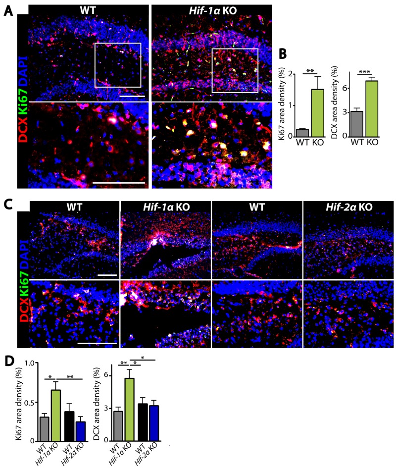
Figure 4. Hif-1α KO mice have increased proliferating immature neurons in the dentate gyrus of hippocampus after MCAO.
(A) Immunostaining images of dentate gyrus of the hippocampal areas in WT or Hif-1α KO mice at d5 post-MCAO for immature neurons and proliferating cells by using antibodies against DCX (red) and Ki67 (green), respectively. (B) Quantification of Ki67 (left) and DCX (right) area densities from A. (C) Immunofluorescent images of dentate gyrus regions in WT, Hif-1α KO or Hif-2α KO at d7 post-MCAO for proliferating neurons as in A. Nuclei are shown in blue and scale bars indicate 100 μm. (D) Quantification of Ki67 and DCX from C. Data in B and D are the mean ± s.e.m. for at least three independent fields examined per mouse, n ≥ 4 mice per group. *, **, and *** denote P < 0.05, 0.01, and 0.001, respectively.

