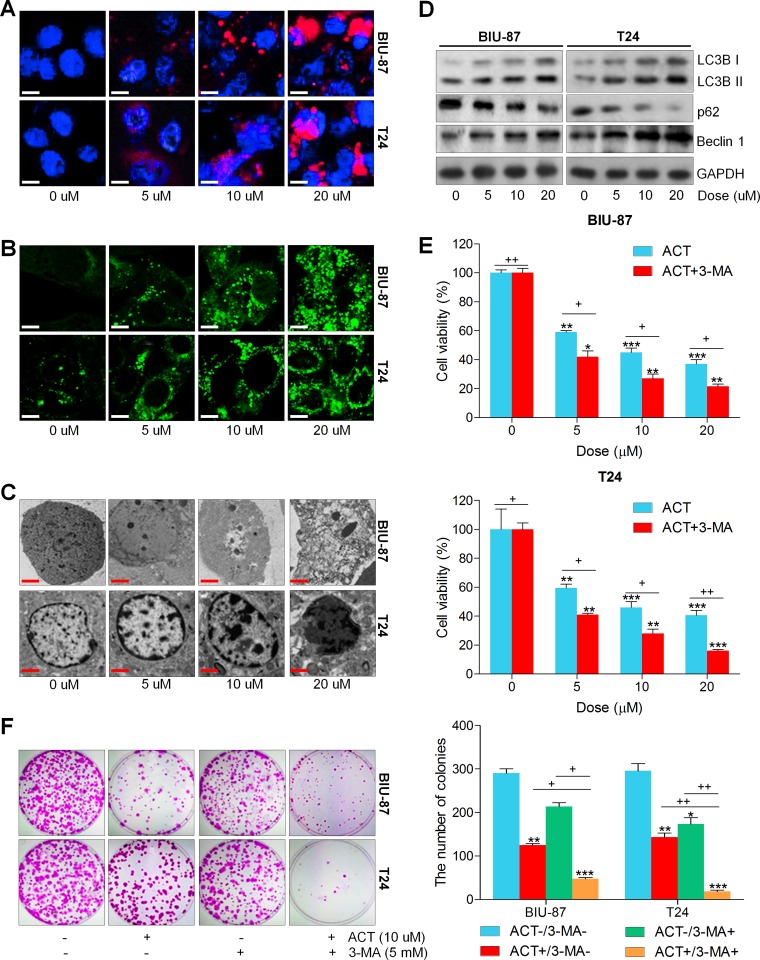
Figure 3. Actein triggers autophagy in human bladder cancer cells.
(A) Human bladder cancer cell lines of BIU-87 and T24 were exposed to ACT for 24 h. Then, all cells were harvested for LysoTracker Red DND-99 (50 nM) analysis to evaluate the cellular acidic compartments, revealing the autolysosomes and lysosomes situations. (B) BIU-87 and T24 cells were pre-transfected with GFP-LC3 plasmid for 24 h, followed by ACT administration for another 24 h at the described concentrations. And confocal microscope was used to observe the images of GFP-LC3 fusion proteins. The fluorescent intensity refers to the formation of autophagosomes. (C) BIU-87 and T24 cells were incubated with ACT for 24 h, and then all cells were collected for transmission electron microscopy (TEM) analysis. (D) Autophagy-related proteins, including LC3BI/II, p62 and Beclin 1, were assessed using western blot analysis. (E, F) BIU-87 and T24 cells were pre-treated with 3-MA (5 mM) for 2 h, followed by various ACT exposure for another 24 h. Then, all cells were collected for MTT and colony formation assays through cologenic analysis. Data are represented as mean ± S.E.M. *p < 0.05, **p < 0.01, ***p < 0.001 versus the untreated group; +p < 0.05 and ++p < 0.01.

