
Figure 3. In vivo expression of nociceptin (Noci) and nociceptin receptor (NOR) in developing human astrocytes.
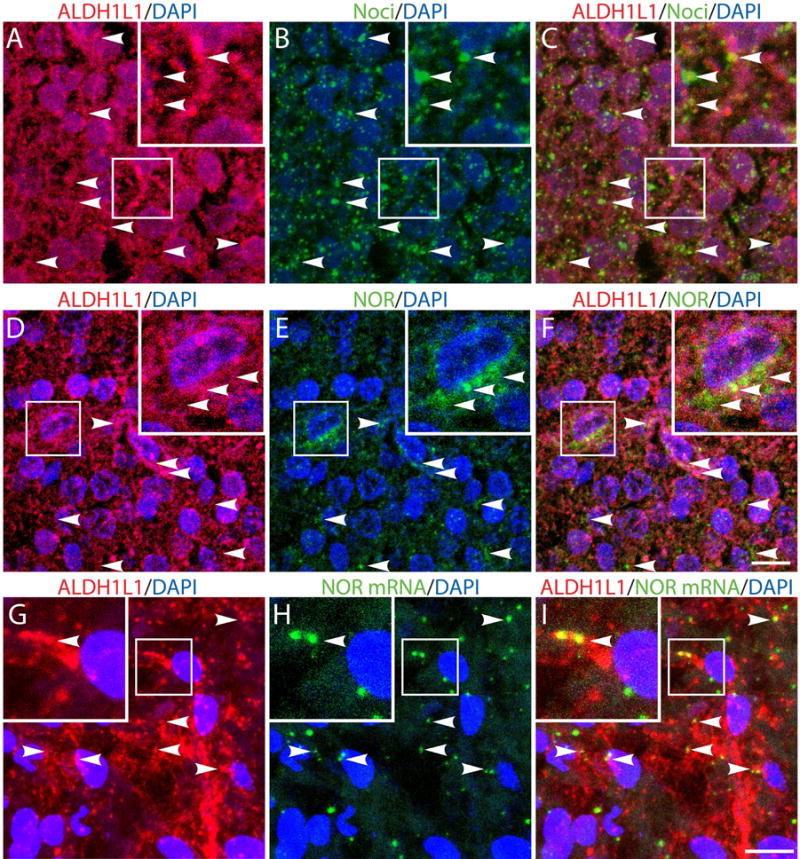
(A-F) Gestational week 23, fetal cortical brain tissue slices were subjected to immunohistochemistry using (A and D) anti-ALDH1L1 together with (B) anti-Noci or (E) anti-NOR antibodies. (G-I) Cortical brain slices were subjected to immunohistochemical staining with (G) anti-ALDH1L1 antibody and to in situ hybridization using (H) a digoxigenin-labeled probe for NOR mRNA, as indicated under “Methods”. Notice the presence of nociceptin (C), NOR protein (F) and NOR mRNA in the ALDH1L1-labeled astrocytes (I). Scale bar: 10μm. Controls for immunocytochemistry using the appropriate normal sera as well as in situ hybridization controls using nonsense deoxyoligonucleotide probes are shown in Supplemental Figures 1 and 2, respectively.