Abstract
AIM
To evaluate the visual outcomes of simultaneous non-topography guided photorefractive keratectomy (PRK) and corneal collagen cross-linking (CXL) in eyes with keratoconus 5y after the procedure.
METHODS
Prospective, interventional, non-randomized, and non-controlled case series design was used. Sixty eyes of 30 patients (16 males and 14 females; age: 21-41y) with mild, non-progressive (stages 1-2) keratoconus were enrolled. Refraction, uncorrected distance visual acuity (UDVA) and corrected distance visual acuity (CDVA), flat and steep keratometry readings, and adverse events were evaluated preoperatively and postoperatively. Data were collected preoperatively and postoperatively at 3mo, 1, 2, 3, 4, and 5y follow-up visits after combined non-topography-guided PRK with CXL was performed. All patients had at least 5y of follow-up.
RESULTS
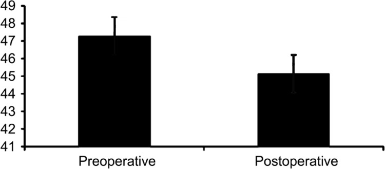
All study parameters showed a statistically significant improvement at 5y over baseline values. The mean follow-up time was 68.20±4.71mo (range: 60-106mo). Patients showed a significant improvement in UDVA from 1.24±0.79 logMAR prior to combined non-TG-PRK+CXL to 0.06±0.15 logMAR postoperatively at the time of their last follow-up visit. CDVA significantly increased from 0.06±0.19 logMAR preoperatively to 0.03±0.12 logMAR postoperatively. A significant decrease in the mean spherical equivalent (SE) refraction was observed from -2.28±1.8 to -0.79±0.93 diopters (D) (P<0.05), and the manifest sphere decreased from -1.62±1.23 to -0.27±0.21 D (P=0.001). The manifest cylinder significantly decreased from -1.73±0.86 to -0.29±0.34 D postoperatively (P=0.001). The mean steep keratometry was 45.13±1.32 vs 47.28±2.12 D preoperatively (P<0.05), and the preoperative mean steepest keratometry (Kmax) 48.6±3.1 was reduced significantly to 46.8±2.9 postoperatively (P<0.05).
CONCLUSION
Combined non-TG-PRK with 15min CXL is an effective and safe option for correcting mild refractive error and improving visual acuity in patients with mild stable keratoconus.
Keywords: non-topography guided, photorefractive keratectomy, corneal collagen cross-linking, keratoconus
INTRODUCTION
Keratoconus is a bilateral, non-inflammatory ectatic disease characterized by central corneal thinning, corneal apical protrusion, and irregular astigmatism[1]–[2]. Classic features of the disease include focal scarring, breaks in Bowman's layer, and iron deposits in the epithelial basement membrane[2]–[3]. Patients may also develop photophobia, glare, and monocular diplopia[4]. As the disease progresses, myopia increases and image quality is further reduced by higher-order ocular aberrations[5]. Eventually, myopia and irregular astigmatism lead to a decrease in visual acuity[6]–[7]. In advanced disease, axial corneal scarring can develop, leading to further impairment of vision[2]. The exact etiology and pathogenesis of keratoconus are not known; however, various genetic and environmental risk factors have been implicated[1]–[2],[8].
The main aims of the keratoconus treatment are to stop the progression of ectasia, improve refractive error and aberrations, and restore the normal shape of the cornea[9]. The management of keratoconus varies with disease severity. Historically, mild keratoconus has been managed with eyeglasses while moderate keratoconus has been treated with contact lenses. Until recently, the only treatment option for keratoconus in patients intolerant to contact lenses was corneal transplantation. Although penetrating keratoplasty is one of the most common types of corneal transplant, this procedure can be complicated by recurrent disease, serious side effects, and a lengthy recovery time.
When excimer laser refractive surgery was first introduced, it was touted as a procedure that could reduce the need for keratoplasty in keratoconus patients. Unfortunately, reports of increased risk of keratectasia after laser-assisted in situ keratomileusis (LASIK) and photorefractive keratectomy (PRK) caused many surgeons to consider excimer laser treatment as a contraindication for keratoconus for more than a decade. Advances in laser technology and approval of corneal collagen cross-linking (CXL) have allowed practitioners to rethink this position[9]. Today, PRK is again considered as an alternative to keratoplasty when used with CXL[10]. CXL uses UVA to activate riboflavin and create covalent bonds between collagen fibrils, resulting in increased biomechanical strength of the cornea[11]–[12]. The goals of simultaneous treatment with PRK/CXL combination treatment are to strengthen the cornea and stop disease progression with CXL and improve vision through laser ablation.
The aim of this study was to report the long-term visual outcomes of patients who had undergone combined non-topography-guided PRK and CXL (non-TG-PRK+CXL) for the treatment of keratoconus.
SUBJECTS AND METHODS
The present case series gathered information in a prospective manner. The included patients had undergone combined non-TG-PRK+CXL between January 2008 and December 2016 at Magrabi Aseer Eye Hospital, Khamis Mushait, Saudi Arabia.
The Institutional Review Board of King Khalid University in Abha, Saudi Arabia approved the study protocol. Written informed consent was obtained from all patients. This study adhered to the tenets of the Declaration of Helsinki.
Inclusion and Exclusion Criteria
Patients between the age of 21 and 41y with keratoconus (stage 1 or 2), which had not progressed for a year before the study, and had undergone combined non-TG-PRK+CXL were included in the study. Other inclusion criteria are as follows: intolerant to contact lens, had a corneal thickness of >400 µm at the thinnest point, and had no other corneal pathological signs.
Pregnant and lactating women, patients with corneal scarring, previous intraocular surgery, incomplete documentation, insufficient postoperative care or follow-up, concomitant eye disease, autoimmune disease, or with a history of herpetic keratitis were excluded from the study.
Outcome Measures
The primary outcome measures for our study were changes in visual acuity and refraction. All adverse events that occurred during the study period were monitored and documented. All measures were recorded preoperatively and postoperatively at all follow-up visits.
Methodology
Patients planned to be included in the study had previously undergone combined non-TG-PRK+CXL (Quest excimer laser platform, NIDEK Co. Ltd., Japan) with conventional ablation profile. The analysis was restricted to the records of patients with topography pattern consistent with keratoconus and an inferior-superior ratio >1.5 on topography mapping and a preoperative corrected distance visual acuity (CDVA) ≥0.3 (logMAR). All had stage 1 or 2 keratoconus as defined based on the Amsler-Krumeich classification (i.e., minimum corneal thickness >400 µm, mean keratometry readings <53.00 D with myopia and/or astigmatism not more than 8.00 D, and no corneal scarring)[13]–[15].
Clinical Evaluation
Pre- and postoperative ophthalmologic evaluation
Standard demographic information, medical and family history were collected from each patient chart. Preoperative evaluation consisted of complete ophthalmic examinations, including autorefractometry and autokeratometry (Nidek Autorefractor Keratometer, Middlesex, UK), corneal topography (OCULUS-Pentacam®, Optikgeräte GmbH, Wetzlar, Germany), and slit-lamp examination of the anterior and posterior segments of the eye. Pre- and postoperative uncorrected distance visual acuity (UDVA) and CDVA, keratometry, and manifest refraction were measured before surgery and at each postoperative visit. All patients were followed postoperatively. The first follow-up visit was at 5d, after which patients were seen at 3mo and yearly for 5y.
Surgical procedures
All procedures were performed by the same surgeon (Al-Amri AM). After topical anesthesia with benoxinate HCl, Benox® 0.4% eye drop (Benox®, Eipico, Egypt) was applied, and the epithelium was removed mechanically using a Beaver surgical blade (8.00 mm in diameter). PRK was performed with Quest excimer laser platform (NIDEK Co. Ltd., USA). Mitomycin 0.2 mg/mL was then applied for 10s, and PRK was immediately followed by CXL.
Hypotonic riboflavin solution (0.1% topical riboflavin sodium phosphate) was added and then ultrasonic pachymetry (Sonogage Corneo-Gage Plus, USA) was performed. If the cornea was <400 µm, additional hypotonic riboflavin solution was administered for 30min until the stroma had swollen to at least 400 µm. The cornea was then exposed to UVA (UV-X 1000; IROC, Zurich, Switzerland) 365 nm light for 15min at an irradiance of 3.0 mW/cm2. During UVA exposure, hypotonic riboflavin solution (0.1% one drop every 2min) was continued every 2min.
Postoperatively, topical antibiotic (moxifloxacin) and corticosteroid (fluorometholone) drops (6 hourly and steroid tapered over 6wk) were administered and continued for 1wk and 6wk, respectively. A therapeutic soft contact lens (Acuvue, USA) (soft contact lens was used in all patients) was placed. The contact lens was removed after epithelial healing. Patients were followed for 3mo postoperatively and had complete examinations yearly for 5y.
Statistical Analysis
Statistical analysis was performed using SPSS version 22 (SPSS Inc., Chicago, IL, USA). P<0.05 was considered statistically significant.
RESULTS
Study Patients
A total of 30 patients between the age of 21 and 41y of age (28.5±5.4y) were included in the study, and almost half of the study population was male (n=16, 53.3%) (Figure 1). All patients had keratoconus in both eyes. The mean duration of follow-up was 68.20±4.71mo (range: 60-106mo), which was done at 3mo and yearly for 5y postoperatively (Table 1). However, the results were collated by comparing clinical parameters at preoperative visit with last follow-up visit (5y).
Figure 1. Gender distribution.
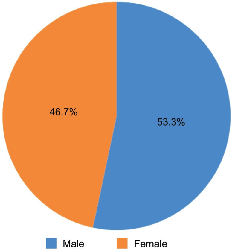
Table 1. Pre- and postoperative follow-up data.
| Parameters | Preoperative | Postoperative |
| Age (a, mean±SD) | 28.5±5.4 | 28.5±5.4 |
| Gender | ||
| M | 16 | 16 |
| F | 14 | 14 |
| UCVA (logMAR) | 1.24±0.79 | 0.06±0.15 |
| CDVA (logMAR) | 0.06±0.19 | 0.03±0.12 |
| Manifest sphere (D) | -1.62±1.23 | -0.27±0.21a |
| Manifest cylinder (D) | -1.73±0.86 | -0.29±0.34a |
| Keratometric astigmatism (D) | -3.21±1.47 | -1.65±0.69 |
| Keratometry (D) | Steep 47.28±2.12 | Steep 45.13±1.32 |
| Flat 43.82±2.8 | Flat 41.6±1.89 | |
| Steepest keratometry (Kmax) | 48.6±3.1 | 46.8 ±2.9 |
| Flat keratometry (Kflat) | 43.82±2.8 | 41.6±1.89 |
| Spherical equivalent refraction (D) | -2.28±1.8 | -0.79±0.93 |
| CT (µm) | 496.1±12.97 | 442.5±15.67 |
UCVA: Uncorrected visual acuity; CDVA: Corrected distance visual acuity; CT: Corneal thickness. aP<0.05, n=30 patients (60 eyes).
mean±SD
Visual Outcome
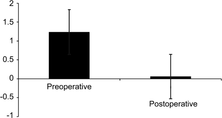
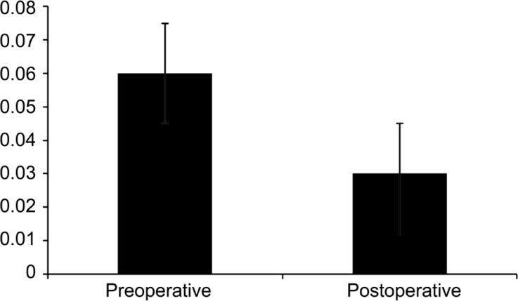
All patients showed a significant (P<0.05) improvement in visual acuity and refraction after surgery at 5-year follow-up visit. Patients showed a significant improvement (P<0.05) in UDVA from 1.24±0.79 logMAR (range 0.7 to 1.7) prior to combined non-TG-PRK+CXL to 0.06±0.15 logMAR (range 0.04 to 0.7) at the time of their last follow-up visit (Figure 2). Seventy-eight percent of patients improved by 2 or more lines of uncorrected distance visual acuity postoperatively. CDVA significantly increased from 0.06±0.19 logMAR preoperatively (range 0.05 to 0.7) to 0.03±0.12 logMAR postoperatively (range 0.04 to 0.6) (P<0.05) (Figure 3). Eighty-one percent of eyes improved by 2 or more lines of CDVA postoperatively. In terms of safety, no eye lost lines of CDVA.
Figure 2. UDVA (logMAR) mean values.
Figure 3. CDVA (logMAR) mean values.
Refractive Outcome
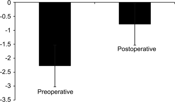
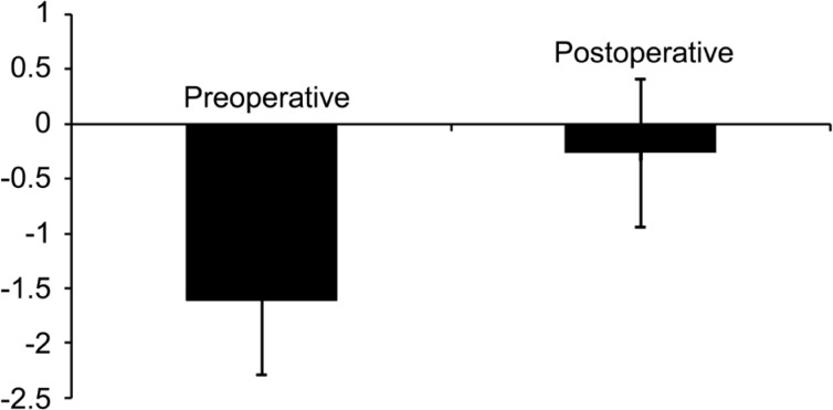
A significant decrease in the mean spherical equivalent (SE) refraction was observed from -2.28±1.8 (range 1.75 to 4.75) to -0.79±0.93 diopters (D) (range 1.75 to 0.75) (P<0.05), and the mean manifest cylinder decreased from -1.73±0.86 preoperatively (range -5.5 to 0) to -0.29±0.34 postoperatively (range 0.50 to 0) (P=0.001) (Figures 4, 5).
Figure 4. SE refraction (D) mean values.
Figure 5. Manifest cylinder (D) mean values.
Topographic Outcome
The mean keratometric values (Ksteep) were significantly reduced at 5-year follow-up visit, from 47.28±2.12 preoperatively to 45.13±1.32 D postoperatively (P<0.05) (range 44.9 to 49.7).
The preoperative mean steepest keratometry (Kmax) 48.6 (SD 3.1, range 47.4 to 52.9) was reduced significantly to 46.8 D (SD 2.9, range 46.2 to 53.4) postoperatively (P<0.05).
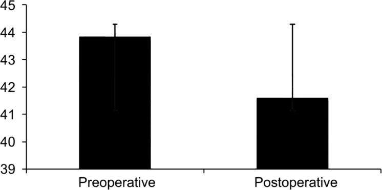
The preoperative mean flat keratometry (Kflat) 43.82±2.8 (range 42.7 to 46.8) was reduced significantly to 41.6±1.89 (range 40.2 to 44.1) postoperatively (P<0.05) (Figures 6, 7).
Figure 6. Keratometry (D) steep mean values.
Figure 7. Keratometry (D) flat mean values.
No evidence of Kmax progression over the follow up period (P>0.05).
Preoperative mean keratometric astigmatism was reduced from -3.21±1.47 D (SD 2.87, range 2.72 to 4.38) preoperatively to -1.65±0.69 postoperatively (SD 2.18, range 1.5 to 2.75) (P=0.05).
Adverse Events
There were no serious intraoperative or postoperative complications or adverse events (AEs) reported in any of the patients. Two patients (4 eyes) developed mild corneal haze. None of the subjects developed infectious keratitis. Only three patients (6 eyes) experienced dry eyes.
DISCUSSION
Conventionally, keratoconus has always been considered a contraindication for PRK; however, clinical studies have shown that CXL alone is also not an effective treatment for keratoconus. Hence, for better results, CXL should be combined with other surgical options as well[16]–[21]. This trend has highlighted the possibility of combining CXL and PRK as a treatment option for patient with stable keratoconus[16]. The two most commonly used PRK+CXL techniques are TG-PRK+CXL and non-TG-PRK+CXL.
Although studies have demonstrated the efficacy and safety of TG-PRK plus CXL for patients with mild to moderate keratoconus, yet the lack of data regarding the long-term stability of TG-PRK plus CXL remained the major limitation of this procedure[4],[9].
Currently, only two studies have evaluated the efficacy of non-TG-PRK+CXL in correcting refractive errors in patients with keratoconus[17],[21]. The findings of these studies suggest that for patients with mild-to-moderate keratoconus, non-TG-PRK+CXL is safe and effective. Fadlallah et al[17] reported that non-TG-PRK after intracorneal ring segment (ICRS) implantation and CXL was found to be an effective and safe option for correcting residual refractive error and improving visual acuity in patients with moderate keratoconus[21], Additionally Fadlallah et al[17] evaluated the safety and clinical outcome of combined non-TG-PRK and CXL for the treatment of mild refractive errors in patients with early stage keratoconus and reported that combined non-TG-PRK and 30min CXL is an effective and safe option for patients with early stable keratoconus[17]. Also, in the present study, all patients were followed postoperatively with the first follow-up visit at 5d, followed by which the patients were seen at 3mo and yearly for 5y. Such prolonged follow-up of patients helped provide important information on both efficacy and safety outcomes. Our study evaluated the efficacy of non-TG-PRK combined with 15min CXL in mild-to-moderate stage of keratoconus with no hyperopic shift and no haze.
The CDVA (0.03±0.12) was found to be similar in both studies, but the UDVA (0.06±0.19) was better in the current paper. The keratometry (flat/steep) and SE were also found to be better with the techniques used in the current paper. However, the small sample size (30 patients, 60 eyes) and single center study are the major limitations of our study.
In conclusion, combined non-TG-PRK+CXL demonstrate good 5-year outcomes in patients with mild, stable keratoconus. Therefore, we recommend conducting future large scale, comparative, randomized trials with extended duration of follow up to establish the long-term stability of this procedure in keratoconus. Findings from such studies could prove helpful in having generalized clinical guidelines and strategies for the management of keratoconus
Acknowledgments
Conflicts of Interest: Al-Amri AM, None.
REFERENCES
- 1.Rabinowitz YS. Keratoconus. Surv Ophthalmol. 1998;42(4):297–319. doi: 10.1016/s0039-6257(97)00119-7. [DOI] [PubMed] [Google Scholar]
- 2.Krachmer JH, Feder RS, Belin MW. Keratoconus and related noninflammatory corneal thinning disorders. Surv Ophthalmol. 1984;28(4):293–322. doi: 10.1016/0039-6257(84)90094-8. [DOI] [PubMed] [Google Scholar]
- 3.Andreassen TT, Simonsen AH, Oxlund H. Biomechanical properties of keratoconus and normal corneas. Exp Eye Res. 1980;31(4):435–441. doi: 10.1016/s0014-4835(80)80027-3. [DOI] [PubMed] [Google Scholar]
- 4.Kanellopoulos AJ, Asimellis G. Revisiting keratoconus diagnosis and progression classification based on evaluation of corneal asymmetry indices, derived from Scheimpflug imaging in keratoconic and suspect cases. Clin Ophthalmol. 2013;7:1539–1548. doi: 10.2147/OPTH.S44741. [DOI] [PMC free article] [PubMed] [Google Scholar]
- 5.Miháltz K, Kovács I, Kránitz K, Erdei G, Németh J, Nagy ZZ. Mechanism of aberration balance and the effect on retinal image quality in keratoconus: optical and visual characteristics of keratoconus. J Cataract Refract Surg. 2011;7(5):914–922. doi: 10.1016/j.jcrs.2010.12.040. [DOI] [PubMed] [Google Scholar]
- 6.Caporossi A, Mazzotta C, Baiocchi S, Caporossi T. Long-term results of riboflavin ultraviolet a cornealcollagen cross-linking for keratoconus in Italy: the Siena eye cross study. Am J Ophthalmol. 2010;149(4):585–593. doi: 10.1016/j.ajo.2009.10.021. [DOI] [PubMed] [Google Scholar]
- 7.Vinciguerra P, Albè E, Trazza S, Seiler T, Epstein D. Intraoperative and postoperative effects of corneal collagen cross-linking on progressive keratoconus. Arch Ophthalmol. 2009;127(10):1258–1265. doi: 10.1001/archophthalmol.2009.205. [DOI] [PubMed] [Google Scholar]
- 8.Spörl E, Huhle M, Kasper M, Seiler T. Increased rigidity of the cornea caused by intrastromal cross-linking. Ophthalmologe. 1997;94(12):902–906. doi: 10.1007/s003470050219. [DOI] [PubMed] [Google Scholar]
- 9.Ormonde S. Refractive surgery for keratoconus. Clin Exp Optom. 2013;96(2):173–182. doi: 10.1111/cxo.12051. [DOI] [PubMed] [Google Scholar]
- 10.Kymionis GD, Kontadakis GA, Kounis GA, Portaliou DM, Karavitaki AE, Magarakis M, Yoo S, Pallikaris IG. Simultaneous topography-guided prk followed by corneal collagen cross-linking for keratoconus. J Refract Surg. 2009;25(9):S807–S811. doi: 10.3928/1081597X-20090813-09. [DOI] [PubMed] [Google Scholar]
- 11.Wollensak G. Crosslinking treatment of progressive keratoconus: new hope. Curr Opin Ophthalmol. 2006;17(4):356–360. doi: 10.1097/01.icu.0000233954.86723.25. [DOI] [PubMed] [Google Scholar]
- 12.Dahl BJ, Spotts E, Truong JQ. Corneal collagen cross-linking: an introduction and literature review. Optometry. 2012;83(1):33–42. doi: 10.1016/j.optm.2011.09.011. [DOI] [PubMed] [Google Scholar]
- 13.Kamiya K, Ishii R, Shimizu K, Igarashi A. Evaluation of corneal elevation, pachymetry and keratometry in keratoconic eyes with respect to the stage of Amsler-Krumeich classification. Br J Ophthalmol. 2014;98(4):459–463. doi: 10.1136/bjophthalmol-2013-304132. [DOI] [PubMed] [Google Scholar]
- 14.Ishii R, Kamiya K, Igarashi A, Shimizu K, Utsumi Y, Kumanomido T. Correlation of corneal elevation with severity of keratoconus by means of anterior and posterior topographic analysis. Cornea. 2012;31(3):253–258. doi: 10.1097/ICO.0B013E31823D1EE0. [DOI] [PubMed] [Google Scholar]
- 15.Toprak I, Yaylali V, Yildirim C. A combination of topographic and pachymetric parameters in keratoconus diagnosis. Cont Lens Anterior Eye. 2015;38(5):357–362. doi: 10.1016/j.clae.2015.04.001. [DOI] [PubMed] [Google Scholar]
- 16.Mastropasqua L. Collagen cross-linking: when and how? A review of the state of the art of the technique and new perspectives. Eye Vis (Lond) 2015;2:19. doi: 10.1186/s40662-015-0030-6. [DOI] [PMC free article] [PubMed] [Google Scholar]
- 17.Fadlallah A, Dirani A, Chelala E, Antonios R, Cherfan G, Jarade E. Non-topography-guided PRK combined with CXL for the correction of refractive errors in patients with early stage keratoconus. J Refract Surg. 2014;30(10):688–693. doi: 10.3928/1081597X-20140903-02. [DOI] [PubMed] [Google Scholar]
- 18.Goldich Y, Marcovich AL, Barkana Y, Avni I, Zadok D. Safety of corneal collagen cross-linking with UV-A and riboflavin in progressive keratoconus. Cornea. 2010;29(4):409–411. doi: 10.1097/ICO.0b013e3181bd9f8c. [DOI] [PubMed] [Google Scholar]
- 19.Vinciguerra P, Albè E, Trazza S, Rosetta P, Vinciguerra R, Seiler T, Epstein D. Refractive, topographic, tomographic, and aberrometric analysis of keratoconic eyes undergoing corneal cross-linking. Ophthalmology. 2009;116(3):369–378. doi: 10.1016/j.ophtha.2008.09.048. [DOI] [PubMed] [Google Scholar]
- 20.Kymionis GD, Portaliou DM, Kounis GA, Limnopoulou AN, Kontadakis GA, Grentzelos MA. Simultaneous topography-guided photorefractive keratectomy followed by corneal collagen cross-linking for keratoconus. Am J Ophthalmol. 2011;152(5):748–755. doi: 10.1016/j.ajo.2011.04.033. [DOI] [PubMed] [Google Scholar]
- 21.Guedj M, Saad A, Audureau E, Gatinel D. Photorefractive keratectomy in patients with suspected keratoconus: five-year follow-up. J Cataract Refract Surg. 2013;39(1):66–73. doi: 10.1016/j.jcrs.2012.08.058. [DOI] [PubMed] [Google Scholar]


