

Abstract
Context:
The identification of root canals plays an important role in successful endodontic diagnosis and treatment. An inappropriate identification of canal resulting in incomplete removal of pulp tissue from the root canals is the main reason for the failure of endodontic treatment in molars. Radiographic imaging is an essential investigative tool in successful endodontics. Cone beam computed tomography (CBCT) the imaging is relatively a new method to visualize the roots in all the three planes.
Aims:
This is a study to assess number of root canals in maxillary and mandibular first and second molars on both the right and left sides using CBCT imaging.
Settings and Design:
A total of 100 CBCT images, which were available as soft copies on the hard drive of the computer in the Department of Oral Medicine and Radiology were considered for the study.
Subjects and Methods:
The axial view sections of the maxillary and mandibular arch of 1 mm thickness were examined with a magnification of 250%. The Images were scrolled down from the cementoenamel junction till the apical foramen and the maximum number of canals in each root were recorded.
Statistical Analysis Used:
The collected data were tabulated (Microsoft Excel 2013) and analyzed by independent t-test using statistical analysis software SPSS.
Results:
Among the maxillary first molars, 72.5% had 4 canals with 76.5% of mesiobuccal root having 2 canals. 49% of maxillary second molar had 3 canals with 53.5% of mesiobuccal root having 1 canal. 67.5% of mandibular first molar had 3 canals with 96.5% of mesial root having 2 canals.
Conclusions:
According to this study, the variations in the number of canals were more with respect to maxillary first molars when compared to the other molars. CBCT can provide the clinician with supplemental information about the different root canal configurations for successful root canal treatment.
Keywords: Cone beam computed tomography, endodontics, molar, root canal
INTRODUCTION
The first molars are the first permanent teeth to erupt, approximately at the age of 6-7 yrs. Since they erupt during the early childhood period, they are more likely to be neglected and are highly prone to decay resulting in pulpal pathology demanding root canal treatment. Furthermore, the molars have a complex morphology with deep pits and fissures favoring food accumulation and progression of decay. The identification of root canals plays an important role in successful endodontic diagnosis and treatment. Incomplete removal of pulp tissue from the root canals is the main reason for the failure of endodontic treatment of molars. This problem is further aggravated by the presence of root canals unnoticed by the clinician, coinciding with anatomical variations or additional canals.
Radiographic imaging is an essential investigative tool for diagnosis and management in endodontics. Two-dimensional views of periapical radiographs reveal only limited aspects regarding the number of canals. Furthermore, there is a high chance of geometric distortion of the anatomical structures in the conventional radiographic methods. These problems can be overcome by utilizing advanced imaging techniques which can provide accurate three-dimensional (3D) details of the teeth and the surrounding dentoalveolar structures.
Cone beam computed tomography (CBCT) is a relatively new mode of imaging to visualize an individual tooth or dentition and its relation to adjacent skeletal tissues and to create 3D images of the area to be examined. It is a method of choice to visualize the number of roots. It provides precise, essentially immediate and accurate 3D radiographic images. The improved detection of the root canals should mean that more of the complex root canal is accessed, disinfected, and filled, which in turn improve the outcome of root canal treatment.[1] The aim of this study was to estimate the number of root canals in maxillary and mandibular molars.
SUBJECTS AND METHODS
The objective of the study was to assess the number of root canals in maxillary and mandibular first and second molars on both the right and left sides using CBCT imaging among the South Indian population. Ethical clearance for the study was obtained from the Institutional ethical committee. A total of 100 CBCT images, which were available as soft copies on the hard drive of the computer in the Department of Oral Medicine and Radiology, taken with New Tom V Gievo under Exposure parameters: 110 kVp, 5.3 mA were selected for the study. Inclusion Criteria were; (1) CBCT images with a field of view covering the entire maxilla and mandibular arch. (2) CBCT images with all the maxillary and mandibular first and second molars. (3) CBCT images without any artefacts. (4) CBCT images with clear visibility of all the root canals. Exclusion Criteria were; (1) CBCT images with artefacts hindering the view of canals, (2) CBCT images with any pathology affecting the jaw, and (3) CBCT images with missing first and second maxillary and mandibular molars.
The axial view sections of the maxillary and mandibular arch of 1 mm thickness were examined with a magnification of 250%. The images were scrolled down from the cementoenamel junction till the apical foramen and the maximum number of canals in each root were recorded [Figure 1]. The collected data were tabulated (Microsoft Excel 2013) and analyzed by independent t-test using statistical analysis software SPSS (SPSS Inc. 1989 2007, Version 16, Team EQX, Chicago, USA).
Figure 1.
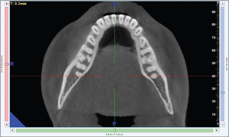
The axial view section of the mandibular arch of 1 mm thickness showing the root canals
RESULTS
In the 100 CBCT images (200 maxillary first molars, 200 maxillary second molars, 200 mandibular first molars, and 200 mandibular second molars) analyzed, altogether 800 teeth were examined. Among the maxillary first molars, 72.5% had 4 canals with 76.5% of mesiobuccal root had 2 canals [Table 1]. 49% of maxillary second molar had 3 canals with 53.5% of mesiobuccal root having 1 canal. 67.5% of mandibular first molar had 3 canals with 96.5% of mesial root having 2 canals [Table 2]. There were no significant changes on comparing the number of canals between right and left side and also between males and females [Tables 3 and 4].
Table 1.
Distribution of number of canals in each root
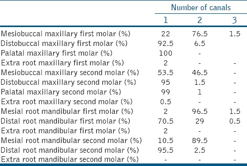
Table 2.
Total number of canals in each tooth
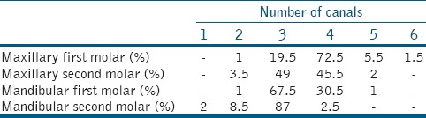
Table 3.
Comparison between males and females
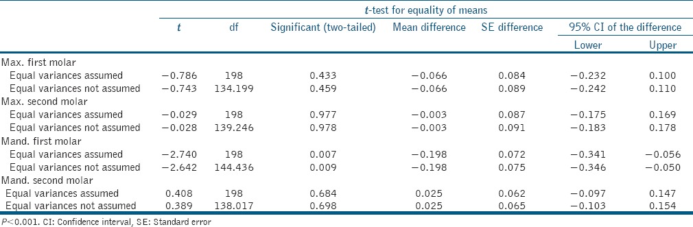
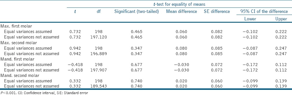
Table 4.
Comparison between right and left side
DISCUSSION
This is a radiographic retrospective study done using New Tom V Gievo CBCT Machine among the 100 subjects. In the 200 upper first molars assessed, the incidence of 2 canals in the mesiobuccal root (76.5%) was greater than the incidence of single canals. Probably because of better visualization using CBCT, studies by Alrahabi and Sohail Zafar.[2] and Lee et al.[3] have mentioned the increased incidence of two canals in the mesiobuccal root similar to our study. However, the percentage of incidence of two canals varies among the different population.
Nearly 53.5% of the mesiobuccal root of the maxillary second molar had a single canal, and 46.5% of mesiobuccal root had two canals. Similar results were obtained by Plotino et al.[4] and Han et al.[5] with an increased incidence of a single canal in the mesiobuccal root. However, the percentage of incidence varied in both the studies probably based on the ethnicity of the population.
In this study, 67.5% of mandibular molars had a total of 3 canals and 30.5% had a total of 4 canals. Similar to our results, a study by Plotino et al.[4] and Demirbuga et al.[6] had also shown an increased incidence of 3 canals in lower first molars. The incidence of 3 canals (87%) in mandibular second molar was seen with a higher percentage than the 4 canals (2.5%) when compared to the number of canals in the mandibular first molar. Similarly, a study by Demirbuga et al.[6] has also revealed an increased incidence of 3 canals in the mandibular second molar. In our study, the number of root canals did not significantly differ between males and females as well as between right and left side indicating a high chance of occurrence of an extra canal if found one side to be present on the other side too.
CBCT is a promising diagnostic modality which provides the clinician with detailed information about the anatomic intricacies and variations which the conventional intraoral radiographs or panoramic images fail to do. Enhanced detection and mapping of the root canal system can result in improved quality of root canal treatment and hence the outcome. However, CBCT is considered as a double-edged sword. Although it has all the mentioned benefits, it also has drawbacks like increased radiation exposure when compared to conventional intraoral periapical radiographs and panoramic radiographs, higher cost, and unavailability in rural areas. The patient's history and clinical examination must justify the use of CBCT by demonstrating that the benefits to the patient outweigh the potential risks.
CONCLUSIONS
This study revealed that variations in the number of canals was more with respect to maxillary first molars when compared to the other molars. Proper attention should be given to the detection of additional canals during root canal treatment of maxillary first molars.
CBCT can provide the clinician with supplemental information about the different root canal configurations for successful root canal treatment. As CBCT imaging also causes increased radiation exposure, if not for the initial treatment; it should be made mandatory before attempting retreatment.
Financial support and sponsorship
Nil.
Conflicts of interest
There are no conflicts of interest.
Acknowledgment
The authors would like to thank A J Institute of Dental Sciences, Mangalore, Karnataka, India.
REFERENCES
- 1.Patel S, Durack C, Abella F, Shemesh H, Roig M, Lemberg K, et al. Cone beam computed tomography in endodontics – A review. Int Endod J. 2015;48:3–15. doi: 10.1111/iej.12270. [DOI] [PubMed] [Google Scholar]
- 2.Alrahabi M, Sohail Zafar M. Evaluation of root canal morphology of maxillary molars using cone beam computed tomography. Pak J Med Sci. 2015;31:426–30. doi: 10.12669/pjms.312.6753. [DOI] [PMC free article] [PubMed] [Google Scholar]
- 3.Lee JH, Kim KD, Lee JK, Park W, Jeong JS, Lee Y, et al. Mesiobuccal root canal anatomy of Korean maxillary first and second molars by cone-beam computed tomography. Oral Surg Oral Med Oral Pathol Oral Radiol Endod. 2011;111:785–91. doi: 10.1016/j.tripleo.2010.11.026. [DOI] [PubMed] [Google Scholar]
- 4.Plotino G, Tocci L, Grande NM, Testarelli L, Messineo D, Ciotti M, et al. Symmetry of root and root canal morphology of maxillary and mandibular molars in a white population: A cone-beam computed tomography study in vivo. J Endod. 2013;39:1545–8. doi: 10.1016/j.joen.2013.09.012. [DOI] [PubMed] [Google Scholar]
- 5.Han X, Yang H, Li G, Yang L, Tian C, Wang Y, et al. A study of the distobuccal root canal orifice of the maxillary second molars in Chinese individuals evaluated by cone-beam computed tomography. J Appl Oral Sci. 2012;20:563–7. doi: 10.1590/S1678-77572012000500012. [DOI] [PMC free article] [PubMed] [Google Scholar]
- 6.Demirbuga S, Sekerci AE, Dinçer AN, Cayabatmaz M, Zorba YO. Use of cone-beam computed tomography to evaluate root and canal morphology of mandibular first and second molars in Turkish individuals. Med Oral Patol Oral Cir Bucal. 2013;18:737–44. doi: 10.4317/medoral.18473. [DOI] [PMC free article] [PubMed] [Google Scholar]