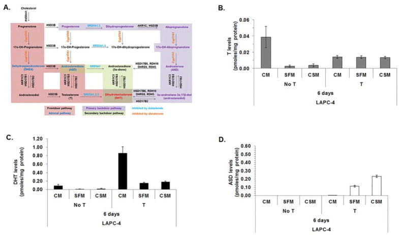
Figure 4. Androgen levels were similar among LAPC-4 cells cultured 6 days in SFM or CSM.
Androgen metabolism pathways for DHT synthesis (A). LAPC-4 cells were analyzed using LC-MS/MS to measure T (B), DHT (C) and ASD (D) levels after LAPC-4 cells were cultured for 6 days in CM, SFM or CSM alone or cultured in CM, SFM or CSM with 1 nM T. Cell pellet and media androgen levels were combined. Cell pellet and media androgen levels were shown in Supplemental Fig. S1. Data were presented as mean pmoles/mg protein +/− SEM. P-values for statistical comparisons among LAPC-4 cells treated with or without T were listed in Supplemental Table S5.

