Abstract
In mammals, spinal cord injury (SCI) leads to dramatic losses in neurons and synaptic connections, and consequently function. Unlike mammals, lampreys are vertebrates that undergo spontaneous regeneration and achieve functional recovery after SCI. Therefore our goal was to determine the complete transcriptional responses that occur after SCI in lampreys and to identify deeply conserved pathways that promote regeneration. We performed RNA-Seq on lamprey spinal cord and brain throughout the course of functional recovery. We describe complex transcriptional responses in the injured spinal cord, and somewhat surprisingly, also in the brain. Transcriptional responses to SCI in lampreys included transcription factor networks that promote peripheral nerve regeneration in mammals such as Atf3 and Jun. Furthermore, a number of highly conserved axon guidance, extracellular matrix, and proliferation genes were also differentially expressed after SCI in lampreys. Strikingly, ~3% of differentially expressed transcripts belonged to the Wnt pathways. These included members of the Wnt and Frizzled gene families, and genes involved in downstream signaling. Pharmacological inhibition of Wnt signaling inhibited functional recovery, confirming a critical role for this pathway. These data indicate that molecular signals present in mammals are also involved in regeneration in lampreys, supporting translational relevance of the model.
Introduction
A fundamental question in regenerative biology is why some organisms can regenerate their central nervous system (CNS), while others cannot1. Unlike mammals, lampreys, fishes, amphibians, and reptiles exhibit robust spontaneous regeneration and functional recovery after SCI2–4. The mammalian peripheral nervous system (PNS) also regenerates after injury5, and intraspinal neurons in mammals can do so when provided a permissive environment, such as a peripheral nerve bridge or growth factors6–9. However, the conserved molecular pathways that promote successful regeneration are unclear10.
We therefore set out to identify deeply conserved pro-regenerative pathways by determining the gene expression changes that occur after SCI in lampreys. The lamprey is a member of an ancient vertebrate lineage that diverged from a common ancestor of humans ~550 million years ago11–13. Despite this evolutionary distance, recent sequencing of the lamprey genome revealed molecular pathways that are conserved with mammals, including genes related to axon guidance and regeneration, synaptic transmission, neural patterning and neurodegeneration12. The organization of the lamprey CNS is highly analogous to human and other jawed vertebrates14,15. Remarkably, lampreys recover locomotor function (e.g. swimming) within 12 weeks after a complete spinal cord transection, which is supported by repair of the spinal lesion, axon regeneration, and synapse formation16–20 (Video S1). To better understand the molecular pathways supporting successful functional recovery, we used RNA-Seq to determine transcriptional profiles in spinal cord of lampreys after SCI. Because many of the regenerating descending axons extend from cell bodies located in the brain, we also profiled the supraspinal transcriptional responses in order to determine their contributions to the recovery process. We identified complex transcriptional responses in both spinal cord and brain throughout the 12 weeks after injury, including a number of expression changes mapping to the Wnt pathway, and used pharmacological blockade of Wnt signaling to demonstrate its critical importance in functional recovery.
Results
Spinal Cord Regeneration in Lampreys
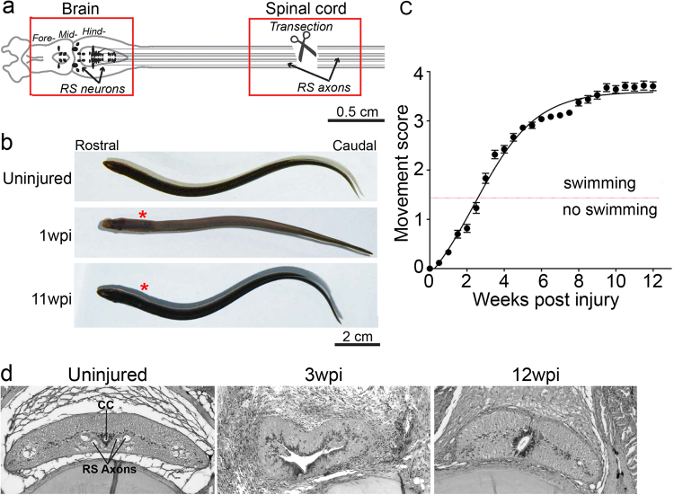
The lamprey CNS contains many homologous structures that are shared with mammals, including a tripartite brain and spinal cord with motor and sensory circuits (Fig. 1a)14,21–23. Within the spinal cord are ~1200 reticulospinal (RS) axons, originating from somata in the midbrain and hindbrain, comprising the major descending pathway for initiating locomotion (Fig. 1a)24. Spinal cord transection severs all axons, leading to paralysis below the lesion that typically lasts 1–2 weeks (Fig. 1a,b; Video S1)16–20. Over the next 10–12 weeks post injury (wpi), lampreys spontaneously recover nearly normal swimming behaviors, which can be described via a quantitative scoring system, where 1 corresponds to an ability to curve into a “C” or “S” shape without translation to forward motion, 2 corresponds to an ability to achieve brief abnormal swimming, 3 corresponds to persistent but abnormal swimming and 4 is normal swimming. (Fig. 1b,c; Video S1) (n = 25–66 animals per time point; R2 = 0.97; t1/2 = 3.43 ± 0.12 weeks)18,25. Animals that reach stage 4 are considered to have achieved full functional recovery. This functional recovery is supported by lesion repair, regeneration of descending and ascending axons, and proliferation of cells that give rise to new neurons and glia16,18,20,26–29. Figure 1d shows the basic cytoarchitecture of the spinal cord during recovery from SCI, as initially shown by Rovainen16. Previously, it has been shown that ~50% of RS axons regenerate beyond the lesion and form synapses with appropriate postsynaptic targets17,18. In the brain, SCI triggers cell death and also regeneration of subpopulations of neurons with descending axons, as well as neurite sprouting from uninjured neurons30–32.
Figure 1.
Robust functional recovery after complete spinal cord transection in lampreys. (a) Diagram of lamprey brain and spinal cord showing location of transection site and tissues collected for molecular profiling (red boxes). Somata of RS neurons (arrows) reside in the midbrain and hindbrain, and their axons (black lines) extend the length of the spinal cord. (b) Images of uninjured lampreys and at 1wpi and 11wpi. Asterisks indicate lesion site. (c) Quantitative scale of functional recovery (i.e. swimming): Movement scores are as described fully in18: 0-paralysis); 1-head wiggle only; 2-brief, abnormal swimming; 3-persistent swimming with abnormal body shape; 4-apparently normal swimming. Data points represent mean ± SEM (n = 27–66 animals per data point). (d) Histological sections of lamprey spinal cord from uninjured, 3wpi, and 12wpi demonstrating cytological changes during the recovery from SCI. CC = central canal, RS axons = reticulospinal axons.
Differential Expression and Enrichment Analysis of Molecular Pathways during Spinal Cord Regeneration and Functional Recovery in Lamprey
Uninjured spinal cord tissues contain many neuronal subtypes (e.g. motoneurons, sensory neurons, interneurons) and glia (e.g. ependymal cells, microglia/macrophages). In addition, we know that injury induces ependymal cells and microglia/macrophages to accumulate at the lesion site28,33–35, though the relative proportions of these and other cell types at each post-injury time point are unknown. Brain tissue contained somata of axotomized neurons with descending axons, as well as other neurons and glia that were not injured directly, but that respond to the injured state. Cell proliferation and neurite sprouting have also been reported in the lamprey brain after SCI28,32. To obtain gene expression profiles accompanying successful spinal cord regeneration and functional recovery in lampreys, we performed RNA-Seq on cDNA libraries generated from spinal cord tissue (1 cm) surrounding the lesion site in controls and at ten time points after SCI, ranging from 6 hours to 12 weeks, and in parallel, from brains of the same animals (Fig. 1a; boxes). RNA-Seq reads were mapped to annotated gene models from the published lamprey genome assembly (https://genome.ucsc.edu/cgi-bin/hgGateway?db=petMar2)12. Standardized expression values for all gene models are reported separately for each time point in brain and spinal cord (see Supplementary Table S1). To confirm homology-based annotations for selected key genes within this study, we performed new BLAST searches for several annotated homologs of human genes that are typically associated with regeneration, proliferation and cell death in mammals. These genes shared ~40–96% amino acid identity and high similarity (50–99% positives, which also include features like charge conservation), with the closest (presumptively orthologous) vertebrate sequence (see Supplementary Table S2).
To determine broad patterns of transcript expression following SCI, we used 2-way hierarchical clustering of expression profiles of spinal cord and brain. In both tissues, the gene expression profiles of the uninjured condition clustered separately from all time points obtained post injury (Fig. 2a,b). In the spinal cord, the clustering of expression profiles reflected progression through the time course of functional recovery, such that acute time points (6hpi to 3dpi) clustered separately from later time points (3 to 12wpi) (Fig. 2a). In contrast, hierarchical clustering of gene expression profiles in brain did not similarly reflect the progression through the time course of functional recovery (Fig. 2b). Notably, expression profiles at 1dpi and 6wpi formed a separate, distinct cluster. Transcriptional profiles at 12wpi in both spinal cord and brain clustered with profiles from other post-injury time points, indicating that altered gene expression occurred even in late stages of functional recovery and not a return to the uninjured state (Fig. 2a,b).
Figure 2.
Differential expression of transcripts after spinal cord injury. (a,b) Dendrograms depict 2-way hierarchical clustering (Ward method) to reveal relationships between transcriptional profiles at experimental time points in spinal cord (a) and brain (b). The number of transcripts that were differentially expressed is shown for spinal cord (c) and brain (d). (e) Line graphs show number of transcripts that are newly differentially expressed at each time point following injury. (f) Venn diagrams show the number of transcripts among differentially expressed transcripts that are shared or unique to spinal cord or brain at each time point. Transcripts corresponding to each panel in F are found in Supplementary Table S3.
Next, we identified transcripts that were differentially expressed after SCI, relative to the uninjured condition, using EBSeq36. These analyses revealed robust and dynamic changes in gene expression in spinal cord and brain throughout the experimental time course (Fig. 2c,d). Notably, a distinct wave of newly differentially expressed transcripts was observed during the first week after SCI, corresponding to the onset of wound healing and tissue morphogenesis, but newly differentially expressed transcripts were detected throughout the course of functional recovery (Fig. 2e). This continued even into late stages of functional recovery (12wpi), at which point 238 and 88 newly differentially expressed transcripts were observed in spinal cord and brain, respectively (Fig. 2e; see Supplementary Table S1). Thus, dynamic changes in gene expression persist throughout the time course of recovery after SCI, even at late stages of behavioral recovery. We observed both shared and tissue-specific responses in expression at all post-injury time points (Fig. 2f; see Supplementary Table S3). This suggests that there are both global transcriptional responses to SCI and more locally tuned responses that reflect intrinsic differences in cell type and varying distances of the spinal cord and brain tissue from the lesion site.
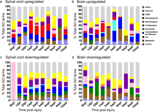
We leveraged functional data from human and mouse to identify likely functions of differentially expressed transcripts using Enrichr, an open bioinformatics platform that utilizes mammalian data37. Analysis of statistically enriched (p < 0.05) Gene Ontology (GO) categories encompassing differentially expressed transcripts indicated complex transcriptional responses in spinal cord and brain at most time points after SCI (Fig. 3a–d; see Supplementary Table S4). Broad functional categories of differentially expressed transcripts were related to immune function, extracellular matrix (ECM) remodeling/deposition, development, neuronal function, proliferation, cell death, cytoskeleton, ion channels, metabolism and transcription/translation (Fig. 3a–d). Previous histological studies of lampreys after SCI showed that cell proliferation occurs in the brain and even more so in the spinal cord, and that neuronal death increases in brain by 3wpi28–31. In agreement with these data, here we found that upregulated transcripts related to cell proliferation were more abundant in spinal cord than brain, while those related to cell death peaked in brain at 3wpi (Fig. 3a,b). Evaluation of differentially expressed transcripts using the Kyoto Encyclopedia of Genes and Genomes (KEGG, www.kegg.jp/kegg/kegg1.html) database indicated similar themes related to immune, ECM, development, neurological, and cytoskeletal processes (see Supplementary Table S5). Taken together, these analyses revealed transcriptional changes in broad functional gene categories after SCI in lampreys that have also been observed in other highly regenerative vertebrate species, such as zebrafish and axolotls, as well as in regenerating mammalian PNS3,38–40.
Figure 3.
Functions of differentially expressed genes. Stacked bar graphs show the percentages (Y-axis) of significantly enriched categories of GO Biological Process (BP) terms encompassing differentially expressed transcripts that are upregulated (a,b) or down-regulated (c,d) in the spinal cord or brain at each time point (p ≤ 0.05). The X-axis indicates the experimental time point. Individual GO terms were manually bundled into larger descriptive groups (see Supplementary Table S4), indicated in the legend.
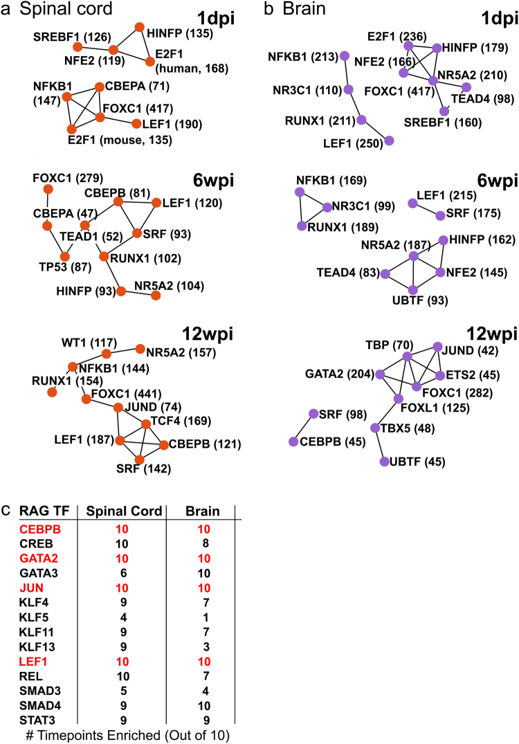
To identify transcription factor (TF) networks that could potentially coordinate molecular responses to SCI in lampreys, we used TRANSFAC/JASPAR to assign the differentially expressed transcripts to TFs known to regulate expression of mammalian homologues. TF networks that regulate the most highly expressed transcripts in uninjured spinal cord and brain are shown in Supplementary Fig. S1. We then analyzed TF networks regulating differentially expressed transcripts after SCI at 1dpi, 6wpi, and 12wpi, corresponding to acute, intermediate, and late phases of functional recovery (Fig. 4). Enriched TF networks differed between tissues at each time point and changed dramatically over time within each tissue (Fig. 4a,b). Notable TFs whose targets were enriched after SCI included: FOXC1, which has been implicated in embryonic development; NFKB1, a master regulator of the immune system and growth factors, and LEF1, a member of the canonical Wnt pathway, which has been implicated in tissue regeneration in several species (Fig. 4a,b)41,42. In addition, 17 TFs with targets enriched amongst differentially expressed genes were previously identified in the mammalian PNS as regeneration-associated genes (RAGs) (Supplemental Table S7), 12 of which are shown in Fig. 4c. Several of these RAGs belong to canonical and non-canonical Wnt signaling pathways (e.g. JUN, LEF1, SMAD) (Fig. 4c)40–42. Interestingly, there were 4 RAGs in the TF networks regulating differentially expressed transcripts in both tissues at all ten post-injury time points: CEBPB, GATA2, JUN, and LEF1 (Fig. 4c-red). Other RAGs with targets enriched amongst differentially expressed transcripts included KLF, SMAD, and STAT family members, which also intersect with Wnt signaling pathways41–43. These data indicate that the lamprey CNS utilizes highly conserved TF networks and signaling pathways that support neural repair and regeneration in the mammalian PNS and in other non-mammalian vertebrates that regenerate after SCI and suggest a role for Wnt signaling40,44–48.
Figure 4.
Transcription factors that are predicted to regulate differentially expressed genes. TRANSFAC/JASPAR was used to infer the transcription factors (TFs) that regulate differentially expressed genes at each time point. TF networks generated by TRANSFAC/JASPAR within Enrichr are shown for spinal cord (a) and brain (b) at 1dpi, 6wpi, and 12wpi. Values shown next to TFs indicate the number of targets that are differentially expressed at that time point. Only the most significant (Fisher’s exact test) TFs are shown for each network. (c) Regeneration associated genes (RAGs) predicted by TRANSFAC/JASPAR to regulate differentially expressed genes. Numbers indicate number of time points when that TF is significantly enriched (P ≤ 0.01, Fisher’s exact test). Red indicates the RAGs whose targets were differentially expressed in both tissues at all 10 post-injury time points, which include targets of the Wnt pathway.
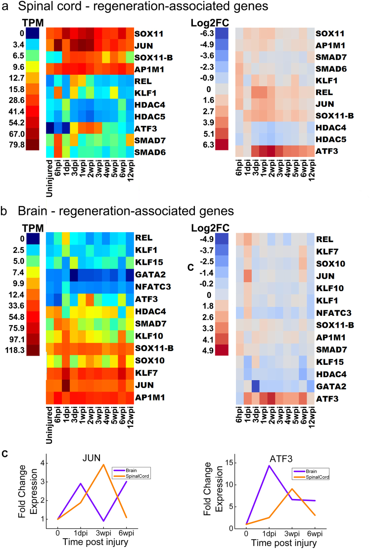
We next examined SCI-induced expression changes in the RAGs themselves (Fig. 5)40,44,45. In the uninjured spinal cord and brain, individual RAGs were expressed at varying levels (Fig. 5a,b, left). SCI induced significant upregulation of most RAGs at one or multiple time points in both spinal cord and brain, beginning as early as 6hpi (Fig. 5a,b, right). As expected, differentially expressed RAGs included many of the specific TFs that were predicted independently in the TRANSFAC analysis, including RAGs that intersect with Wnt signaling: JUN, KLF, REL, and SMAD family members (Fig. 5a,b). Interestingly, ATF3, a member of the CREB TF family that promotes axon regeneration in mammalian PNS, as well as central axon branches in dorsal root ganglion neurons, was the most robustly induced RAG in both spinal cord and brain after SCI (Fig. 5a,b)40,49,50. In addition, homologs of JUN, SOX11, SMAD and REL, which along with ATF3, are positioned centrally within the core TF networks associated with mammalian PNS injury responses, were highly upregulated in spinal cord (Fig. 5)40. In the brain, several RAGs were significantly upregulated specifically at 1dpi and 6wpi (Fig. 5b) reflecting broader patterns identified through hierarchical clustering (Fig. 2a). The temporal expression profiles for JUN and ATF3 were further validated using qPCR, including the upregulation of JUN at 1dpi and 6wpi in brain (Fig. 5c). Together, these data show activation of highly conserved molecular pathways in the lamprey after SCI that also accompany regeneration in the mammalian PNS.
Figure 5.
Temporal expression patterns of regeneration associated genes (RAGs). Heat maps generated from RNA-Seq data showing expression of genes that were differentially expressed at least once during the experimental time course for spinal cord (a) and brain (b). Left panels show expression data and right panels show log2-fold changes (FC) relative to the uninjured state. TPM = transcripts per million reads. Lamprey gene IDs and corresponding gene symbols are found in Supplementary Table S2. (c) qPCR data showing expression changes for JUN and ATF at 1dpi, 3wpi, and 6wpi, compared to uninjured controls (time 0).
We also examined gene expression profiles for other gene families commonly studied in the context of PNS or CNS regeneration, including axon growth and guidance (Supplementary Fig. S2), extracellular matrix (Supplementary Fig. S3), cell proliferation/death (Supplementary Fig. S4), ion channels (Supplementary Fig. S5), and immune function (Supplementary Fig. S6). Broadly speaking, many axonal growth-promoting transcripts were upregulated after SCI (e.g. SNAP25, activin receptor ACVR1), while growth-restricting transcripts were downregulated (e.g. RHOB, SLIT2/3, ROBO2/3) (Supplementary Fig. S2). We also observed an upregulation of neurofilaments (NEFH/L) and synapsin (SYN2/3) gene family members in the brain, as well as differential expression of semaphorins and their receptors (SEMA3/4/5, PLXN) in both tissues, which is in agreement with previously published lamprey SCI studies that used in situ hybridization to detect expression changes (Supplementary Fig. S2)32,51–53. Transcripts associated with ECM deposition and remodeling were generally upregulated [e.g. collagens (COL), laminins (LAM), ADAMs, TIMPs] (Supplementary Fig. S3). Reflecting ongoing tissue repair, many cell proliferation related transcripts were highly upregulated after SCI in both spinal cord and brain (Supplementary Fig. S4)16,18. Interestingly, synuclein (SNCA) (Supplementary Fig. S4), a gene whose overexpression or mutation is linked to neurodegeneration in Parkinson’s disease, is upregulated in the spinal cord after SCI54,55. This increased expression may contribute to the accumulation of synuclein protein that occurs in a subset of lamprey neurons after SCI and leads to neurodegeneration, which we previously reported31,56. Several ion channel transcripts were also differentially expressed after SCI, likely reflecting substantial changes in neuronal excitability during regeneration and functional recovery (Supplementary Fig. S5). In mammals, the immune system has both beneficial and detrimental effects after SCI57,58. In lamprey, robust immune system responses were observed, with cytokines (e.g. IL8) and chemokines (e.g. CXCR4) and prostaglandins, upregulated acutely after SCI (Supplementary Fig. S6). However, this does not inhibit the robust regeneration and functional recovery that is normally achieved in these animals (see Fig. 1). Notably, upregulation of prostaglandins after traumatic brain injury in zebrafish has been previously shown to enhance tissue repair and recovery59. Though this is far from conclusive, these data are consistent with a permissive or positive role for immune responses in recovery from SCI in lampreys.
Wnt Signaling Pathways are differentially expressed after SCI and Necessary for Functional Recovery
Wnt pathways are involved in a variety of biological processes that may be relevant to SCI, including body plan patterning, contact-dependent signaling, cell proliferation, tissue development and regeneration, stem cell self renewal, and axon guidance41–43,60–62. There are 19 mammalian Wnt genes, many of which are conserved across species (including invertebrates) and interact promiscuously with their multimeric receptors involving Frizzled and LRRP proteins. Porcupine (PORCN) is an O-acyltransferase required for Wnt palmitoylation, maturation, and subsequent secretion63,64. Due to the important role of Wnts in various disease settings, including cancer, several small molecules have been identified that target PORCN or other Wnt pathway members60,63,64. The role of Wnt signaling after SCI in mammals is complex and still controversial given the observation of highly variable results among different rat and mouse models42,43,65–67. However, Wnts appear to be pro-regenerative after SCI in zebrafish and after tail injury in salamander41,48,68,69. In fact, Voss and colleagues recently demonstrated that incubation in C59, a drug that inhibits PORCN, blocked salamander tail regeneration63,69.
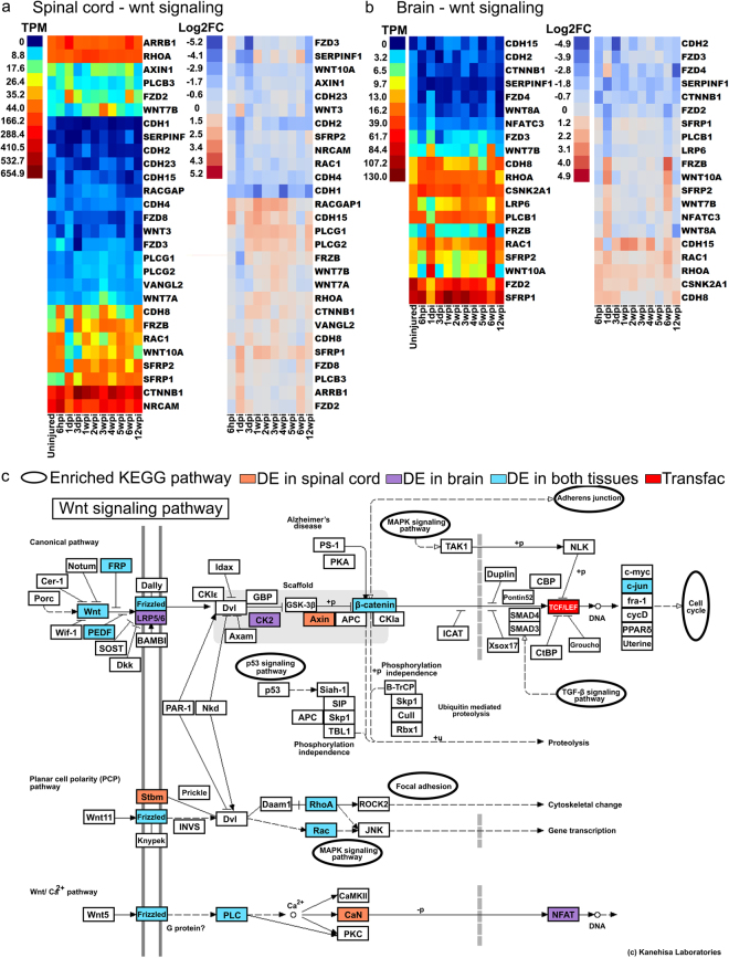
As discussed above, some of the RAGs (e.g. JUN) and other differentially expressed transcripts belong to or intersect with Wnt signaling pathways, both canonical (beta-catenin dependent) or non-canonical (beta-catenin independent) pathways. We identified a total of 121 and 101 differentially expressed transcripts related to Wnt signaling in spinal cord and brain, respectively (Supplementary Table S6). Amongst these were transcripts encoding for a number of Wnt and Frizzled family members, as well as proteins involved in intracellular Wnt signaling (Fig. 6a,b). KEGG analysis revealed how extensively the Wnt signaling pathways were represented amongst the differentially expressed transcripts in spinal cord and brain, as well as TCF/LEF1 that was identified by TRANSFAC/JASPR in the transcription factor networks (Fig. 6c). The number of Wnt related transcripts peaked acutely within the first week post-injury in both spinal cord and brain with additional increases at later time points (Fig. 7a). In light of these data, we decided to directly assess whether Wnt signaling has any effect on functional recovery after SCI in lampreys. We applied a single dose of the PORCN inhibitor C59 (10 μM) or vehicle to the spinal cord at the time and site of transection. Since porcupine is required for Wnt palmitoylation, secretion, and therefore activity, C59 potently inhibits Wnt signaling63. We then scored swimming behavior for 12 weeks, as in Fig. 1c. At 12wpi, when lampreys typically achieve full recovery, the animals that received C59 achieved an average movement score of 1.6 ± 0.6 and thus remained non-swimming or exhibited only brief uncoordinated swimming, while vehicle-treated animals achieved an average score of 3.33 ± 0.2 and swam in a coordinated sinusoidal wave (Fig. 7b).
Figure 6.
Differential expression of Wnt signaling pathway genes after spinal cord injury. (a,b) Canonical and non-canonical Wnt signaling pathways are shown. Genes that were differentially expressed at least once during the experimental time course are indicated in red (up) or blue (down) regulated. Heat maps showing expression of genes that were differentially expressed at least once during the experimental time course for spinal cord and brain. Left panels show raw expression data and right panels show log2-fold changes (FC) relative to the uninjured state. TPM = transcripts per million reads. Lamprey gene IDs and corresponding gene symbols are found in Supplementary Table S2. (c) KEGG analysis of the Wnt signaling pathways showing transcripts identified as differentially expressed in spinal cord (orange), brain (purple) or both (blue). KEGG pathway map 04310 is adapted here from http://www.kegg.jp/kegg/kegg1.html. The KEGG database has been described previously93–95.
Figure 7.
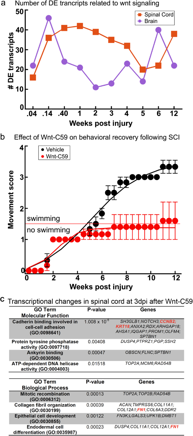
Blocking Wnt signaling inhibits functional recovery after complete spinal cord transection in lampreys. (a) Number of differentially expressed transcripts related to Wnt signaling over time. Lamprey gene IDs and corresponding gene symbols are shown in Supplementary Table S6. (b) Functional recovery (swimming) is inhibited in animals that were treated with Wnt-C59 (red), as compared to animals that were treated with vehicle (black) at the time and site of transection. Functional recovery was scored as in Fig. 1. Data points represent mean ± SEM (n = 5–6 animals per time point). (c) Transcriptional responses in spinal cord at 3dpi after C59 treatment. Gene ontology (GO) analysis is consistent with changes in cell adhesion, differentiation and proliferation. Gene symbols in red indicate direct targets of the Wnt pathway. See also Supplementary Table S6.
To determine the molecular basis of these functional effects, and to confirm the efficacy of C59 in lampreys, we performed RNA-Seq on C59- and vehicle-treated lampreys at 3dpi, a period of rapid and dynamic change in the transcription of Wnt-responsive genes (Fig. 7a). Gene ontology analysis revealed significant expression changes in spinal cord transcripts that were related to cadherin and ankyrin binding; DNA replication and mitotic recombination; collagen and fibrin organization; and cell development and differentiation, all of which are affected by Wnt signaling (Fig. 7c)70,71. Individual transcripts that were differentially expressed in spinal cord after C59 treatment included direct targets of the canonical Wnt signaling pathway, such as cadherin (CDH2), claudin (Cldn19), cyclin (CCNB2), fibronectin (FN1), and keratins (KRT6a/8/18) (Fig. 7c; Supplementary Table S6). In brain, the differentially expressed transcripts included additional targets of Wnt signaling, such as axin (Axin1), Jagged (JAG2), JUN, keratins (KRT7/10), matrix metalloproteinase (MMP9), and T-box transcription factors (Tbr1; TBX20) (Supplementary Table S6). These data indicate that Wnt signaling plays a positive and crucial role early on during the process of functional recovery after SCI in lampreys, likely through effects on cell adhesion and proliferation.
Discussion
Large-scale gene expression analyses are rapidly advancing our understanding of molecular responses in vertebrates to nervous system injury38,40,72. For example, a microarray study of mammalian cortical neurons revealed developmentally regulated genes promoting axon outgrowth and an RNA-Seq study showed that in comparison to mammalian CNS neurons, PNS neurons had greater expression of pro-regenerative genes (i.e. growth factors)44,73. Molecular responses to SCI in zebrafish, which recover after SCI, resembled injury responses in the mammalian PNS5,38,40. Similarly, in frogs and salamanders, SCI induced rapid molecular responses that support neural regeneration39,74. Despite these advances in knowledge, there are a number of fundamental questions that still need to be addressed. First, what are the most conserved molecular mechanisms that support spinal cord regeneration and functional recovery2? This requires unbiased molecular profiling of CNS regeneration in a basal vertebrate that recovers after SCI, such as the lamprey. Second, how do the molecular mechanisms that support functional CNS regeneration change over time? This requires a denser temporal sampling after SCI than has been reported previously, spanning the entire time course of recovery. Third, how extensive are the molecular responses to SCI in the brain, which contains the cell bodies of injured neurons with descending axons? This requires parallel analyses of supraspinal molecular responses after SCI, which has been greatly understudied. While recent studies in mammals have yielded exciting potential strategies such as manipulating microtubule dynamics, enhancing successful regeneration and functional recovery after SCI in mammals remains challenging, in part because our understanding of the conserved molecular pathways that promote these processes successfully in other vertebrate species and biological settings is incomplete10.
Here, we addressed these three questions by performing the first RNA-Seq analysis on spinal cord and brain during regeneration after SCI in the sea lamprey, a vertebrate that provides a critical perspective on the deep ancestry of all living vertebrates. The lamprey has long been appreciated to have the amazing capacity to regenerate and recover function after complete spinal cord transection. However, a relative paucity of molecular tools in the lamprey has largely limited studies of the molecular mechanisms underlying these abilities to a candidate gene approach33,51,52,75–77. The increasing ease of performing RNA-Seq, which uses direct sequencing without prior sequence information, is aiding a greater understanding of molecular responses in species with historically limited genomic resources such as lamprey12,78,79.
Results from this study reveal several key findings. First, these data reinforce lessons from the recent publication of the lamprey genome, which is that the lamprey CNS expresses homologs of a large number of mammalian CNS genes, indicating a high degree of molecular conservation across vertebrates12. Second, SCI in lamprey induces expression of many transcripts associated with regeneration in the mammalian PNS, illustrating the power of this organism as a model for identifying and studying highly conserved, fundamental, pro-regenerative molecular pathways3,38–40,44. Third, SCI induces rapid, robust, and long-lasting changes in gene expression in the brain, implicating supraspinal responses as a major component of anatomical and functional recovery. Fourth, late stages of functional recovery occur in a novel molecular context and are not a simple restoration to the uninjured transcriptional program. Fifth, Wnt signaling is necessary for functional recovery in the lamprey after SCI, justifying future studies aimed at elucidating the required aspects of the Wnt pathway.
One of the most surprising findings from this study is the robust and complex transcriptional responses occurring in the lamprey brain after SCI. Previous studies in lamprey revealed SCI-induced changes in expression levels of genes related to axon growth and guidance within the RS neuron somata located in the brain and in the tips of regenerating axons33,51,52,76,77,80. Here, we confirmed and extended these observations to demonstrate global supraspinal changes in gene expression throughout the course of functional recovery. As data increasingly indicate that it is possible to promote functional recovery in persons in the chronic phase of SCI, understanding supraspinal molecular responses that are consistent with neuroplasticity and regeneration, such as those described here, will be of increasing importance81,82.
Multiple independent bioinformatics analyses revealed that RAGs, many of which are members of the canonical and non-canonical Wnt signaling pathways, are differentially expressed after SCI in lamprey. ATF3 was the most highly induced RAG in both tissues (Fig. 5). In mammals, ATF3 enhances neurite outgrowth, partly by promoting effects of JUN49,83. Microarray analyses also revealed that ATF3 is upregulated after SCI in zebrafish38. Both ATF3 and JUN were identified as synergistic hubs in TF networks that promote regeneration of mammalian dorsal root ganglion neurons, peripheral neurons with high regenerative potential40. In mammals, Wnts plays a role in axon growth during development. However, the role of Wnts after SCI is still unclear because different injury models and use of multiple rodent species have resulted in conflicting data suggestive of both positive and negative roles41–43,65–67. As mentioned earlier, Wnts promote regeneration and recovery from injury in several non-mammalian vertebrates, including zebrafish, salamanders48,68,69 and now lampreys. Interestingly, Wnts were also recently shown to be differentially expressed during head regeneration in a hemichordate, suggesting that their functional roles are deeply evolutionarily conserved84. Future experiments are needed to determine the mechanism by which Wnt signaling promotes functional recovery, including an analysis of the affected cellular and molecular pathways.
Despite these important new insights, we acknowledge several limitations to this study. The current lamprey reference transcriptome is incomplete, and future iterations will incorporate germline-specific regions and more diverse transcriptomic datasets12. Also, the tools for functional genetic analyses in lamprey are not as well developed as in other SCI models. However, gene overexpression, morpholino and CRISPR/Cas9 knockdowns, as well as pharmacological agents, have already been used successfully to manipulate gene expression in lampreys, providing multiple avenues for additional functional analyses53,56,85–89. Furthermore, although we present here a complex molecular profile of transcripts expressed in the entire brain and spinal cord, which contains a wide array of cell types, future analyses will be greatly facilitated by determining the molecular programs that are specific to individual cell types or brain regions. Lastly, we used an inhibitor that targets secretion of all Wnts, so the precise components of this pathway needed to support regeneration after SCI requires further inquiry. Despite these considerations, this study provides broad mechanistic insights into conserved molecular pathways, including Wnt signaling, that promote anatomical and functional recovery after SCI in vertebrates.
Methods
Experiments were performed on late larval sea lampreys (Petromyzon marinus, 10–13 cm; ~5–7 years old), in accordance with institutional IACUC regulations and included in experimental protocols approved by The Marine Biological Laboratory and The Feinstein Institute for Medical Research. Spinal cord transections, behavioral recovery scoring, and tissue histochemistry were performed as described previously18,27. To block Wnt signaling, Wnt-C59, (Selleckchem; Houston, TX) was added (10μM in 0.1% DMSO/PBS) at the time and site of spinal injury via a small piece of Gelfoam (Pfizer, NY, NY). Animals undergoing spinal cord injury and treated with vehicle (0.1% DMSO/PBS) were used as controls. These manipulations slowed the trajectory of functional recovery (Fig. 7b-vehicle), compared to recovery without any manipulations (Fig. 1c), which is due to insertion of the Gelfoam.
RNA-Seq library preparation and sequencing
Brains (whole brains without olfactory lobes) and spinal cords (1 cm surrounding the lesion), were harvested from uninjured lampreys, and at 10 time points from 6 hours to 12 weeks post injury (Fig. 1a). Tissue was stored in RNALater and then homogenized, pooled by time point (n = 6 animals per time point), and total RNA (RIN > 8.4) extracted using Trizol. RNA-Seq libraries were created using TruSeq RNA Sample Prep Kit v2 (Illumina Inc., San Diego, CA), + Ribo-Zero Gold (Epicentre). RNA-Seq libraries (~250 bp) were sequenced using 100 bp paired end (pe) reads on the Illumina HiSeq Platform at the Genomics Core, Icahn School of Medicine at Mount Sinai (New York, NY).
Statistical analysis of RNA-Seq data
The average number of RNA-Seq reads per timepoint was 135,742,182 in spinal cord and 139,024,579 in brain. Total RNA-Seq reads for all time points was 1,493,164,006 for the spinal cord and 1,529,270,374 for the brain. RNA-Seq reads were mapped against gene models from the publicly available lamprey genome (Ensembl; Pmarinus_7.0)12. Gene expression levels were estimated for all lamprey gene models separately for all time points, and tissues and were quantified from each time point using RNA-Seq by Expectation-Maximization (RSEM)90. The expression values (TPM) for all 24,272 gene models are provided in Table S1. Within the lamprey genome, more than one gene may be the presumptive homolog of a specific human or mouse gene and may represent split gene models or gene duplication events (recent and ancient)12,91. For preparation of figures, a single homolog was selected that maximized transcript length, expression (transcripts per million, TPM), and amino acid sequence identity with the corresponding mammalian homolog. BLAST results for a sample of transcripts are shown in Table S2. For clarity, data presented in the figures focus only on the annotated transcripts, unless otherwise specified. Following estimation of expression values, EBSeq was used to identify differentially expressed genes throughout the experimental time course36,92. Fold change estimates and corresponding Bayesian statistics are presented in Table S1. Genes were considered of interest for further analysis if they were determined to be differentially expressed by EBSeq with a posterior probability of differential expression (PPDE) of ≥0.95. JMP Statistical software (SAS) was used to create heat maps and to perform hierarchical clustering. To illustrate relative changes in transcript expression (Figs 5 and 6, Supplementary Figures S2–S6), fold change (FC) relative to uninjured values shown is the −Log2(PostFC) transformation of values generated by EBSeq. Deep sequencing and processed data sets were deposited in Gene Expression Omnibus.
Enrichment analysis of RNA-Seq data
Genes were analyzed using the program Enrichr37. Enrichr uses official human and mouse gene symbols for its input data, and symbols used in our analyses were assigned on the basis of the published set of lamprey gene annotations12. GO terms that were concatenated for simplicity into larger related categories or categorized as “Other” in Fig. 3 are available in Table S4.
Electronic supplementary material
Acknowledgements
We wish to thank AML Labs, Clara Wilson, Ismael Rodriquez, Dylan Lukes, and Nataliya Timoshevskaya for technical support. We wish to thank Joel Smith, Avis Cohen (Univ. Maryland), and S. Randal Voss (Univ. Kentucky) for early discussions, Betty Diamond and K.J. Tracey (Feinstein), E.T. Walters (UT Houston) and Phil Popovich (The Ohio State University) for comments on the manuscript. We gratefully acknowledge support from the National Institutes of Health (R03NS078519 to OB; R01GM104123 to JJS; R01NS078165 to JRM), The Feinstein Institute for Medical Research and The Marine Biological Laboratory, including the Charles Evans Foundation Research Award, the Albert and Ellen Grass Foundation Faculty Research Award, and The Eugene and Millicent Bell Fellowship Fund in Tissue Engineering.
Author Contributions
P.E.H. established bioinformatics pipeline, performed data analysis, prepared the figures and wrote the manuscript. A.P. performed surgeries, collected tissue samples, participated in data analysis, and edited the manuscript text and figures. A.A. designed and performed experiments. S.A.B., C.W., and J.R.H. performed data analysis. J.D.B., J.J.S., J.R.M. and O.B. conceived the project, designed experiments, performed data analysis, discussed interpretation of data, prepared the figures and wrote the manuscript.
Competing Interests
The authors declare that they have no competing interests.
Footnotes
Paige E. Herman and Angelos Papatheodorou contributed equally to this work.
Electronic supplementary material
Supplementary information accompanies this paper at 10.1038/s41598-017-18757-1.
Publisher's note: Springer Nature remains neutral with regard to jurisdictional claims in published maps and institutional affiliations.
Contributor Information
Jennifer R. Morgan, Email: jmorgan@mbl.edu
Ona Bloom, Email: obloom@northwell.edu.
References
- 1.Smith J, et al. Regeneration in the era of functional genomics and gene network analysis. Biol Bull. 2011;221:18–34. doi: 10.1086/BBLv221n1p18. [DOI] [PMC free article] [PubMed] [Google Scholar]
- 2.Tanaka, E. M. & Ferretti, P. Considering the evolution of regeneration in the central nervous system. Nat Rev Neurosci10, 713–723, doi:nrn2707 (2009). [DOI] [PubMed]
- 3.Rasmussen JP, Sagasti A. Learning to swim, again: Axon regeneration in fish. Exp Neurol. 2016 doi: 10.1016/j.expneurol.2016.02.022. [DOI] [PubMed] [Google Scholar]
- 4.Morgan, J. R. & Shifman, J. M. In Textbook of Neural Repair and Rehabilitation Vol. 1 (eds M. E. Selzer et al.) Ch. 13, 329–338, 10.1017/cbo9780511995583 (Cambridge University Press, 2014).
- 5.Michaelevski I, et al. Signaling to transcription networks in the neuronal retrograde injury response. Sci Signal. 2010;3:ra53. doi: 10.1126/scisignal.2000952. [DOI] [PMC free article] [PubMed] [Google Scholar]
- 6.Kadoya K, et al. Combined intrinsic and extrinsic neuronal mechanisms facilitate bridging axonal regeneration one year after spinal cord injury. Neuron. 2009;64:165–172. doi: 10.1016/j.neuron.2009.09.016. [DOI] [PMC free article] [PubMed] [Google Scholar]
- 7.Tuszynski MH, et al. Neural stem cell dissemination after grafting to CNS injury sites. Cell. 2014;156:388–389. doi: 10.1016/j.cell.2014.01.016. [DOI] [PubMed] [Google Scholar]
- 8.David S, Aguayo AJ. Axonal elongation into peripheral nervous system “bridges” after central nervous system injury in adult rats. Science. 1981;214:931–933. doi: 10.1126/science.6171034. [DOI] [PubMed] [Google Scholar]
- 9.Lu P, et al. Long-distance axonal growth from human induced pluripotent stem cells after spinal cord injury. Neuron. 2014;83:789–796. doi: 10.1016/j.neuron.2014.07.014. [DOI] [PMC free article] [PubMed] [Google Scholar]
- 10.Niekerk Van EA, Tuszynski MH, Lu P, Dulin JN. Molecular and Cellular Mechanisms of Axonal Regeneration After Spinal Cord Injury. Mol Cell Proteomics. 2016;15:394–408. doi: 10.1074/mcp.R115.053751. [DOI] [PMC free article] [PubMed] [Google Scholar]
- 11.Murakami Y, Watanabe A. Development of the central and peripheral nervous systems in the lamprey. Dev Growth Differ. 2009;51:197–205. doi: 10.1111/j.1440-169X.2009.01087.x. [DOI] [PubMed] [Google Scholar]
- 12.Smith JJ, et al. Sequencing of the sea lamprey (Petromyzon marinus) genome provides insights into vertebrate evolution. Nat Genet. 2013;45(415–421):421e411–412. doi: 10.1038/ng.2568. [DOI] [PMC free article] [PubMed] [Google Scholar]
- 13.Shimfeld S, Donoghue P. Evolutionary crossroads in developmental biology: cyclostomes (lamprey and hagfish) Development. 2012;139:2091–2099. doi: 10.1242/dev.074716. [DOI] [PubMed] [Google Scholar]
- 14.Robertson B, et al. The lamprey blueprint of the mammalian nervous system. Prog Brain Res. 2014;212:337–349. doi: 10.1016/B978-0-444-63488-7.00016-1. [DOI] [PubMed] [Google Scholar]
- 15.Grillner S, Robertson B. The Basal Ganglia Over 500 Million Years. Curr Biol. 2016;26:R1088–R1100. doi: 10.1016/j.cub.2016.06.041. [DOI] [PubMed] [Google Scholar]
- 16.Rovainen CM. Regeneration of Muller and Mauthner axons after spinal transection in larval lampreys. J Comp Neurol. 1976;168:545–554. doi: 10.1002/cne.901680407. [DOI] [PubMed] [Google Scholar]
- 17.Cohen AH, Mackler SA, Selzer ME. Functional regeneration following spinal transection demonstrated in the isolated spinal cord of the larval sea lamprey. Proc Natl Acad Sci USA. 1986;83:2763–2766. doi: 10.1073/pnas.83.8.2763. [DOI] [PMC free article] [PubMed] [Google Scholar]
- 18.Oliphint PA, et al. Regenerated synapses in lamprey spinal cord are sparse and small even after functional recovery from injury. J Comp Neurol. 2010;518:2854–2872. doi: 10.1002/cne.22368. [DOI] [PMC free article] [PubMed] [Google Scholar]
- 19.McClellan AD. Locomotor recovery in spinal-transected lamprey: regenerated spinal coordinating neurons and mechanosensory inputs couple locomotor activity across a spinal lesion. Neuroscience. 1990;35:675–685. doi: 10.1016/0306-4522(90)90338-5. [DOI] [PubMed] [Google Scholar]
- 20.Selzer ME. Mechanisms of functional recovery and regeneration after spinal cord transection in larval sea lamprey. J Physiol. 1978;277:395–408. doi: 10.1113/jphysiol.1978.sp012280. [DOI] [PMC free article] [PubMed] [Google Scholar]
- 21.Rovainen CM. Neurobiology of lampreys. Physiol Rev. 1979;59:1007–1077. doi: 10.1152/physrev.1979.59.4.1007. [DOI] [PubMed] [Google Scholar]
- 22.Grillner S, Robertson B, Stephenson-Jones M. The evolutionary origin of the vertebrate basal ganglia and its role in action selection. J Physiol. 2013;591:5425–5431. doi: 10.1113/jphysiol.2012.246660. [DOI] [PMC free article] [PubMed] [Google Scholar]
- 23.Sugahara F, et al. Evidence from cyclostomes for complex regionalization of the ancestral vertebrate brain. Nature. 2016;531:97–100. doi: 10.1038/nature16518. [DOI] [PubMed] [Google Scholar]
- 24.Dubuc R, et al. Initiation of locomotion in lampreys. Brain Res Rev. 2008;57:172–182. doi: 10.1016/j.brainresrev.2007.07.016. [DOI] [PubMed] [Google Scholar]
- 25.Davis GR, Jr., Troxel MT, Kohler VJ, Grossmann EM, McClellan AD. Time course of locomotor recovery and functional regeneration in spinal-transected lamprey: kinematics and electromyography. Exp Brain Res. 1993;97:83–95. doi: 10.1007/BF00228819. [DOI] [PubMed] [Google Scholar]
- 26.Wood MR, Cohen MJ. Synaptic regeneration in identified neurons of the lamprey spinal cords. Science. 1979;206:344–347. doi: 10.1126/science.482943. [DOI] [PubMed] [Google Scholar]
- 27.Davis GR, Jr., McClellan AD. Time course of anatomical regeneration of descending brainstem neurons and behavioral recovery in spinal-transected lamprey. Brain Res. 1993;602:131–137. doi: 10.1016/0006-8993(93)90252-I. [DOI] [PubMed] [Google Scholar]
- 28.Zhang G, Vidal Pizarro I, Swain GP, Kang SH, Selzer ME. Neurogenesis in the lamprey central nervous system following spinal cord transection. J Comp Neurol. 2014;522:1316–1332. doi: 10.1002/cne.23485. [DOI] [PMC free article] [PubMed] [Google Scholar]
- 29.Fernandez-Lopez B, Barreiro-Iglesias A, Rodicio MC. Anatomical recovery of the spinal glutamatergic system following a complete spinal cord injury in lampreys. Sci Rep. 2016;6:37786. doi: 10.1038/srep37786. [DOI] [PMC free article] [PubMed] [Google Scholar]
- 30.Shifman MI, Zhang G, Selzer ME. Delayed death of identified reticulospinal neurons after spinal cord injury in lampreys. J Comp Neurol. 2008;510:269–282. doi: 10.1002/cne.21789. [DOI] [PubMed] [Google Scholar]
- 31.Busch DJ, Morgan JR. Synuclein accumulation is associated with cell-specific neuronal death after spinal cord injury. J Comp Neurol. 2012;520:1751–1771. doi: 10.1002/cne.23011. [DOI] [PubMed] [Google Scholar]
- 32.Lau BY, et al. Increased synapsin expression and neurite sprouting in lamprey brain after spinal cord injury. Exp Neurol. 2011;228:283–293. doi: 10.1016/j.expneurol.2011.02.003. [DOI] [PubMed] [Google Scholar]
- 33.Shifman MI, Yumul RE, Laramore C, Selzer ME. Expression of the repulsive guidance molecule RGM and its receptor neogenin after spinal cord injury in sea lamprey. Exp Neurol. 2009;217:242–251. doi: 10.1016/j.expneurol.2009.02.011. [DOI] [PMC free article] [PubMed] [Google Scholar]
- 34.Lau BY, Fogerson SM, Walsh RB, Morgan JR. Cyclic AMP promotes axon regeneration, lesion repair and neuronal survival in lampreys after spinal cord injury. Exp Neurol. 2013 doi: 10.1016/j.expneurol.2013.09.004. [DOI] [PubMed] [Google Scholar]
- 35.Buchanan, J. T. & Cohen, A. H. Activities of identified interneurons, motoneurons, and muscle fibers during fictive swimming in the lamprey and effects of reticulospinal and dorsal cell stimulation. Journal of Neurophysiology47, 948–960, http://jn.physiology.org/content/47/5/948.full.pdf (1982). [DOI] [PubMed]
- 36.Leng N, et al. EBSeq: an empirical Bayes hierarchical model for inference in RNA-seq experiments. Bioinformatics. 2013;29:1035–1043. doi: 10.1093/bioinformatics/btt087. [DOI] [PMC free article] [PubMed] [Google Scholar]
- 37.Chen EY, et al. Enrichr: interactive and collaborative HTML5 gene list enrichment analysis tool. BMC bioinformatics. 2013;14:128. doi: 10.1186/1471-2105-14-128. [DOI] [PMC free article] [PubMed] [Google Scholar]
- 38.Hui SP, et al. Genome wide expression profiling during spinal cord regeneration identifies comprehensive cellular responses in zebrafish. PLoS One. 2014;9:e84212. doi: 10.1371/journal.pone.0084212. [DOI] [PMC free article] [PubMed] [Google Scholar]
- 39.Monaghan JR, et al. Early gene expression during natural spinal cord regeneration in the salamander Ambystoma mexicanum. J Neurochem. 2007;101:27–40. doi: 10.1111/j.1471-4159.2006.04344.x. [DOI] [PubMed] [Google Scholar]
- 40.Chandran V, et al. A Systems-Level Analysis of the Peripheral Nerve Intrinsic Axonal Growth Program. Neuron. 2016;89:956–970. doi: 10.1016/j.neuron.2016.01.034. [DOI] [PMC free article] [PubMed] [Google Scholar]
- 41.Onishi K, Hollis E, Zou Y. Axon guidance and injury-lessons from Wnts and Wnt signaling. Curr Opin Neurobiol. 2014;27:232–240. doi: 10.1016/j.conb.2014.05.005. [DOI] [PMC free article] [PubMed] [Google Scholar]
- 42.Lambert C, Cisternas P, Inestrosa NC. Role of Wnt Signaling in Central Nervous System Injury. Mol Neurobiol. 2016;53:2297–2311. doi: 10.1007/s12035-015-9138-x. [DOI] [PubMed] [Google Scholar]
- 43.Hollis ER, 2nd, Zou Y. Expression of the Wnt signaling system in central nervous system axon guidance and regeneration. Front Mol Neurosci. 2012;5:5. doi: 10.3389/fnmol.2012.00005. [DOI] [PMC free article] [PubMed] [Google Scholar]
- 44.Lerch JK, et al. Isoform diversity and regulation in peripheral and central neurons revealed through RNA-Seq. PLoS One. 2012;7:e30417. doi: 10.1371/journal.pone.0030417. [DOI] [PMC free article] [PubMed] [Google Scholar]
- 45.Smith RP, et al. Transcriptional profiling of intrinsic PNS factors in the postnatal mouse. Mol Cell Neurosci. 2011;46:32–44. doi: 10.1016/j.mcn.2010.07.015. [DOI] [PMC free article] [PubMed] [Google Scholar]
- 46.Lerch JK, Martinez-Ondaro YR, Bixby JL, Lemmon VP. cJun promotes CNS axon growth. Mol Cell Neurosci. 2014;59:97–105. doi: 10.1016/j.mcn.2014.02.002. [DOI] [PMC free article] [PubMed] [Google Scholar]
- 47.Blackmore MG, et al. Kruppel-like Factor 7 engineered for transcriptional activation promotes axon regeneration in the adult corticospinal tract. Proc Natl Acad Sci USA. 2012;109:7517–7522. doi: 10.1073/pnas.1120684109. [DOI] [PMC free article] [PubMed] [Google Scholar]
- 48.Strand NS, et al. Wnt/beta-catenin signaling promotes regeneration after adult zebrafish spinal cord injury. Biochem Biophys Res Commun. 2016;477:952–956. doi: 10.1016/j.bbrc.2016.07.006. [DOI] [PubMed] [Google Scholar]
- 49.Fagoe ND, Attwell CL, Kouwenhoven D, Verhaagen J, Mason MR. Overexpression of ATF3 or the combination of ATF3, c-Jun, STAT3 and Smad1 promotes regeneration of the central axon branch of sensory neurons but without synergistic effects. Hum Mol Genet. 2015;24:6788–6800. doi: 10.1093/hmg/ddv383. [DOI] [PubMed] [Google Scholar]
- 50.Greer JE, McGinn MJ, Povlishock JT. Diffuse traumatic axonal injury in the mouse induces atrophy, c-Jun activation, and axonal outgrowth in the axotomized neuronal population. J Neurosci. 2011;31:5089–5105. doi: 10.1523/JNEUROSCI.5103-10.2011. [DOI] [PMC free article] [PubMed] [Google Scholar]
- 51.Jacobs, A. J. et al. Recovery of neurofilament expression selectively in regenerating reticulospinal neurons. J Neurosci17, 5206–5220, http://www.jneurosci.org/content/17/13/5206.full-text.pdf (1997). [DOI] [PMC free article] [PubMed]
- 52.Shifman MI, Selzer ME. Differential expression of class 3 and 4 semaphorins and netrin in the lamprey spinal cord during regeneration. J Comp Neurol. 2007;501:631–646. doi: 10.1002/cne.21283. [DOI] [PMC free article] [PubMed] [Google Scholar]
- 53.Zhang G, Jin LQ, Hu J, Rodemer W, Selzer ME. Antisense Morpholino Oligonucleotides Reduce Neurofilament Synthesis and Inhibit Axon Regeneration in Lamprey Reticulospinal Neurons. PLoS One. 2015;10:e0137670. doi: 10.1371/journal.pone.0137670. [DOI] [PMC free article] [PubMed] [Google Scholar]
- 54.Trojanowski JQ, Lee VM. Parkinson’s disease and related neurodegenerative synucleinopathies linked to progressive accumulations of synuclein aggregates in brain. Parkinsonism Relat Disord. 2001;7:247–251. doi: 10.1016/S1353-8020(00)00065-1. [DOI] [PubMed] [Google Scholar]
- 55.Spillantini MG, Crowther RA, Jakes R, Hasegawa M, Goedert M. alpha-Synuclein in filamentous inclusions of Lewy bodies from Parkinson’s disease and dementia with lewy bodies. Proc Natl Acad Sci USA. 1998;95:6469–6473. doi: 10.1073/pnas.95.11.6469. [DOI] [PMC free article] [PubMed] [Google Scholar]
- 56.Fogerson SM, et al. Reducing synuclein accumulation improves neuronal survival after spinal cord injury. Exp Neurol. 2016;278:105–115. doi: 10.1016/j.expneurol.2016.02.004. [DOI] [PMC free article] [PubMed] [Google Scholar]
- 57.Benowitz LI, Popovich PG. Inflammation and axon regeneration. Curr Opin Neurol. 2011;24:577–583. doi: 10.1097/WCO.0b013e32834c208d. [DOI] [PubMed] [Google Scholar]
- 58.Schwab JM, Zhang Y, Kopp MA, Brommer B, Popovich PG. The paradox of chronic neuroinflammation, systemic immune suppression, autoimmunity after traumatic chronic spinal cord injury. Exp Neurol. 2014;258:121–129. doi: 10.1016/j.expneurol.2014.04.023. [DOI] [PMC free article] [PubMed] [Google Scholar]
- 59.Kyritsis N, et al. Acute inflammation initiates the regenerative response in the adult zebrafish brain. Science. 2012;338:1353–1356. doi: 10.1126/science.1228773. [DOI] [PubMed] [Google Scholar]
- 60.Clevers H, Nusse R. Wnt/beta-catenin signaling and disease. Cell. 2012;149:1192–1205. doi: 10.1016/j.cell.2012.05.012. [DOI] [PubMed] [Google Scholar]
- 61.Cadigan KM, Nusse R. Wnt signaling: a common theme in animal development. Genes Dev. 1997;11:3286–3305. doi: 10.1101/gad.11.24.3286. [DOI] [PubMed] [Google Scholar]
- 62.MacDonald BT, Tamai K, He X. Wnt/beta-catenin signaling: components, mechanisms, and diseases. Dev Cell. 2009;17:9–26. doi: 10.1016/j.devcel.2009.06.016. [DOI] [PMC free article] [PubMed] [Google Scholar]
- 63.Proffitt KD, et al. Pharmacological inhibition of the Wnt acyltransferase PORCN prevents growth of WNT-driven mammary cancer. Cancer Res. 2013;73:502–507. doi: 10.1158/0008-5472.CAN-12-2258. [DOI] [PubMed] [Google Scholar]
- 64.You L, et al. Development of a triazole class of highly potent Porcn inhibitors. Bioorg Med Chem Lett. 2016;26:5891–5895. doi: 10.1016/j.bmcl.2016.11.012. [DOI] [PMC free article] [PubMed] [Google Scholar]
- 65.Liu Y, et al. Repulsive Wnt signaling inhibits axon regeneration after CNS injury. J Neurosci. 2008;28:8376–8382. doi: 10.1523/JNEUROSCI.1939-08.2008. [DOI] [PMC free article] [PubMed] [Google Scholar]
- 66.Miyashita T, et al. Wnt-Ryk signaling mediates axon growth inhibition and limits functional recovery after spinal cord injury. J Neurotrauma. 2009;26:955–964. doi: 10.1089/neu.2008.0776. [DOI] [PubMed] [Google Scholar]
- 67.Hollis ER, 2nd, Zou Y. Reinduced Wnt signaling limits regenerative potential of sensory axons in the spinal cord following conditioning lesion. Proc Natl Acad Sci USA. 2012;109:14663–14668. doi: 10.1073/pnas.1206218109. [DOI] [PMC free article] [PubMed] [Google Scholar]
- 68.Briona LK, Poulain FE, Mosimann C, Dorsky RI. Wnt/ss-catenin signaling is required for radial glial neurogenesis following spinal cord injury. Dev Biol. 2015;403:15–21. doi: 10.1016/j.ydbio.2015.03.025. [DOI] [PMC free article] [PubMed] [Google Scholar]
- 69.Ponomareva LV, Athippozhy A, Thorson JS, Voss SR. Using Ambystoma mexicanum (Mexican axolotl) embryos, chemical genetics, and microarray analysis to identify signaling pathways associated with tissue regeneration. Comp Biochem Physiol C Toxicol Pharmacol. 2015;178:128–135. doi: 10.1016/j.cbpc.2015.06.004. [DOI] [PMC free article] [PubMed] [Google Scholar]
- 70.Durak O, et al. Ankyrin-G regulates neurogenesis and Wnt signaling by altering the subcellular localization of beta-catenin. Molecular psychiatry. 2015;20:388–397. doi: 10.1038/mp.2014.42. [DOI] [PMC free article] [PubMed] [Google Scholar]
- 71.Heuberger J, Birchmeier W. Interplay of cadherin-mediated cell adhesion and canonical Wnt signaling. Cold Spring Harb Perspect Biol. 2010;2:a002915. doi: 10.1101/cshperspect.a002915. [DOI] [PMC free article] [PubMed] [Google Scholar]
- 72.Blackmore MG. Molecular control of axon growth: insights from comparative gene profiling and high-throughput screening. Int Rev Neurobiol. 2012;105:39–70. doi: 10.1016/B978-0-12-398309-1.00004-4. [DOI] [PubMed] [Google Scholar]
- 73.Blackmore MG, et al. High content screening of cortical neurons identifies novel regulators of axon growth. Mol Cell Neurosci. 2010;44:43–54. doi: 10.1016/j.mcn.2010.02.002. [DOI] [PMC free article] [PubMed] [Google Scholar]
- 74.Lee-Liu D, et al. Genome-wide expression profile of the response to spinal cord injury in Xenopus laevis reveals extensive differences between regenerative and non-regenerative stages. Neural Dev. 2014;9:12. doi: 10.1186/1749-8104-9-12. [DOI] [PMC free article] [PubMed] [Google Scholar]
- 75.Jin LQ, et al. Axon regeneration in the absence of growth cones: acceleration by cyclic AMP. J Comp Neurol. 2009;515:295–312. doi: 10.1002/cne.22057. [DOI] [PubMed] [Google Scholar]
- 76.Shifman MI, Selzer ME. Semaphorins and their receptors in lamprey CNS: Cloning, phylogenetic analysis, and developmental changes during metamorphosis. J Comp Neurol. 2006;497:115–132. doi: 10.1002/cne.20990. [DOI] [PubMed] [Google Scholar]
- 77.Shifman MI, Selzer ME. Expression of the netrin receptor UNC-5 in lamprey brain: modulation by spinal cord transection. Neurorehabil Neural Repair. 2000;14:49–58. doi: 10.1177/154596830001400106. [DOI] [PubMed] [Google Scholar]
- 78.Wang Z, Gerstein M, Snyder M. RNA-Seq: a revolutionary tool for transcriptomics. Nat Rev Genet. 2009;10:57–63. doi: 10.1038/nrg2484. [DOI] [PMC free article] [PubMed] [Google Scholar]
- 79.Metzker ML. Sequencing technologies - the next generation. Nat Rev Genet. 2010;11:31–46. doi: 10.1038/nrg2626. [DOI] [PubMed] [Google Scholar]
- 80.Jin LQ, Pennise CR, Rodemer W, Jahn KS, Selzer ME. Protein synthetic machinery and mRNA in regenerating tips of spinal cord axons in lamprey. J Comp Neurol. 2016;524:3614–3640. doi: 10.1002/cne.24020. [DOI] [PMC free article] [PubMed] [Google Scholar]
- 81.Donati AR, et al. Long-Term Training with a Brain-Machine Interface-Based Gait Protocol Induces Partial Neurological Recovery in Paraplegic Patients. Sci Rep. 2016;6:30383. doi: 10.1038/srep30383. [DOI] [PMC free article] [PubMed] [Google Scholar]
- 82.Rejc E, Angeli CA, Bryant N, Harkema S. Effects of stand and step training with epidural stimulation on motor function for standing in chronic complete paraplegics. J Neurotrauma. 2016 doi: 10.1089/neu.2016.4516. [DOI] [PMC free article] [PubMed] [Google Scholar]
- 83.Seijffers R, Allchorne AJ, Woolf CJ. The transcription factor ATF-3 promotes neurite outgrowth. Mol Cell Neurosci. 2006;32:143–154. doi: 10.1016/j.mcn.2006.03.005. [DOI] [PubMed] [Google Scholar]
- 84.Luttrell SM, Gotting K, Ross E, Alvarado AS, Swalla BJ. Head regeneration in hemichordates is not a strict recapitulation of development. Dev Dyn. 2016;245:1159–1175. doi: 10.1002/dvdy.24457. [DOI] [PMC free article] [PubMed] [Google Scholar]
- 85.Bryant SA, Herdy JR, Amemiya CT, Smith JJ. Characterization of Somatically-Eliminated Genes During Development of the Sea Lamprey (Petromyzon marinus) Mol Biol Evol. 2016 doi: 10.1093/molbev/msw104. [DOI] [PMC free article] [PubMed] [Google Scholar]
- 86.Hall GF, Yao J, Lee G. Human tau becomes phosphorylated and forms filamentous deposits when overexpressed in lamprey central neurons in situ. Proc Natl Acad Sci USA. 1997;94:4733–4738. doi: 10.1073/pnas.94.9.4733. [DOI] [PMC free article] [PubMed] [Google Scholar]
- 87.Square T, et al. CRISPR/Cas9-mediated mutagenesis in the sea lamprey Petromyzon marinus: a powerful tool for understanding ancestral gene functions in vertebrates. Development. 2015;142:4180–4187. doi: 10.1242/dev.125609. [DOI] [PMC free article] [PubMed] [Google Scholar]
- 88.Zu Y, et al. Biallelic editing of a lamprey genome using the CRISPR/Cas9 system. Sci Rep. 2016;6:23496. doi: 10.1038/srep23496. [DOI] [PMC free article] [PubMed] [Google Scholar]
- 89.McCauley DW, Bronner-Fraser M. Importance of SoxE in neural crest development and the evolution of the pharynx. Nature. 2006;441:750–752. doi: 10.1038/nature04691. [DOI] [PubMed] [Google Scholar]
- 90.Li B, Dewey CN. RSEM: accurate transcript quantification from RNA-Seq data with or without a reference genome. BMC bioinformatics. 2011;12:323. doi: 10.1186/1471-2105-12-323. [DOI] [PMC free article] [PubMed] [Google Scholar]
- 91.Timoshevskiy VA, Herdy JR, Keinath MC, Smith JJ. Cellular and Molecular Features of Developmentally Programmed Genome Rearrangement in a Vertebrate (Sea Lamprey: Petromyzon marinus) PLoS Genet. 2016;12:e1006103. doi: 10.1371/journal.pgen.1006103. [DOI] [PMC free article] [PubMed] [Google Scholar]
- 92.Li B, Ruotti V, Stewart RM, Thomson JA, Dewey CN. RNA-Seq gene expression estimation with read mapping uncertainty. Bioinformatics. 2010;26:493–500. doi: 10.1093/bioinformatics/btp692. [DOI] [PMC free article] [PubMed] [Google Scholar]
- 93.Kanehisa Furumichi M, Tanabe M, Sato Y, Morishima K. KEGG: new perspectives on genomes, pathways, diseases and drugs. Nucleic Acids Res. 2017;45:D353–D361. doi: 10.1093/nar/gkw1092. [DOI] [PMC free article] [PubMed] [Google Scholar]
- 94.Kanehisa M, Sato Y, Kawashima M, Furumichi M, Tanabe M. KEGG as a reference resource for gene and protein annotation. Nucleic Acids Res. 2016;44:D457–D462. doi: 10.1093/nar/gkv1070. [DOI] [PMC free article] [PubMed] [Google Scholar]
- 95.Kanehisa M, Goto S. KEGG: Kyoto Encyclopedia of Genes and Genomes. Nucleic Acids Res. 2000;28:27–30. doi: 10.1093/nar/28.1.27. [DOI] [PMC free article] [PubMed] [Google Scholar]
Associated Data
This section collects any data citations, data availability statements, or supplementary materials included in this article.


