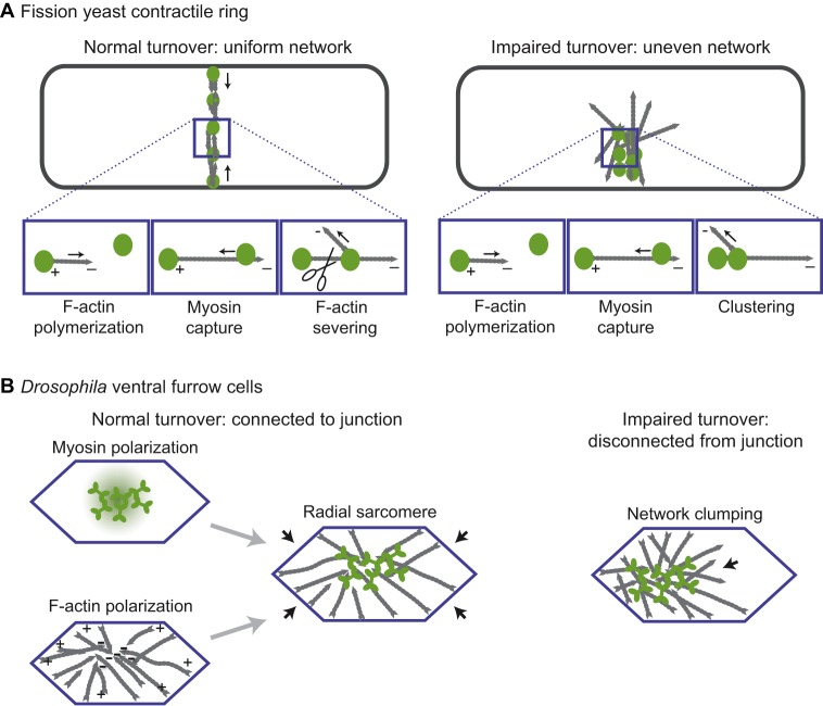
Fig. 2.
Actomyosin network organization in the fission yeast contractile ring and in Drosophila ventral furrow cells. (A) (Left) Organization of a normal contractile ring in fission yeast. The panels beneath show actin polymerizing from a node structure that contains myosin and an F-actin elongating formin, capturing a second myosin node, and being severed to maintain the correct density of actin. F-actin is in gray and myosin oligomers are in green. There is not a net polarity to this network, but within the network myosin captures polymerizing F-actin near the minus end and pulls on it. (Right) A contractile ring with actin turnover inhibited. Myosin nodes aggregate because they are never detached from each other. (B) (Left) The actomyosin network in a normal Drosophila ventral furrow cell is organized in a manner that resembles a sarcomere, but is radially arranged. The myosin (green) is activated at the center of the apical cell surface. Actin organization is depicted in gray. The organization of each component is depicted separately and together. (Right) The actin network in a Drosophila ventral furrow cell that has impaired turnover. The network aggregates and separates from junctions (blue) on one side of the cell (arrow).

