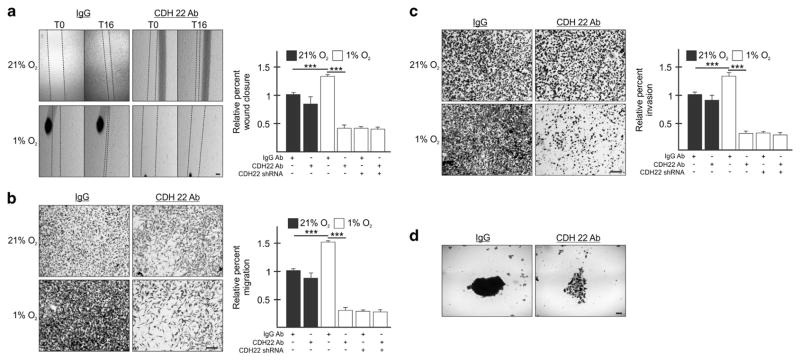
Figure 5.
Blocking CDH22 impairs hypoxic MDA-MB-231 cell migration, invasion and spheroid formation. (a) Parental cells or cells stably expressing shRNA targeting CDH22 (clone KD3.1) incubated with a control non-targeting antibody (IgG) or an antibody targeting the extracellular domain of CDH22 were exposed to normoxia (21% O2) or hypoxia (1% O2) for 24 h followed by wound generation. Representative images at 0 h (T0) and 16 h (T16) after wound generation. (b, c) Transwell migration (b) and invasion (c) assays of parental cells or cells stably expressing shRNA targeting CDH22 incubated with control or anti-CDH22 antibodies and exposed to normoxia or hypoxia for 24 h. Representative images of transwell inserts 16 h after seeding and stained with crystal violet. (d) Light micrographs of spheroids composed of parental cells incubated with control or anti-CDH22 antibodies. Data are presented relative to normoxic Ctrl as mean ±s.e.m., n ≥3, ***P<0.001, using a one-way ANOVA followed by Tukey’s HSD test. Scale bar, 100 μm.

