Abstract
Full-spectrum utilization of diffusive solar energy by a photocatalyst for environmental remediation and fuel generation has long been pursued. In contrast to tremendous efforts in the UV-to-VIS light regime of the solar spectrum, the NIR and IR areas have been barely addressed although they represent about 50% of the solar flux. Here we put forward a biomimetic photocatalyst blueprint that emulates the growth pattern of a natural plant—a peapod—to address this issue. This design is exemplified via unidirectionally seeding core-shell Au@Nb nanoparticles in the cavity of semiconducting HxK1−xNbO3 nanoscrolls. The biomimicry of this nanopeapod (NPP) configuration promotes near-field plasmon–plasmon coupling between bimetallic Au@Nb nanoantennas (the peas), endowing the UV-active HxK1−xNbO3 semiconductor (the pods) with strong VIS and NIR light harvesting abilities. Moreover, the characteristic 3D metal-semiconductor junction of the Au@Nb@HxK1−xNbO3 NPPs favors the transfer of plasmonic hot carriers to trigger dye photodegradation and water photoelectrolysis as proofs-of-concept. Such broadband solar spectral response renders the Au@Nb@HxK1−xNbO3 NPPs highly promising for widespread photoactive devices.
Although near-infrared light makes up a large portion of the solar spectrum, harvesting it for photocatalytic applications remains challenging. Here the authors deposit unidirectional Au@Nb core-shell nanoparticles into tubular HxK1–xNbO3 nanoscrolls and report cooperative full solar spectrum absorption.
Introduction
For millennia, nature routinely has fed humanity with useful energy in either food supplies or carbon-based fuels by solar-to-chemical conversion, namely photosynthesis using sunlight1–4. In particular, these carbohydrates are produced from water and carbon dioxide. The sustainability and environmental benignity in the context of photosynthesis have inspired scientists and engineers to emulate so-called artificial photosynthesis or photocatalysis using man-made materials5–12. One of the major endeavors in the field addresses the sunlight-harvesting ability of the photocatalyst, preferentially ranging from ultraviolet (UV) to visible (VIS) light—accounting for nearly 50% of the solar spectrum—to photogenerate electron/hole pairs (e−/h+) for fuel production13–17. Although near-infrared (NIR) light is responsible for an additional 50% of the solar flux, quite to our surprise, further exploitation on its charge-carrier-generation efficacy is currently in its infancy18–21. In the light of its enormous percentage in sunlight, charge-carrier generation in the NIR spectral region is of obvious significance.
To this end, coupling of a photocatalyst with plasmonic nanoantennas stands out since the size and shape of metallic nanostructures can be readily used to tailor the light-harvesting ability7,22,23. Knight et al.24 demonstrated for the first time that an array of rod-shaped Au nanoantennas allows NIR light to trigger a current response on a silicon-based photodiode, even though the wavelength (1250–1600 nm) is well below the semiconductor band gap. The NIR-promoted e−/h+ pairs arise exclusively from non-radiative damping of surface plasmons on Au. More importantly, they become accessible to a Si diode with the aid of a Schottky junction at the metal-to-semiconductor interface, in which the energy barrier is much smaller for those energetic or hot electrons than the band gap of Si. The significance of these results lies in corroborating the propensity of NIR light to induce useful energetic e−/h+ pairs in photoactive devices.
Recently, those hot charge carriers prompted by NIR-excited plasmons were reported not only to generate electricity24–27 but also to support chemical fuel generation20,21,28, thereby addressing a paradigm in photocatalysis. Hence, Mubeen et al.28 reported on the evidence of hydrogen (H2) gas evolution on a photoexcited dense Au nanorod (NR) array that was sprinkled atop with a bifunctional TiO2 semiconductor and laterally with a cobalt-based oxygen-evolution catalyst (Co-OEC) in the presence of a sacrificial agent. Furthermore, Zheng et al.21 introduced an alternative of strewing the tips of Au NRs merely with Pt in lieu of TiO2 and Co-OEC. The dual functionality of Pt, on the one hand, includes serving as an electron sink to prevent hot electrons to drift back to the Au NRs. This is also the asset and function of a Schottky junction. Moreover, the catalytic effect of Pt allows H2 generation in the presence of a photohole scavenger. These results validate the persistence of energetic electrons in producing H2 upon photoexcitation of Au NR with visible-NIR light radiation. However, the direct exposure of the plasmonic metals to the electrolyte can result in undesirable corrosion and/or dissolution of the Au NR during reaction22,23,29. As an alternative, we here suggest a metal-semiconductor composite structure with the plasmonic nanoantennas embedded in the semiconductor, emerging as a breakthrough to retrieve the aforementioned depleted photoactivity. Even more important, the concomitant 3D heterojunction of such metal-semiconductor composite can also support hot-electron injection due to the substantially increased metal-to-semiconductor interface27,30.
In this contribution, we implanted Au@Nb core-shell nanoparticles (CS-NP) into the cavity of tubular protonated metaniobate (HxK1−xNbO3) nanoscrolls (NSs). In particular, this infiltration emulates the design of a natural plant, namely, peapods. Thus, Au@Nb CS-NPs are unidirectionally deposited yet mutually isolated with a break of few nanometers in between. In addition to the passivation against photocorrosion and the enhanced Schottky junction area, the biomimicry of this peapod design endow the Au@Nb@HxK1−xNbO3 nanopeapod (NPP) photocatalyst with an exceptional broadband photoabsorption ability that eminently matches with the solar spectrum. In particular, the outmost HxK1−xNbO3 semiconductor NS (the pod) is responsible for UV light absorption, whereas the inner Au@Nb CS-NPs (the peas) account for visible light absorption via surface plasmon resonance (SPR). Most significantly, this nanopeapod design gives rise to a strong near-field interantenna coupling that additionally creates the excellent photoresponse of Au@Nb@HxK1−xNbO3 NPPs to NIR light. To the best of our knowledge, this peculiar NPP design is the first alternative pattern—in addition to the geometry and size—to effectively expand the resonant plasmon wavelength of Au nanoantennas over the full solar spectrum. The resulting effective NIR-induced dye decolorization and water cleavage in the absence of an irreversible whole scavenger substantiate the relevance and potential of the peapod-structured photocatalyst.
Results
Synthesis and characterization of Au@Nb@HxK1−xNbO3 nanopeapods
The peapod design is foremost characterized by the tubular morphology of the semiconductor. Moreover, the passivation of the encapsulated plasmonic metal is a most relevant aspect to avoid its dissolution due to photocorrosion processes. To this regard, oxide-based materials known for high stability—particularly, TiO2, α-Fe2O3 or niobates—are promising semiconductors that were already shown to form tubular nanostructures31–33. Hereof, HxK1−xNbO3 is favored due to its delicate band-edge position straddling most redox reactions of interest, and due to the d-orbital nature of its conduction band rendering abundant density of states (DOSs) for a rapid electron uptake34,35.
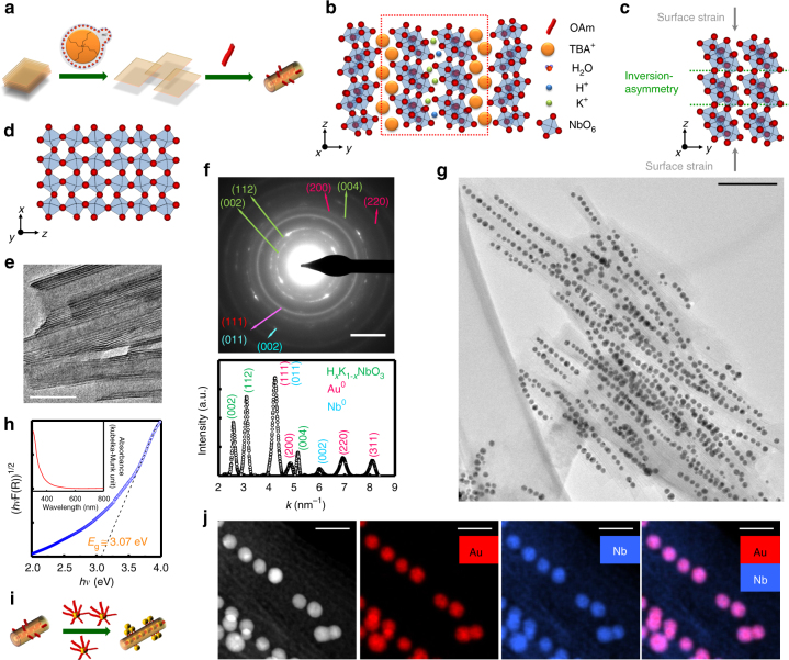
Preparing HxK1−xNbO3 directly from either HNbO3 or KNbO3 in an anisotropic tubular shape, however, is not straightforward as both have a cubic structure36,37. Such cubic symmetry typically leads to the formation of an isotropic crystallite growth most often resulting in cubes or octahedra6,36. To fabricate tubular HxK1−xNbO3 either by protonating of KNbO3 or by exchange of protons (H+) in HNbO3 by potassium (K+) ions seems highly unlikely. Here we have addressed this issue by using the orthorhombic potassium hexaniobate (K4Nb6O17) as the starting material, which already has a lamellar structure and which is thus ideal as a template38–40. Hence, the synthesis started with the K4Nb6O17 template that was subsequently subjected to a cascade of reactions mainly including an interlamellar ion exchange (i.e., HxK4−xNb6O17) to degenerate its structural integrity followed by an exfoliation process of individual lamella by bulky tetrabutylammonium (TBA+) ions (Fig. 1a–c). The resulting isolated molecular sheets then undergo automatic curling due to an intrinsic lattice asymmetry inside each lamella and finally forms structured tubular NSs38. Hereof, the last two steps using a strongly hydrated tetrabutylammonium hydroxide and an organic media are the key to promote the desired phase transformation, in which the dissociation of coordinated water molecules from TBA+ was retarded that otherwise promote the exfoliated HxK4−xNb6O17 lamella to undergo hydrolysis at high temperature according to Equation (1)36,40.
| 1 |
It is worth mentioning that the HxK4−xNb6O17 sheets consist of a central layer of [NbO6] octahedra shearing corners in the [100] direction and alternating edges and corners in the [001] direction (Fig. 1c)36,37. Such corner-shearing [NbO6] octahedra are also an inherent structural feature of the HxK1−xNbO3 perovskite (Fig. 1d) serving as a backbone and facilitating the phase transformation. Such templated crystallization can be regarded as topotactic reaction, in which the template is subjected to a phase transition without sacrificing its morphology (Fig. 1e). Selected area electron diffraction (SAED) patterns (Fig. 1f, top) and azimuthally averaged SAED patterns (Fig. 1f, bottom) of an Au@Nb@HxK1−xNbO3 NPP ensemble show (002), (112), and (004) reflections belonging to the HxK1−xNbO3 perovskite (space group Im-3, a = 7.65 Å). In addition, reflections of pure Au (face-centered-cubic structure, space group Fm-3m, a = 4.08 Å) and Nb (body-centered cubic structure, space group Im-3m, a = 3.32 Å) are visible. Geometric features of the finally formed HxK1−xNbO3 NSs are delineated by transmission electron microscopy (TEM) with an orifice diameter of about d = 20 nm and an outer scroll diameter in a range of D = 35–70 nm (Fig. 1e), while the length of the NSs lies between 0.1 and 1.0 μm (Fig. 1g). The photoabsorption, which is another crucial property with regard to photocatalysis application, was characterized through diffuse reflectance spectroscopy (DRS). Hence, a featured absorption edge at 390 nm (Fig. 1h, inset) results in a band gap of 3.07 eV (Fig. 1h) for the HxK1−xNbO3 NSs, which is in good agreement with the values reported in the literature41,42. A slight decrement of the band gap is presumably related to weakened Nb–O–Nb bonds originating from the curling of the niobate layers to form the NSs43.
Fig. 1.
Stepwise formation and characterization of Au@Nb@HxK1−xNbO3 NPPs. a Schematic and b, c crystallographic illustrations of the morphological evolution of templated HxK4−xNb6O17 crystallites to lamellar and tubular colloids. d Crystal structure of cubic HNbO3 perovskite. e TEM image (scale bar: 70 nm) of tubular HxK1−xNbO3 NSs, in which the hollow cavity functions as the host to breed the Au@Nb CS-NPs. f SAED pattern (top, scale bar: 3 nm), corresponding azimuthally averaged SEAD pattern (bottom) and g TEM image (scale bar: 100 nm) of the Au@Nb@HxK1−xNbO3 NPPs. h Absorption spectrum (inset) and derived Tauc plot of the HxK1−xNbO3 NSs. i Schematic highlights of the biomimicry of the Au@Nb@HxK1−xNbO3 NPPs, wherein the Au@Nb CS-NPs as the “peas” are unidirectionally loaded into the tubular cavity of nanoscale HxK1−xNbO3 as the “pods”. j HAADF-STEM image and EDXS elemental maps (scale bars: 20 nm) of the Au (red), Nb (blue), and combined Au/Nb distribution prove the structure of the Au@Nb@HxK1−xNbO3 NPPs, in which the Au@Nb CS-NPs (lilac) are unidirectionally located inside the HxK1−xNbO3 NSs (blue). Abbreviations used: OAm, oleylamine; TBA+, tetrabutylammonium cation; NS, nanoscroll; CS-NPs, core-shell nanoparticles; NPP, nanopeapod
To date, there is a variety of technologies for fabricating 1D Au peapodded nanostructures, including vapor–liquid–solid (VLS) growth and atomic layer deposition (ALD)44–46. However, the Rayleigh perturbation at the tempering step in those approaches generally generates NPPs with an interparticle spacing (dgap) of nearly 100 nm which fairly exceeds the diameter of the Au particles (dAu) and which leads to a ratio of interdistance over diameter (dgap/dAu) >1. Such giant spacing hinders the surface plasmon polaritons (SPPs) on photoexcited Au NPs from electric near-field coupling47–50. As a consequence, a waning photoresponse at excitation wavelengths remote from the characteristic SPR was reported44–46. In order to retrieve the fading plasmon coupling, this article tackles this challenge by taking a dissolved Au precursor in place of a solid Au catalyst to exclude the necessity of any thermal treatment51. Moreover, such solution-processed Au precursors can readily be capillarily engulfed into the cavity of the preformed HxK1−xNbO3 NSs. Afterwards, the employment of a reducing agent (i.e., oleyamine) allows the Au NPs to seed directly inside the semiconductor NSs (Fig. 1i). Figure 1g, j summarize TEM, high-angle annular dark-field scanning TEM (HAADF-STEM) and energy dispersive X-ray spectroscopy (EDXS) results that clearly depict the morphology and the Au and Nb distribution of the as-prepared NPPs. The TEM images (Fig. 1g, j) highlight the uniformity and filling efficacy while the unidirectionally aligned bright spots in HAADF-STEM images and the distinct elemental contrast in EDXS mappings (Fig. 1j) point to the arrangement of the Au NPs and the niobate NSs in the Au@HxK1−xNbO3 NPPs, respectively.
Characterization of the Au@Nb core-shell nanoparticles
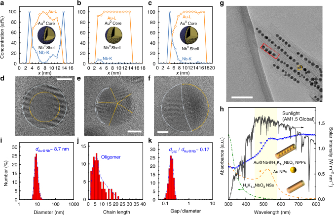
The high yield of the Au@HxK1−xNbO3 NPPs distinctly indicates a strong contrast between Au nucleation rates outside and inside the niobate NSs. This discrepancy originates from the presence of oleyamine (OAm) at the earliest scroll formation stage (Fig. 1a)40,51. Hence, OAm can saturate the cavity of the NSs and accelerates the growth rate of inner Au nanopeas. Surprisingly, EDXS (Fig. 1j) of those inner Au nanopeas reveals an exceptional effect of this preliminary treatment. Namely, OAm intercepts the niobium (Nb5+) leakage from HxK4−xNb6O17 during the phase transition to the final HxK1−xNbO3 that contains a lower stoichiometric content of Nb36. The excess Nb5+ in turn is deposited as Nb0 atop the nucleated Au NPs (element maps in Fig. 1j; concentration profiles across single NPs in Fig. 2a–c) via underpotential deposition (UPD), resulting in the exceptional Au@Nb@HxK1−xNbO3 multi-core-shell peapod structure (Supplementary Figs 1–3, see Supplementary Methods and Supplementary Note 1 for details). Au@Nb CS-NPs with different Nb distributions are observed by high-resolution (HR) TEM (Fig. 2d–f) and the corresponding Nb-line profiles across single CS-NPs (Fig. 2a–c). Figure 2d shows a CS-NP with single-crystalline structure and a continuous Nb shell with a thickness of 1–2 nm (Fig. 2a). We note that HxK1−xNbO3 NSs cannot be seen in the HRTEM images due to their low contrast compared to the CS-NP contrast, but they can be well recognized in TEM overview images (Fig. 2g).
Fig. 2.
CS-NP configuration and configuration-dependent surface plasmon resonances of the Au@Nb@HxK1−xNbO3 NPPs. a–c EDXS line profiles (element contributions from the HxK1−xNbO3 NSs were subtracted) and d–f corresponding HRTEM images reveal the CS-NP configuration of the Au@Nb@HxK1−xNbO3 NPPs. Orange and white dashed lines in the HRTEM images delineate core and shell boundaries (lines are guide to the eye) of single-crystalline Au@Nb CS-NPs with a continuous Nb shell (a, d) (scale bar: 4 nm); multiply twinned Au NPs with thin incomplete Nb shells (b, e) (scale bar: 3 nm); Au@Nb CS-NPs with a partial Nb shell (c, f) (scale bar: 2 nm). g TEM overview image (scale bar: 50 nm) of the Au@Nb@HxK1−xNbO3 NPPs showing the Au@Nb CS-NPs located within (red frame) and on (orange frame) the HxK1−xNbO3 NSs. h Absorption spectrum of the Au@Nb@HxK1−xNbO3 NPPs (half-filled circles). AM 1.5G solar spectrum (solid line), naked Au NPs (dashed line), and hollow HxK1−xNbO3 NSs counterparts (dash-dotted line) are plotted also for comparison. i–k Pattern features of the Au@Nb CS-NPs with i particle size, j length of continuous CS-NP chains (in number of CS-NPs per chain) and k interparticle distance in units of Au@Nb particle size, respectively. Experimental data determined from different TEM images and fits of these data (blue dashed lines). Abbreviations used: dAu@Nb, diameter of bimetallic Au@Nb nanoparticle; dgap, interparticle distance between adjacent Au@Nb nanoparticles of the Au@Nb@HxK1−xNbO3 NPP; NSs, nanoscrolls; NPs, nanoparticles; NPPs, nanopeapods
CS-NPs within HxK1−xNbO3 NSs (red frame in Fig. 2g) are typically covered by a continuous Nb shell as demonstrated by Fig. 2a, d. The formation of a pure Nb shell (instead of Nb oxide) is further supported by its structure, which corresponds to bulk cubic Nb (Supplementary Fig. 4). Inhomogeneous Nb distributions are found for CS-NPs at the outer surface of the HxK1−xNbO3 NSs. For example, the CS-NP in Fig. 2e is only partially covered by a Nb shell (Fig. 2b). The Au core of this NP exhibits a multiply twinned structure with a decahedral shape along its fivefold symmetry axis (Supplementary Fig. 5). The CS-NP in Fig. 2c, f forms a Nb bridge at the interface toward the niobate NSs. The position of this CS-NP is marked by an orange frame in Fig. 2g. The Bragg reflections of the CS-NPs are also present in the SAED pattern (Fig. 1f), where Au (200), (220), and (311) diffraction rings with a uniform intensity support the conclusions from the HRTEM analysis. The intensity of the Nb reflections is in general weak due to the small Nb amount contained in the CS-NPs but the Nb (002) reflection can be clearly recognized in the azimuthally averaged SAED pattern (Fig. 1f, bottom). These analyses show exemplary features of bimetallic CS-NPs prepared by an UPD process, which refers to the deposition of a metal film with the thickness up to few atomic layers on a foreign metal substrate at a potential much more positive than that for the deposition on the same metal surface52. Accordingly, UPD is the most likely mechanism for the formation of metallic Nb (standard reduction potential –0.64 V vs. SHE) by OAm in the presence of Au NPs.
Broadband light harvesting ability
One of the important merits of the unique Au@Nb CS-NP formation is that the Nb shell can superimpose its characteristic SPR absorption upon that of the Au core to modulate the optical properties of the Au@Nb CS-NPs53. As a consequence, the Au@Nb@HxK1−xNbO3 NPPs produce an absorption spectrum (Fig. 2h), exhibiting a somewhat flatter nature at wavelengths beyond 400 nm (featured SPR absorption of Nb) yet below 530 nm (featured SPR wavelength of Au)22,23,54. Intriguingly, a similar scenario is also observed at wavelengths beyond the SPR frequencies of both the Au core and the Nb shell, namely, an intensive and continuous bathochromic spectral feature emerges in the red-NIR region (Fig. 2h). Evidently, this does not originate from the Au@Nb CS-NPs. One should therefore take another key component in Au@Nb@HxK1−xNbO3 NPPs into account, viz. is the niobate NSs. Bringing the metallic nanoantennas into contact with a dielectric NS, unlike the metal–metal interaction, generally leads to a redshift of the SPR22,23,53. This is illustrated by the SPR absorption of the Au@Nb@HxK1−xNbO3 NPPs in comparison to the uncovered Au core (yellowish tinted region in Fig. 2h). The absorption band in the red-NIR region; however, cannot yet be reasonably explained.
Taken together, we could neither call the individual ingredients nor an overall combination in a NPP to account for its peculiar red-NIR absorption. In other words, only the biomimicry of the peapod design, and especially, its specific configurational traits, that differ significantly from known peapod structures, are responsible for this absorption band (Fig. 2i–k)44–46. Thus, the diameter of the Au@Nb CS-NPs is highly monodisperse with an average of 9 nm (Fig. 2i), and the CS-NPs are intensively accumulated ranging from dimers to most often observed heptamers (Fig. 2j) that show uniform interparticulate distances of about 2 nm (Fig. 2k). Even more important, this interparticular distance is small in relation to the Au@Nb CS-NPs size and well below the theoretically suggested threshold to induce a desirable electric near-field coupling47. Such near-field coupling effect resembles the longitudinal SPR (LSPR) mode of an anisotropic Au nanoantenna and promotes an additional absorption band at wavelengths longer than the intrinsic transverse-mode SPR (TSPR) frequency (530 nm) of the Au NPs47–50. The uniaxially aligned Au@Nb CS-NPs result in a field enhancement particularly along the chain and lead to a LSPR-analogous absorption overwhelming nearly the entire red-NIR regime up to 800 nm.
Photocatalytic dye decomposition
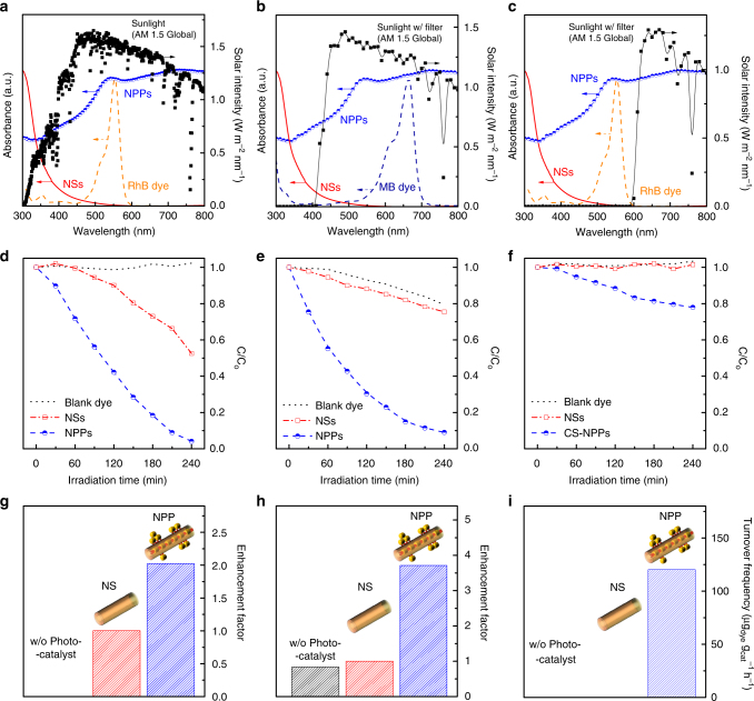
So far, we have specified the configuration-dependent SPR effect of the Au@Nb@HxK1−xNbO3 NPPs endowed with a broadband light harvesting ability. Next, photocatalytic dye decomposition was carried out to substantiate the effect of comprehensive photon accumulation to enhance the useful charge carrier concentration for chemical reactions (Fig. 3). The experiments were carefully designed by using a UV cutoff filter and colored glass filters (λ > 610 nm) (Fig. 3a–c) to distinguish the plasmon-initiated carriers from those originating from the semiconductor. Moreover, methylene blue (MB) and Rhodamine B (RhB) were employed as the probing molecules to avoid the photosensitized dye perturbing the evaluation (see Methods section for details)34,55,56.
Fig. 3.
Broadband photocatalytic activity of the Au@Nb@HxK1−xNbO3 NPPs. a–c Spectra of simulated sunlight (a) further equipped with either an UV cutoff filter (b) or a colored glass filter (c) that work as irradiation sources for photocatalytic dye degradation. Absorption spectra of the employed HxK1−xNbO3 NSs (solid line) and the Au@Nb@HxK1−xNbO3 NPPs (half-filled circles) as photocatalysts; methylene blue (MB) and rhodamine B (RhB) as probing species (dashed line) are plotted alongside for reference. d–f Photocatalytic decoloring of RhB (d, f), and MB (e) in the absence (dotted line) or in the presence of either niobate NSs (empty squares) or Au@Nb@HxK1−xNbO3 NPPs (half-filled circles). g–i Comparison of the wavelength-dependent photoactivity between HxK1−xNbO3 NSs and Au@Nb@HxK1−xNbO3 NPPs under illumination with full solar light (g), visible-NIR integral light (h), and red-NIR integral light (i), respectively. Abbreviations used: MB, methylene blue; RhB, Rhodamine B; NSs, nanoscrolls; NPPs, nanopeapods; NIR, near-infrared
In all cases, the presence of the Au@Nb CS-NPs substantially accelerates the HxK1−xNbO3 NS semiconductor to bleach the dye solutions (Fig. 3d–f). The highest decoloring efficiency is observed in the case of the Au@Nb@HxK1−xNbO3 NPPs under full solar illumination (Fig. 3d). Evidently, the charge carriers involved in the chemical conversion only partly arise from the niobate NSs upon UV excitation. Moreover, the Au@Nb CS-NPs offer extra carriers upon SPR that is responsible for the acceleration (Fig. 3g–i). Taken together, the plasmonic nanoantennas boost the carrier quantity in either populating the energetic electrons originally residing on the metal to the adjacent semiconductor (direct electron transfer, DET) or in resonantly transferring its plasmonic energy to generate more e−/h+ pairs on the semiconductor (resonant energy transfer, RET)22,23,57,58. Clearly, the enhancement in carrier concentration on the Au@Nb@HxK1−xNbO3 NPPs arises mostly from a DET in lieu of a RET process in terms of a minute spectral overlap between the Au NPs and the HxK1−xNbO3 NSs (Fig. 2h). In a DET process, electrons of a plasmonic metal receive energy from an interacting photon followed by traversing a metal-semiconductor interface—the Schottky interface to migrate into the CB of the semiconductor. On this basis, the internal quantum transmission probability (ηi) for electron injection can be formulated by a modified Fowler equation59,60,
| 2 |
where hν is the incident photon energy, and where qΦb is the barrier height of the Schottky junction. The barrier height is established by electronic alignment between the Fermi level of the plasmonic metal and the flat-band potential (Vfb) of the coupled semiconductor. Herein, the work function of the Au@Nb CS-NPs is located in a spectrum with Au (~5.1 eV) and Nb (~4.3 eV) standing at the ends61. Given the insignificant fraction of a thin Nb shell in the Au@Nb CS-NPs (Fig. 2), the work function is primarily dictated by Au with a value close to 5.1 eV. Vfb of the HxK1−xNbO3 semiconductor was estimated by the equation reported by Scaife et al.62 for oxides that do not contain metal cations with partially filled d-orbital as follows,
| 3 |
where Eg is the bandgap of the semiconductor. Consequently, a Vfb of –0.13 V (vs. NHE) for the HxK1−xNbO3 NSs and a barrier height of ~0.7 eV for the Schottky junction between the Au@Nb CS-NPs and the HxK1−xNbO3 NSs were derived. Surprisingly, this energy barrier is nearly one quadrant of the bandgap of the HxK1−xNbO3 semiconductor, which is the key to allow photons that are invisible to the niobate, to produce active carriers to be used in chemical reactions. More significantly, the strong peapod-specific T- and L-SPR phenomena (Fig. 2h) permit visible- and NIR-photons to efficiently lend electrons on Au@Nb CS-NPs sufficient energy to enter the HxK1−xNbO3 NSs and to contribute to the dye degradation63. This prima facie evidence is given by the steady and efficient photobleaching courses (Fig. 3e, f).
Photoelectrochemical water splitting
In addition to the fundamental dye degradation study, the Au@Nb@HxK1−xNbO3 NPPs were examined in view of the photoelectrochemical (PEC) water splitting to corroborate the ability of the SPR-initiated photoinduced carriers in transferring solar energy into useful chemical fuels. In such artificial photosynthesis process (i.e., 2H2O → 2H2 + O2), highly valuable H2 molecules are produced under sunlight3, which highlights the merit of employing this reaction for evaluation. The evaluation of the respective photon energy storage efficiency is deduced from the overall reaction rate, which was mostly expressed by the photoinduced current flowing from the Au@Nb@HxK1−xNbO3 photoelectrode (Methods section). It is to be noted that an argon-purged aqueous solution containing 0.5 M Na2SO4 was used as the electrolyte and no sacrificial agent was present.
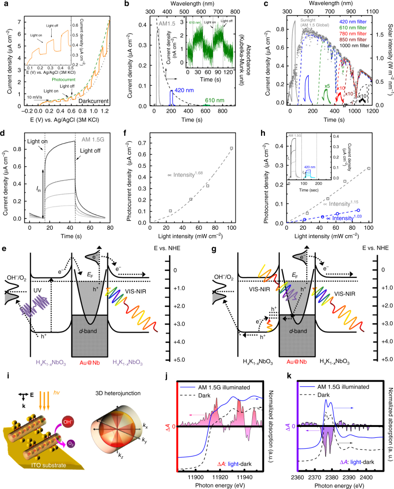
The specific photocurrent-potential response of the Au@Nb@HxK1−xNbO3 photoelectrode was first characterized under chopped full solar illumination at a fluence of 100 mW cm−2 (Fig. 4a). The anodic photocurrent was first perceived at about 0.1 V (Fig. 4a) and raises steadily to 0.9 μA cm−2 at 1 V (i.e., potentials on the Ag/AgCl scale). The enhancement upon anodic polarization stems from the reinforced band bending within the space-charge layer of the outmost HxK1−xNbO3 NSs which effectively rectifies the carrier transport (Supplementary Fig. 6a, b, see Supplementary Note 2 for details)64. This anodically biased photocurrent doubles the performance of the HxK1−xNbO3 photoelectrode under the same conditions (Fig. 4b, c). Even more important, the photocurrent enhancement multiplies significantly upon modulating the irradiation wavelength. The enhancement factor calculated as the photocurrent of the Au@Nb@HxK1−xNbO3 NPP photoelectrode divided by that of the HxK1−xNbO3 NS photoelectrode is threefold below the integral of VIS-NIR light illumination and reaches one order of magnitude below the broadband red-NIR light illumination. Most surprisingly, the maximum improvement is achieved under NIR light irradiation alone, even though its photon energy (<1.8 eV) is far below that of visible (2–3 eV) and UV (>3 eV) photons. Moreover, the characteristic photocurrent-time response of the Au@Nb@HxK1−xNbO3 photoelectrode was monitored (at 1 V vs. Ag/AgCl), in which the photocurrent shows an intriguing transient behavior (Fig. 4d). In the light-on period, the photocurrent demonstrates an initial shoot (Iin) followed by a steady raise mimicking the rectangular signal that is expected in an ideal case64. This variation implies a non-equilibrium between the carrier generation rate under light casting and its consumption rate upon reacting with water molecules. Several probable mechanisms may account for that and are inspected in detail below.
Fig. 4.
Photoelectrochemical water splitting with the Au@Nb@HxK1−xNbO3 NPP photoelectrode. a Current-potential characteristics of the Au@Nb@HxK1−xNbO3 photoelectrode in 0.5 M Na2SO4 solution (pH 6.8) under chopped (orange line) and continuous (green dashed line) AM 1.5G simulated sunlight illumination. The dark current is plotted alongside (dark dotted line). b, c Wavelength-dependent photocurrent-time plots (baseline subtracted) of a HxK1−xNbO3 NS (b) and a Au@Nb@HxK1−xNbO3 NPP photoelectrodes (c) under irradiation of AM 1.5G simulated sunlight (gray line) equipped with different long-pass filters (colored solid lines) in the presence of an anodic bias of 1 V (vs. Ag/AgCl). Absorption spectra of the HxK1−xNbO3 NSs (black dashed line in b) and AM 1.5G simulated sunlight (filled squares in c) illumination after passing a UV cutoff filter (blue dashed line in c) or different colored glass filters (green, red, brown, and black dashed line in c) are plotted alongside for comparison. b Photocurrent-time plot (baseline subtracted) of the HxK1−xNbO3 NS photoelectrode under integral red-NIR illumination in the presence of an anodic bias of 1 V (vs. Ag/AgCl). d Fluence-dependent photocurrent-time plots (baseline subtracted) of the Au@Nb@HxK1−xNbO3 NPP photoelectrode. e Postulated charge transfer on the Au@Nb@HxK1−xNbO3 NPP heterojunction starts with the photoabsorption process. f Iin as a function of simulated sunlight intensity of the Au@Nb@HxK1−xNbO3 NPP photoelectrode. g Postulated charge transfer on the Au@Nb@HxK1−xNbO3 NPP heterojunction via a two sequential one-photon absorption process. h Iin as a function of the intensity of either simulated sunlight (gray line) or integral VIS-NIR light (blue line) for the HxK1−xNbO3 NS photoelectrode. h Fluence-dependent photocurrent-time plots (baseline subtracted) of the HxK1−xNbO3 NS photoelectrode. i Schematic illustration of the 3D metal-semiconductor heterojunction of the Au@Nb@HxK1−xNbO3 NPP photoelectrode. j, k Normalized Au (j) and Nb (k) L3-edge XAS spectra of the Au@Nb@HxK1−xNbO3 NPPs collected in the presence and absence of AM 1.5G simulated sunlight. The difference in absorbance (∆A) between the aforementioned two conditions is plotted alongside. Abbreviations used: NS, nanoscroll; NPP, nanopeapod; VIS, visible, NIR, near-infrared, Iin, initial photocurrent shoot at the light-on instant; EF, Fermi level; E, electric field, k, wavenumber; ITO, tin-doped indium oxide
Considering the thermodynamically (△rG0 = + 472 KJ mol−1) and kinetically (four-electron oxidation: 2H2O → 2O2 + 4 H+ + 4e−) unfavorable cleavage of water, the poor water oxidation on the Au@Nb@HxK1−xNbO3 NPP photoelectrode can influence the PEC performance. For this reason, 1 M Na(COOH) was added to the electrolyte to modify the overall reaction mechanism. The oxidation of formate is comparably easy (one-electron oxidation: [HCOO]− → CO2 + H+ + e−)65. As a consequence, the photocurrent is nearly doubled in the presence of Na(COOH) (Supplementary Fig. 7a–c, see Supplementary Note 2 for details). Nonetheless, the independence of the photocurrent transient implies that the reaction kinetics of the chemical reaction is not the major contribution. In other words, the charge-carrier generation kinetics is responsible for the phenomenon. This finding encourages us to postulate the following mechanism for the different photocurrent response (Fig. 4d). Under broadband sunlight illumination, charge carriers occur simultaneously on the HxK1−xNbO3 NS semiconductor and the Au@Nb CS-NP nanoantenna upon UV and integral VIS-NIR light excitation (Fig. 4e). The SPR-mediated energetic electrons on the Au@Nb CS-NPs become the charge carriers on the HxK1−xNbO3 NS semiconductor via the DET process57,58. An external bias then dictates these charges, including the metal-injected and the semiconductor-originating electrons—to move to the Pt electrode for water reduction to H23. Meanwhile, the photogenerated hole on the HxK1−xNbO3 NS semiconductor is scavenged by the surrounding aqueous medium to generate O2 on the Au@Nb@HxK1−xNbO3 NPP photoanode (Supplementary Figs. 8 and 9, see Supplementary Note 2 for details). Finally, a second UV photon produces the next e−/h+ pair on the HxK1−xNbO3 NS semiconductor followed by an electron transfer to a Au@Nb CS-NP to compensate the accumulated hole and to complete the overall process66,67.
The above process suggests a linear dependency of the photocurrent on the intensity of incident simulated sunlight (see Supplementary Note 3 for details), which, however, is in significant contrast to the superlinear behavior shown in Fig. 4f. The quasi-quadratic correlation indicates a nonlinear effect in the process of the photohole compensation. In brief, the electron vacancies on the Au@Nb CS-NPs are first subjected to an excitation process to a lower energy state in the d-band on the Au@Nb CS-NPs upon electron transition of the subsequently incident photon (Fig. 4g). This view is strongly supported by Jiang et al.68 who reported on a similar phenomenon regarding the photoluminescence of anisotropically coupled Au NPs. The overall process—designated as two sequential one-photon absorption in the literature—starts with a first VIS-/NIR-photon inducing a sp → sp intraband transition to create an electron vacancy below the Fermi level of certain plasmonic nanostructure, which is in analogy to the SPR-mediated hot-electron generation process on the Au@Nb CS-NPs in our study. The second photon then excites an electron from the d- to the sp-band via an interband transition to transfer the hole to the d-band. According to Jiang et al.68, the d-band hole recombines with the electron in the sp-band, giving rise to photoluminescence. In contrast, the photocurrent response in our study indicates that the hole is retrieved by the coupled HxK1−xNbO3 NS semiconductor, and thereafter it participates in water oxidation in preference to charge recombination.
In this hole recovery, energy states associated to HxK1−xNbO3 defects at the interface between the Au@Nb CS-NPs and the HxK1−xNbO3 NSs play an important role. The surface states originate mostly from the stepwise nano-texturization of the HxK1−xNbO3 NSs, as evidenced by the absorption spectra (i.e., the inefficient absorbance at wavelengths beyond the band gap) and the photocurrent-time plot (i.e., the photocurrent response under sub-bandgap integral VIS-NIR light irradiation). In general, these mid-gap electronic states are identified with a shallow energy position close to the valence band (VB) edge, which favors them to serve as a hole relay to facilitate the extraction58,64. Afterwards, a thermal transition brings the hole from this trapped state to the VB of the HxK1−xNbO3 NS semiconductor, where it drifts to the surface and reacts with water molecules to generate O2. This cascade-type charge delivery explains the superlinearity of the transient photocurrent response of the Au@Nb@HxK1−xNbO3 NPP photoelectrode on the light intensity. Noteworthy, the probability of the intraband transition predominates that of the interband transition in the very beginning of the light-on phase, because the electron density in the sp band is much higher than that of the photogenerated sp band vacancy. Afterwards, the quantitative surge in the sp-band hole in turn reinforces the probability of the interband transition (Supplementary Fig. 10a–f, see Supplementary Note 3 for details), which is responsible for the current rise following Iin in the photocurrent-time plot (Fig. 4d).
Despite of the photohole transfer subjected to the above described cascade mechanism, the photocurrent response is yet clearly visible under NIR light illumination (Fig. 4c). This implies a significantly mitigated recombination of antipodal charge carriers. In particular, such carrier loss in the volume of the HxK1−xNbO3 NSs is efficiently suppressed, as evidenced by the quasi-linear dependency demonstrated by the HxK1−xNbO3 photoelectrode without plasmonic Au@Nb nanoantennas (Fig. 4h). This can be attributed to the depletion region developed due to the applied bias that nearly overwhelmes the complete niobate NS (Supplementary Fig. 6a, b, see Supplementary Note 2 for details). Likewise, the carrier decay in the SPR excitation process is significantly quenched, which most likely relates to rapid spatial separation at the instant of the hot-electron generation. One should keep in mind that the isolation of the hot electron with an energy exceeding the Schottky-barrier height has to reach the metal-semiconductor interface to allow the DET process59. This propagation of a hot electron requests a momentum change (∆k) that is harvested from the wavevector (k-vector) of the incident photon with additional support of a phonon or an imperfection69. Such defect-assisted propagation suggests that the energetic electrons are preferentially formed at the margin of the sub-10-nm-sized Au@Nb CS-NPs due to the enormous imperfection area available at the boundary of the HxK1−xNbO3 NSs and the plasmonic CS-NPs. Herein, a considerable bridge area offered by the peapod-specific 3D metal-semiconductor interface greatly favors the HxK1−xNbO3 semiconductor to immediately collect those spatially localized hot electrons (Fig. 4i)30,35. Moreover, an energy dissipation due to electron collision is also circumvented and results in a high-DET efficiency. To this concern, X-ray absorption spectroscopy (XAS) at the Au and Nb L3-edge—describing the empty Au 5d/6s-p hybridized orbitals and the unoccupied Nb 4d states being most responsible for the CB nature of the HxK1−xNbO3—was employed to study the performance. At first sight, a clearly increased absorbance is evidenced by Au L3-edge XAS after simultaneously irradiation with simulated sunlight, which indicates an additional electron transition from the Au 2p to the s-p-d hybridization orbitals by soft X-rays (Fig. 4j). This finding implies that additional electron vacancies are formed in this hybridized band. Concurrently, Nb L3-edge XAS shows a decreased absorbance that suggests reduced empty states in the Nb 4d orbital being available to X-ray excited electrons from the Nb 2p state (Fig. 4k). Otherwise, the CB of HxK1−xNbO3 is occupied. Such inversed absorbance of the Au and the Nb L3-edge XAS reveals substantial evidence for the conversion of SPR-mediated hot electrons into the CB of HxK1−xNbO3 and validates their spatial isolation from the geminate photoholes70. Overall, the synergism between the rapid DET process and the cascade hole delivery is illustrated by the constant photocurrent flow at the incidence of modulated sunlight.
In summary, an alternative promising configuration of a plasmonic photocatalyst design is introduced emulating the growth pattern of a natural plant—a peapod. Spherical Au@Nb core-shell plasmonic nanoantennas with a nanometric break in between are unidirectionally deposited inside the cavity of a tubular HxK1−xNbO3 semiconductor. In particular, the biomimicry of this peapod design induces a strong interantenna electric near-field coupling that endows the Au@Nb@HxK1−xNbO3 nanopeapod photocatalyst with a NIR photon harvesting ability. More importantly, the collected NIR photons are exclusively transferred into energetic charge carriers via a direct electron transfer (DET) process to promote highly relevant chemical reactions, such as the degradation of organic dyes or water splitting (see Supplementary Note 4 for details).
For both organic dye degradation and water splitting, the photoactivity of the Au@Nb@HxK1−xNbO3 nanopeapod photocatalyst outperforms a photocatalyst without Au@Nb nanoantenna significantly. The enhancement stems mostly from the synergistic effect between (i) a rapid DET process due to the appreciable interfacial area provided by a peapod-specific 3D metal-semiconductor junction boosting hot-electron collection, and (ii) a cascade hole delivery supported by a sequential two-photon-assisted hole excitation process with rapid hole injection into the HxK1−xNbO3 semiconductor that is mediated by a mid-gap relay state. Taken together, the results suggest that the biomimetic pattern holds a great promise as a key design for widespread photoactive devices involving the use of NIR photons. This includes phototheranostic agents, optical sensors, plasmonic waveguides, broadband solar cells and so forth.
Methods
Fabrication of the HxK1−xNbO3 nanoscrolls
Templated K4Nb6O17 was prepared by a solid-state reaction, in which reagent-grade K2CO3 and Nb2O5 (99.99%) with a molar ratio of 1.1:1.5 were first ground together and heated in an alumina crucible at 900 °C for 1 h before continuing to 1050 °C for another 24 h. A slight excess of K2CO3 (10 mol-%) was employed to compensate for any material volatilized during heating. The cooling product was washed three times with distilled water and acetone respectively and dried in the oven overnight. The as-prepared K4Nb6O17 was next immersed in a warm (60 °C) and concentrated (3 mol/L) HCl solution for at least 4 days to obtain the proton-exchanged hexaniobates (HxK4−xNb6O17). After the acid-treatment, the product was centrifuged and washed with distilled water and acetone, three times in each case, and dried overnight. Templated phase transformation from HxK4−xNb6O17 to HxK1−xNbO3 and concurrent NS formation were carried out based on a solvothermal process reported by Adireddy et al.40 with some modifications. Experimental conditions were described below, the reaction solution was prepared by putting HxK4−xNb6O17 (0.5 g), tetrabutylammonium hydroxide hydrate (TBAOH·30H2O, 0.75 g, 0.95 mmol) and oleylamine (OAm, 25 mL, ~75 mmol) together in 40 mL toluene. This mixture was magnetically stirred at ambient temperature for 1 h before transferring into a Teflon-lined stainless steel autoclave. The autoclave was then maintained at 220 °C (ramping rate = 2 °C/min) for 6 h and followed by naturally cooling. The resulting HxK1−xNbO3 NSs were collected by centrifugation, washed with ethanol several times and then dried overnight.
Synthesis of the Au@Nb@HxK1−xNbO3 nanopeapods
In a standard synthesis, the preformed HxK1−xNbO3 NSs (20 mg), HAuCl4·4H2O (10 mg, ~0.025 mmol), oleic acid (OAc, 160 μL, 0.5 mmol), and OAm (165 μL, 0.5 mmol) were added to 3 mL hexane. The solution was then vigorously stirred and heated to nearly 60 °C for 24 h before cooling to room temperature. The color of the reacting mixture gradually turned from white to purple, suggesting the formation of Au@Nb CS-NPs inside HxK1−xNbO3 NSs. The obtained composites were purified by a repetitive dispersion/precipitation cycle with ethanol and finally dispersed in either deionized water or hexane for subsequent photo(electro)catalytic examinations.
Analytical techniques
Samples for transmission electron microscopy (TEM) were prepared by casting one drop of hexane solution with Au@Nb@HxK1−xNbO3 NPPs onto a 300 mesh copper grid with carbon support film. The size, the morphology and the spatial distribution of Au@Nb CS-NPs and HxK1−xNbO3 NSs in Au@Nb@HxK1−xNbO3 NPPs were investigated by high-resolution (HR) TEM performed with an aberration-corrected FEI Titan3 80–300 microscope operated at 300 kV. Moreover, a Philips CM200 FEG/ST microscope operated at 200 kV was utilized to perform selected area electron diffraction (SAED) for crystal structure and phase identification. SAED patterns of NPPs often show only few and rather diffuse reflections on Debye-Scherrer rings at large reciprocal lattice distances. A way to prevent the loss of valuable diffraction information is achieved by azimuthally averaging the intensity of the SAED pattern (for short radial scans), which is obtained by a 2π intensity integration of each spatial frequency in the initial SAED pattern. Radial scans can be used to investigate the structure on NPs similar to XRD patterns. Lattice parameters and crystal structure of NPP material are determined from radial scans by using the peak positions obtained by fitting a Voigt function to each individual peak profile after subtraction a linearly interpolated background. Alternatively, HRTEM images were evaluated by calculating the two-dimensional Fourier transform, denoted as diffractogram, which yields information on the crystal structure (lattice parameters and crystal symmetry) of single nanoparticles. Crystal structure analyses was performed by comparing the experimental diffractograms and calculated diffraction patterns with Miller indices, where the latter were obtained by using the JEMS software. The absorption spectra of HxK1−xNbO3 NSs and Au@Nb@HxK1−xNbO3 NPPs were measured using a Varian Cary 100 Spectrometer and further analyzed by means of Kubelka-Munk transformation. For comparison, the ligand-stabilized Au NPs were additionally prepared in the same procedure as the fabrication of Au@Nb@HxK1−xNbO3 NPPs but devoid of HxK1−xNbO3 NSs. The as-obtained Au NPs were stored in hexane to collect an extinction spectrum.
Photocatalytic dye decomposition measurements
The photocatalytic activities of Au@Nb@HxK1−xNbO3 NPPs and HxK1−xNbO3 NSs were evaluated based on the enhancement in degradation rate of Rhodamine B (RhB) and methylene blue (MB) at their presence. The reaction was carried out in a Pyrex reaction cell containing a suspension of the photocatalyst powder (50 mg) in 100 mL aqueous dye solution with continuous agitation using a magnetic stirrer. The employed dye concentration was 5.44 mg/L while a threefold diluted solution was used in evaluating the photoactivity under red-NIR light illumination. Before illumination, the suspensions were magnetically stirred in the dark for 1 h to ensure the establishment of an adsorption/desorption equilibrium of dyes on the photocatalyst surface. Afterwards, the reactions were conducted under irradiation of simulated AM 1.5 G solar light (100 mW/cm) using a LOT Class ABA solar simulator (type LS0805) as the light source. For the measurements under visible-NIR or red-NIR light, an additional UV cutoff (λ > 420 nm) or colored glass filter (λ > 610 nm) was used, respectively. The UV cutoff filter was employed to distinguish carriers produced by the UV-active HxK1−xNbO3 semiconductor from the plasmon-initiated carriers. Moreover, the colored glass filter was deployed to subdivide the SPR-initiated carrier formation into the visible-responsive transverse mode and the red-NIR-sensitive longitudinal mode, respectively. Since a charge transfer via the photosensitized dye may perturb reliable evaluation of the aspired SPR-boosted photon-to-carrier conversion efficiency in a dye degradation study, special attention needed to be paid to the probing molecules55. Herein, MB was chosen owing to the energetic mismatch between the lowest unoccupied molecular orbital (LUMO) of MB and the CB edge of the HxK1−xNbO3 semiconductor34,56. Moreover, RhB was selected to investigate the activity of LSPR-promoted carriers and to avoid competitive photon absorption by the dye as evidenced in the absorption spectra showing that a colored glass filter can efficiently exclude eventual self-absorption of RhB (Fig. 3c). The experiments were carried out at room temperature in air. At each 30 min, 2 mL of aliquot was collected from the reactor. The catalysts were separated by centrifugation and the dye solutions were analyzed by UV–Vis spectrophotometry. The concentration evolution of the dye solution was monitored based on the intensity variation in the featured absorption peak of dyes (λabs of RhB and MB are 554 and 664 nm, respectively).
Photoelectrochemical water splitting characterizations
The measurement was carried out in an undivided either two- or a three-electrode cell configuration, employing either Au@Nb@HxK1−xNbO3 NPPs or HxK1−xNbO3 NSs as the working electrode subjected to the irradiation, a Pt coil or Pt foil as the counter electrode and an additional silver/silver chloride reference electrode (Ag/AgCl in 3 M KCl, 0.207 V vs. NHE) was applied for the three-electrode setup. The photoelectrodes were fabricated upon drop-casting 100 μL Au@Nb@HxK1−xNbO3 NPP/HxK1−xNbO3 NS suspension (20 mg NPP/NS in 1 mL hexane) onto an indium tin oxide (ITO) conducting substrate without post-thermal and -chemical treatments. A potentiostat (CH Instruments, CHI 627D) was used for monitoring the photocurrent behaviors as a function of either potential or time, respectively. A simulated AM 1.5G solar light was used as the irradiation source and the light intensity was calibrated to 100 mW/cm (1 sun). The photocurrent-potential plots were obtained with a potential window from –0.25 to +1.3 V (on the Ag/AgCl scale) at a potential sweeping rate of 10 mV/s. The photocurrent-time plots were recorded at 1 V (vs. Ag/AgCl) with different irradiation wavelengths. For the wavelength-dependent measurement, a UV cutoff filter (λ > 420 nm) was employed to eliminate UV light, and different colored glasses (λ > 610 nm) were used to progressively filter the integral UV to visible light. The incident light was casting on the photoelectrode from the back side through a quartz window and the electrolyte. Argon-purged aqueous solutions containing either 0.5 M Na2SO4 or 0.5 M Na2SO4 and 1 M Na(COOH) were used as the electrolytes.
Data availability
The data that support the findings of this study are available from the corresponding author upon reasonable request.
Electronic supplementary material
Acknowledgements
The authors gratefully thank the German Research Society (DFG), Germany, for funding of equipment. We also acknowledge the Ministry of Science and Technology (MOST) and the National Synchrotron Radiation Research Center (NSRRC), Taiwan, for financial support under contracts MOST 103–2112-M-213–001-MY2, 105–2221-E-259–024-MY3, and 105–2221-E-259–026. Y.-C.C. acknowledges support from the German Academic Exchange Service (DAAD) for scholarship (No. A/13/92805). The valuable comments of Y.F. Huang are also appreciated.
Author contributions
Y.-C.C. fabricated the samples, performed the experiments, analyzed the data and wrote the manuscript. R.P. and D.G. performed the TEM, EDX, and SAED characterization and analyzed the results. Y.-G.L. conceived and designed XAS characterizations. Y.-K.H. conceived and designed the PEC analyses and interpreted the data. C.F. conceived, designed and supervised the study, interpreted the data and wrote the manuscript. All authors reviewed and approved the manuscript.
Competing interests
The authors declare no competing financial interests.
Footnotes
Ying-Chu Chen and Yu-Kuei Hsu contributed equally to this work.
Electronic supplementary material
Supplementary Information accompanies this paper at 10.1038/s41467-017-02676-w.
Publisher's note: Springer Nature remains neutral with regard to jurisdictional claims in published maps and institutional affiliations.
References
- 1.Barber J. Photosynthetic energy conversion: natural and artificial. Chem. Soc. Rev. 2009;38:185–196. doi: 10.1039/B802262N. [DOI] [PubMed] [Google Scholar]
- 2.Long SP, Marshall-Colon A, Zhu XG. Meeting the global food demand of the future by engineering crop photosynthesis and yield potential. Cell. 2015;161:56–66. doi: 10.1016/j.cell.2015.03.019. [DOI] [PubMed] [Google Scholar]
- 3.Tachibana Y, Vayssieres Y, Durrant JR. Artificial photosynthesis for solar water-splitting. Nat. Photon. 2012;6:511–518. doi: 10.1038/nphoton.2012.175. [DOI] [Google Scholar]
- 4.Roy SC, Varghese OK, Paulose M, Grimes CA. Toward solar fuels: photocatalytic conversion of carbon dioxide to hydrocarbons. ACS Nano. 2010;4:1259–1278. doi: 10.1021/nn9015423. [DOI] [PubMed] [Google Scholar]
- 5.Faunce TA, et al. Energy and environment policy case for a global project on artificial photosynthesis. Energy Environ. Sci. 2013;6:695–698. doi: 10.1039/c3ee00063j. [DOI] [Google Scholar]
- 6.Chen YC, et al. β-SnWO4 photocatalyst showing controlled morphological transition of cubes to spikecubes. ACS Catal. 2016;6:2357–2367. doi: 10.1021/acscatal.5b02444. [DOI] [Google Scholar]
- 7.Linic S, Aslam U, Boerigter C, Morabito M. Photochemical transformations on plasmonic metal nanoparticles. Nat. Mater. 2015;14:567–576. doi: 10.1038/nmat4281. [DOI] [PubMed] [Google Scholar]
- 8.Lin YG, et al. Hierarchically porous Ca-containing MnO2 nanorod bundles with superior photoelectrochemical activity. ChemCatChem. 2014;6:1684–1690. doi: 10.1002/cctc.201400012. [DOI] [Google Scholar]
- 9.Wang Q, et al. Scalable water splitting on particulate photocatalyst sheets with a solar-to-hydrogen energy conversion effiency exceeding 1% Nat. Mater. 2016;15:611–615. doi: 10.1038/nmat4589. [DOI] [PubMed] [Google Scholar]
- 10.Lin YG, et al. Cobalt-phosphate-assisted photoelectrochemical water oxidation by arrays of molybdenum-doped zinc oxide nanorods. ChemSusChem. 2014;7:2748–2754. doi: 10.1002/cssc.201402025. [DOI] [PubMed] [Google Scholar]
- 11.Tu WG, Zhou Y, Zou ZG. Photocatalytic conversion of CO2 into renewable hydrocarbon fuels: state-of-the-art accomplishment, challenges, and prospects. Adv. Mater. 2014;26:4607–4626. doi: 10.1002/adma.201400087. [DOI] [PubMed] [Google Scholar]
- 12.Hsu YK, Fu SY, Chen MH, Chen YC, Lin YG. Facile synthesis of Pt nanoparticles/ZnO nanorod arrays for photoelectrochemical water splitting. Electrochim. Acta. 2014;120:1–5. doi: 10.1016/j.electacta.2013.12.095. [DOI] [Google Scholar]
- 13.Ghosh S, et al. Conducting polymer nanostructures for photocatalysis under visible light. Nat. Mater. 2015;14:505–511. doi: 10.1038/nmat4220. [DOI] [PubMed] [Google Scholar]
- 14.Schultz DM, Yoon TP. Solar synthesis: prospects in visible light photocatalysis. Science. 2014;343:1239176. doi: 10.1126/science.1239176. [DOI] [PMC free article] [PubMed] [Google Scholar]
- 15.Oshikiri T, Ueno K, Misawa H. Plasmon-induced ammonia synthesis through nitrogen photofixation with visible light irradiation. Angew. Chem. Int. Ed. 2014;53:9802–9805. doi: 10.1002/anie.201404748. [DOI] [PubMed] [Google Scholar]
- 16.Moniz SJA, Shevlin SA, Martin DJ, Guo ZX, Tang JW. Visible-light driven heterojunction photocatalysts for water splitting—a critical review. Energy Environ. Sci. 2015;8:731–759. doi: 10.1039/C4EE03271C. [DOI] [Google Scholar]
- 17.Hsu YK, Chen YC, Lin YG. Novel ZnO/Fe2O3 core-shell nanowires for photoelectrochemical water splitting. ACS Appl. Mater. Inter. 2015;7:14157–14162. doi: 10.1021/acsami.5b03921. [DOI] [PubMed] [Google Scholar]
- 18.Sang Y, et al. From UV to near-infrared, WS2 nanosheet: a novel photocatalyst for full solar light spectrum photodegradation. Adv. Mater. 2015;27:363–369. doi: 10.1002/adma.201403264. [DOI] [PubMed] [Google Scholar]
- 19.Zhang X, et al. Visible/near-infrared-light-induced H2 production over g‑C3N4 cosensitized by organic dye and zinc phthalocyanine derivative. ACS Catal. 2015;5:504–510. doi: 10.1021/cs5016468. [DOI] [Google Scholar]
- 20.Nishijima Y, et al. Near-infrared plasmon-assisted water oxidation. J. Phys. Chem. Lett. 2012;3:1248–1252. doi: 10.1021/jz3003316. [DOI] [PubMed] [Google Scholar]
- 21.Zheng Z, Tachikawa T, Majima T. Single-particle study of Pt-modified Au nanorods for plasmon-enhanced hydrogen generation in visible to near-infrared region. J. Am. Chem. Soc. 2014;136:6870–6873. doi: 10.1021/ja502704n. [DOI] [PubMed] [Google Scholar]
- 22.Zhang X, Chen YL, Liu RS, Tsai DP. Plasmonic photocatalysis. Rep. Prog. Phys. 2013;76:046401. doi: 10.1088/0034-4885/76/4/046401. [DOI] [PubMed] [Google Scholar]
- 23.Zhang P, Wang T, Gong JL. Mechanistic understanding of the plasmonic enhancement for solar water splitting. Adv. Mater. 2015;27:5328–5342. doi: 10.1002/adma.201500888. [DOI] [PubMed] [Google Scholar]
- 24.Knight MW, Sobhani H, Nordlander P, Halas NJ. Photodetection with active optical antennas. Science. 2011;332:702–704. doi: 10.1126/science.1203056. [DOI] [PubMed] [Google Scholar]
- 25.Pescaglini A, et al. Hot-electron injection in Au nanorod−ZnO nanowire hybrid device for near-infrared photodetection. Nano Lett. 2014;14:6202–6209. doi: 10.1021/nl5024854. [DOI] [PubMed] [Google Scholar]
- 26.Seo E, Ko SJ, Min SH, Kim JY, Kim BS. Plasmonic transition via interparticle coupling of Au@Ag core−shell nanostructures sheathed in double hydrophilic block copolymer for high-performance polymer solar cell. Chem. Mater. 2015;27:4789–4798. doi: 10.1021/acs.chemmater.5b01591. [DOI] [Google Scholar]
- 27.Fang Z, et al. Graphene-antenna sandwich photodetector. Nano Lett. 2012;12:3808–3813. doi: 10.1021/nl301774e. [DOI] [PubMed] [Google Scholar]
- 28.Mubeen S, et al. An autonomous photosynthetic device in which all charge carriers derive from surface plasmons. Nat. Nanotechnol. 2013;8:247–251. doi: 10.1038/nnano.2013.18. [DOI] [PubMed] [Google Scholar]
- 29.Kazuma E, Sakai N, Tatsuma T. Nanoimaging of localized plasmon-induced charge separation. Chem. Commun. 2011;47:5777–5779. doi: 10.1039/c1cc10936g. [DOI] [PubMed] [Google Scholar]
- 30.Knight MW, et al. Embedding plasmonic nanostructure diodes enhances hot electron emission. Nano Lett. 2013;13:1687–1692. doi: 10.1021/nl400196z. [DOI] [PubMed] [Google Scholar]
- 31.Roy P, Berger S, Schmuki P. TiO2 nanotubes: synthesis and applications. Angew. Chem. Int. Ed. 2011;50:2904–2939. doi: 10.1002/anie.201001374. [DOI] [PubMed] [Google Scholar]
- 32.Lin YG, Hsu YK, Lin YC, Chen YC. Hierarchical Fe2O3 nanotube/nickel foam electrodes for electrochemical energy storage. Electrochim. Acta. 2016;216:287–294. doi: 10.1016/j.electacta.2016.09.033. [DOI] [Google Scholar]
- 33.Maeda K, Eguchi M, Youngblood WJ, Mallouk TE. Niobium oxide nanoscrolls as building blocks for dye-sensitized hydrogen production from water under visible light irradiation. Chem. Mater. 2008;20:6770–6778. doi: 10.1021/cm801807b. [DOI] [Google Scholar]
- 34.Kudo A, Miseki Y. Heterogeneous photocatalyst materials for water splitting. Chem. Soc. Rev. 2009;38:253–278. doi: 10.1039/B800489G. [DOI] [PubMed] [Google Scholar]
- 35.Clavero C. Plasmon-induced hot-electron generation at nanoparticle/metal-oxide interfaces for photovoltaic and photocatalytic devices. Nat. Photon. 2014;8:95–103. doi: 10.1038/nphoton.2013.238. [DOI] [Google Scholar]
- 36.Pribosic I, Makovec D, Drofenik M. Formation of nanoneedles and nanoplatelets of KNbO3 perovskite during templated crystallization of the precursor gel. Chem. Mater. 2005;17:2953–2958. doi: 10.1021/cm050079c. [DOI] [Google Scholar]
- 37.Kong XG, et al. Transformation of potassium Lindquist hexaniobate to various potassium niobates: solvothermal synthesis and structural evolution mechanism. Dalton Trans. 2013;42:7699–7709. doi: 10.1039/c3dt00062a. [DOI] [PubMed] [Google Scholar]
- 38.Saupe GB, et al. Nanoscale tubules formed by exfoliation of potassium hexaniobate. Chem. Mater. 2000;12:1556–1562. doi: 10.1021/cm981136n. [DOI] [Google Scholar]
- 39.Kobayashi Y, Hata H, Salama M, Mallouk TE. Scrolled sheet precursor route to niobium and tantalum oxide nanotubes. Nano Lett. 2007;7:2142–2145. doi: 10.1021/nl0708260. [DOI] [Google Scholar]
- 40.Adireddy S, Yao Y, He F, Wiley JB. Rapid solvothermal fabrication of hexaniobate nanoscrolls. Mater. Res. Bull. 2013;48:3236–3241. doi: 10.1016/j.materresbull.2013.04.090. [DOI] [Google Scholar]
- 41.Zhang TT, et al. Insights into the structure–photoreactivity relationships in well-defined perovskite ferroelectric KNbO3 nanowires. Chem. Sci. 2015;6:4118–4123. doi: 10.1039/C5SC00766F. [DOI] [PMC free article] [PubMed] [Google Scholar]
- 42.Zhang TT, et al. Organic pollutant photodecomposition by Ag/KNbO3 nanocomposites: a combined experimental and theoretical study. J. Phys. Chem. C. 2016;120:2777–2786. doi: 10.1021/acs.jpcc.5b11297. [DOI] [Google Scholar]
- 43.Sarahan MC, et al. K4Nb6O17-derived photocatalysts for hydrogen evolution from water: nanoscrolls versus nanosheets. J. Solid State Chem. 2008;181:1678–1683. doi: 10.1016/j.jssc.2008.06.021. [DOI] [Google Scholar]
- 44.Hu MS, et al. Photosensitive gold-nanoparticle-embedded dielectric nanowires. Nat. Mater. 2006;5:102–106. doi: 10.1038/nmat1564. [DOI] [PubMed] [Google Scholar]
- 45.Hsieh CH, Chou LJ, Lin GR, Bando Y, Golberg D. Nanophotonic switch: gold-in-Ga2O3 peapod nanowires. Nano Lett. 2008;8:3081–3085. doi: 10.1021/nl0731567. [DOI] [PubMed] [Google Scholar]
- 46.Qin Y, Liu L, Yang R, Gosele U, Knez M. General assembly method for linear metal nanoparticle chains embedded in nanotubes. Nano Lett. 2008;8:3221–3225. doi: 10.1021/nl801548h. [DOI] [PubMed] [Google Scholar]
- 47.Harris N, Arnold MD, Blaber MG, Ford MJ. Plasmonic resonances of closely coupled gold nanosphere chains. J. Phys. Chem. C. 2009;113:2784–2791. doi: 10.1021/jp8083869. [DOI] [Google Scholar]
- 48.Atay T, Song JH, Nurmikko AV. Strongly interacting plasmon nanoparticle pairs: from dipole-dipole interaction to conductively coupled regime. Nano Lett. 2004;4:1627–1631. doi: 10.1021/nl049215n. [DOI] [Google Scholar]
- 49.Zhong Z, Pakovskyy S, Bouvrette P, Luong JHT, Gedanken A. The surface chemistry of Au colloids and their interactions with functional amino acids. J. Phys. Chem. B. 2004;108:446–4052. [Google Scholar]
- 50.Thomas KG, Barazzouk S, Ipe BI, Joseph STS, Kamat PV. Uniaxial plasmon coupling through longitudinal self-assembly of gold nanorods. J. Phys. Chem. B. 2004;108:13066–13068. doi: 10.1021/jp049167v. [DOI] [Google Scholar]
- 51.Adireddy S, et al. Peapod-type nanocomposites through the in situ growth of gold nanoparticles within preformed hexaniobate nanoscrolls. Angew. Chem. Int. Ed. 2014;53:4614–4617. doi: 10.1002/anie.201310834. [DOI] [PubMed] [Google Scholar]
- 52.Kolb DM, Przasnyski M, Gerischer H. Underpotential deposition of metals and work function differences. Electro. Chem. Inter. Electrochem. 1974;54:25–38. doi: 10.1016/S0022-0728(74)80377-3. [DOI] [Google Scholar]
- 53.Cortie MB, McDonagh AM. Synthesis and optical properties of hybrid and alloy plasmonic nanoparticles. Chem. Rev. 2011;111:3713–3735. doi: 10.1021/cr1002529. [DOI] [PubMed] [Google Scholar]
- 54.Creighton JA, Eadont DG. Ultraviolet-visible absorption spectra of the colloidal metallic elements. J. Chem. Soc. Faraday Trans. 1991;87:3881–3891. doi: 10.1039/FT9918703881. [DOI] [Google Scholar]
- 55.Yi ZG, et al. An orthophosphate semiconductor with photooxidation properties under visible-light irradiation. Nat. Mater. 2010;9:559–564. doi: 10.1038/nmat2780. [DOI] [PubMed] [Google Scholar]
- 56.Shen JS, Yu T, Xie JW, Jiang YB. Photoluminescence of CdTe nanocrystals modulated by methylene blue and DNA. A label-free luminescent signaling nanohybrid platform. Phys. Chem. Chem. Phys. 2009;11:5062–5069. doi: 10.1039/b900053d. [DOI] [PubMed] [Google Scholar]
- 57.Cushing SK, et al. Photocatalytic activity enhanced by plasmonic resonant energy transfer from metal to semiconductor. J. Am. Chem. Soc. 2012;134:15033–15041. doi: 10.1021/ja305603t. [DOI] [PubMed] [Google Scholar]
- 58.Li JT, et al. Solar hydrogen generation by a CdS-Au-TiO2 sandwich nanorod array enhanced with Au nanoparticle as electron relay and plasmonic photosensitizer. J. Am. Chem. Soc. 2014;136:8438–8449. doi: 10.1021/ja503508g. [DOI] [PubMed] [Google Scholar]
- 59.Fowler RH. The analysis of photoelectric sensitivity curves for clean metals at various temperatures. Phys. Rev. 1931;38:45–56. doi: 10.1103/PhysRev.38.45. [DOI] [Google Scholar]
- 60.Sze, S. M. & Ng, K. K. Physics of Semiconductor Devices (Wiley, Hoboken, NJ, USA, 2007).
- 61.Hölzl, J., Schulte, F. K. & Wagner, H. Solid Surface Physics (Springer, Berlin, Germany, 1979).
- 62.Scaife DE. Oxide semiconductors in photoelectrochemical conversion of solar energy. Sol. Energy. 1980;25:41–54. doi: 10.1016/0038-092X(80)90405-3. [DOI] [Google Scholar]
- 63.Li BX, et al. (Gold core)@(ceria shell) nanostructures for plasmon-enhanced catalytic reactions under visible light. ACS Nano. 2014;8:8152–8162. doi: 10.1021/nn502303h. [DOI] [PubMed] [Google Scholar]
- 64.Salvador P. Kinetic approach to the photocurrent transients in water photoelectrolysis at n-titanium dioxide electrodes. 1. Analysis of the ratio of the instantaneous to steady-state photocurrent. J. Phys. Chem. 1985;89:3863–3869. doi: 10.1021/j100264a019. [DOI] [Google Scholar]
- 65.Hsu YK, Chen YC, Lin YG, Chen LC, Chen KH. Birnessite-type manganese oxides nanosheets with hole acceptor assisted photoelectrochemical activity in response to visible light. J. Mater. Chem. 2012;22:2733–2739. doi: 10.1039/C1JM14355G. [DOI] [Google Scholar]
- 66.Hirakawa T, Kamat PV. Charge separation and catalytic activity of Ag@TiO2 core-shell composite clusters under UV-Irradiation. J. Am. Chem. Soc. 2005;127:3928–3934. doi: 10.1021/ja042925a. [DOI] [PubMed] [Google Scholar]
- 67.Jakob M, Levanon H, Kamat PV. Charge distribution between UV-irradiated TiO2 and gold nanoparticles: determination of shift in the fermi level. Nano Lett. 2003;3:353–358. doi: 10.1021/nl0340071. [DOI] [Google Scholar]
- 68.Jiang XF, et al. Excitation nature of two-photon photoluminescence of gold nanorods and coupled gold nanoparticles studied by two-pulse emission modulation spectroscopy. J. Phys. Chem. Lett. 2013;4:1634–1638. doi: 10.1021/jz400582h. [DOI] [PubMed] [Google Scholar]
- 69.Shalaev VM, Douketis C, Stuckless JT, Moskovits M. Light-induced kinetic effects in solids. Phys. Rev. B. 1996;53:11388–11402. doi: 10.1103/PhysRevB.53.11388. [DOI] [PubMed] [Google Scholar]
- 70.Hung SF, et al. Iridium oxide-assisted plasmon-induced hot carriers: improvement on kinetics and thermodynamics of hot carriers. Adv. Energy Mater. 2016;6:1501339. doi: 10.1002/aenm.201501339. [DOI] [Google Scholar]
Associated Data
This section collects any data citations, data availability statements, or supplementary materials included in this article.
Supplementary Materials
Data Availability Statement
The data that support the findings of this study are available from the corresponding author upon reasonable request.


