Sir,
Advanced surface ablation like photorefractive keratectomy (PRK) represents an excellent alternative to laser in situ keratomileusis (LASIK) for the correction of low to moderate myopia and low to moderate astigmatism.1 However, PRK is associated with high risk for keratocyte activation in the corneal stroma, which may lead to visually significant corneal opacification (haze) and regression of the refractive outcomes.2 Although a number of signaling mechanisms have been identified in the pathophysiology of myofibroblast formation and scarring during corneal wound healing, with transforming growth factor-ß (TGF-ß) and interleukin-6 (IL-6) being key elements in this process,3, 4, 5 the complexity of the mechanisms underlying the corneal haze formation after PRK has not been fully investigated yet.
In our study, we used adult pigmented male mice (C57BL/6, 6–10 weeks of age), which were anaesthetized, followed by mechanical abrasion of the central 1.5 mm of corneal epithelium. Six mice (N=6) were used in each experiment. Excimer laser ablation was performed to a depth of 30 μm (50% stromal thickness) using the phototherapeutic mode of a 500 Hz scanning spot excimer laser (WaveLight Concerto, WaveLight, Erlangen, Germany) with a spot size of 0.8 mm. The ablation zone was limited to a diameter of 1.2 mm using an aperture mask placed on the cornea. Volume of 100 μl of TGF-ß2 (1 mg/ml) (Sigma-Aldrich, St Louis, MO 63103, USA) was applied to mouse cornea directly after excimer laser ablation. The mice were killed at the following times: at 1 day, 3 days, 5 days and 7 days after surface ablation. Ofloxacin ointment (Floxal ointment, Bausch and Lomb, Steinhausen, Switzerland) was applied daily to the cornea until complete closure of the epithelium. Animals that were killed immediately with and without mechanical abrasion served as controls. Light microscopic analysis was facilitated after eyes were rapidly enucleated and fixed in 2% paraformaldehyde for 2 h followed by dehydration and paraffin embedding, with the aid of a digitalized Axiovision microscope (Carl Zeiss Microscopy, Jena, Germany). Isolation of mRNA and real-time polymerase chain reaction was also performed.
All animal experiments adhered to the ARRIVE guidelines and have been carried out in accordance with the UK Animals (Scientific Procedures) Act, 1986 and associated guidelines (EU Directive 2010/63/EU for animal experiments).
Wild-type mice treated with excimer laser ablation and TGF-ß (1 mg/ml) demonstrated beginning stromal haze 3 days after laser treatment (Figure 1e). At postoperative day 5, circular corneal neovascularization, massive haze, and absence of re-epithelialization were observed (Figure 1f). On postoperative day 7 they showed corneal scarring with deep stromal vascularization in the absence of epithelialization (Figure 1g). Controls that received excimer laser ablation alone (Figures 1b–d) displayed complete corneal epithelial healing, with only minimal stromal haze at postoperative day 7 (Figure 1d). Untreated controls showed a regular cornea (Figure 1a).
Figure 1.
(a) Untreated wild-type mouse eye with regular aspect. (b–d) Wild-type mice treated with deep stromal ablation alone. White vectors in b indicate the area of corneal epithelial defect. White arrow in d shows residual corneal haze. (e–g) Wild-type mice treated with deep stromal ablation and TGF-ß. White vectors indicate the area of corneal epithelial defect in e, while the white arrow in the same figure shows beginning corneal vascularization. White arrows in h show marked central corneal scarring with extensive corneal vascularization. (h) Treated wild-type mouse (one eye with deep stromal ablation alone and the partner eye with deep stromal ablation and TGF-ß.
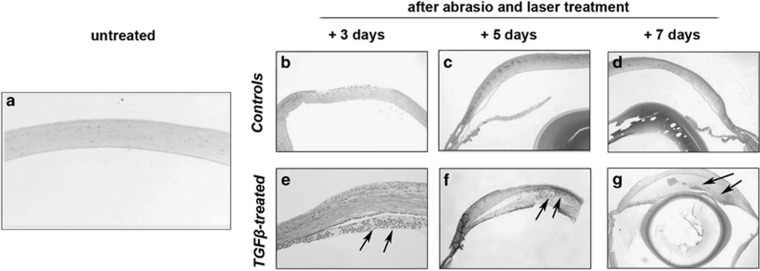
Histological analysis revealed that treatment of wild-type mice with TGF-ß induced corneal edema, marked corneal angiogenesis, as well as an anterior chamber reaction with massive infiltrates and fibrin production (Figures 2e–g). Controls showed normal anterior segment (Figures 2b–d).
Figure 2.
(a) Histological section of an untreated control wild-type mouse. Normal anterior segment. (b–g) Histological section of a wild-type mouse 3, 5, and 7 days after deep stromal ablation (b–d) and deep stromal ablation with TGF-ß treatment (e–g). Note the anterior chamber reaction with massive infiltrates and fibrin production in e–g as indicated by the black arrows.
Semi-quantitative real-time PCR of caspase and cytokine activity showed that abrasion alone markedly upregulated the expression of Caspase-1 in wild-type mice. Deep stromal ablation also markedly upregulated Caspase-1, starting at postoperative 3, and continuing at postoperative day 5 and 7. Similarly, IL-1ß showed distinct upregulation after abrasion alone, and massive upregulation after deep stromal ablation, which peaked at postoperative day 1. The expression of caspase-2, IL-6 and TNF alpha did not vary significantly after abrasion and after deep stromal ablation in wild-type mice.
Our experiments showed that TGF-ß induced significant corneal haze and promoted angiogenesis in vivo following deep excimer laser ablation in mice. Its expression increased significantly between postoperative day 1 and 5. Interestingly, IL-6 deficient mice showed marked corneal haze and neovascularization following PRK in the presence and absence of exogenous TGF-ß, indicating that IL-6 has rather a suppressive role in the inflammatory process occurring during excimer laser-mediated corneal wound healing in vivo. This presumed inflammatory-constraining role may interfere with the signaling pathway underlying the IL-6 and TGF-ß interaction and requires further investigation. Modulation of IL-6 in the cornea might therefore be a means to influence the corneal wound healing response in vivo. Moreover, both TGF-ß and IL-6 appear to exert an angiogenetic effect, which may have significant implications in corneal wound healing after laser refractive procedures.
This study has certain limitations. The relatively low number of animals used in each experiment did not allow a valid statistical analysis between the subgroups. Moreover, the excimer laser ablation alone might have contributed to some extent to the formation of corneal neovascularisation. Further studies are required to validate these results and elucidate the complex interactions between IL-6 and TGF-ß in corneal wound healing after laser ablation procedures.
Acknowledgments
Author contributions
Study concept and design (FH, LMI); performance of experiments (FH, LMI); data collection, analysis and interpretation (FH, ZG, RA, ME, LMI); drafting of the manuscript (FH, ZG, RA, EM, LMI); critical revision of the manuscript (FH, ZG, RA, ME, LMI).
Footnotes
The authors declare no conflict of interest.
References
- O'Connor J, O'Keeffe M, Condon PI. Twelve-year follow-up of photorefractive keratectomy for low to moderate myopia. J Refract Surg 2006; 22(9): 871–877. [DOI] [PubMed] [Google Scholar]
- Amm M, Wetzel W, Winter M, Uthoff D, Duncker GI. Histopathological comparison of photorefractive keratectomy and laser in situ keratomileusis in rabbits. J Refract Surg 1996; 12(7): 758–766. [DOI] [PubMed] [Google Scholar]
- Ebihara N, Matsuda A, Nakamura S, Matsuda H, Murakami A. Role of the IL-6 classic- and trans-signaling pathways in corneal sterile inflammation and wound healing. Invest Ophthalmol Vis Sci 2011; 52(12): 8549–8557. [DOI] [PubMed] [Google Scholar]
- Saika S. TGF-beta signal transduction in corneal wound healing as a therapeutic target. Cornea 2004; 23(8 Suppl): S25–S30. [DOI] [PubMed] [Google Scholar]
- Wilson SE, Liu JJ, Mohan RR. Stromal-epithelial interactions in the cornea. Prog Retin Eye Res 1999; 18(3): 293–309. [DOI] [PubMed] [Google Scholar]


