

Abstract
BACKGROUND:
The term decreased occlusal vertical dimension refers to the reduced distance between two anatomical points while the teeth are in a state of occlusion. The development of this situation is about some parafunctional activities of the masticatory system.
AIM:
To evaluate the value of decreased occlusal vertical dimension in cases with temporomandibular disorder and to follow up the influence of corrective treatment with occlusal splints and definitive prosthetic construction upon the elimination of clinical symptoms.
MATERIAL AND METHODS:
Eight cases with decreased occlusal vertical dimension accompanied with temporomandibular disorders were treated with an occlusal splint, as part of reversible occlusal treatment. After reducing, or complete elimination of the symptoms related to problems of decreased occlusal vertical dimension, the definitive prosthetic therapy was performed.
RESULTS:
The mean value of decreased occlusal vertical dimension in our patients is 8.5 mm, and the mean value of therapy time with an occlusal splint in these patients was 3.5 months.
CONCLUSION:
Occlusal splint is a part of reversible occlusal therapy in cases with decreased occlusal vertical dimension. After reducing the symptoms related to decreased occlusal vertical dimension definitive prosthetic therapy can be done.
Keywords: Occlusal vertical dimension, Temporomandibular disorder, Occlusal splint
Introduction
Decreased occlusal vertical dimension (dOVD) is irregularity between upper and lower jaw about a vertical plane. dOVD has decreased the distance between two anatomical points. Those points are very often arbitrary checked dots on the top of the nose and the top of the mentum while the teeth are in a state of intercuspidation. In the development of this disorder has to pass the longer time in collaboration with some factors such as uncontrolled gnashing and tightening of the teeth, than psychological stress and finally trauma on jaw bones. Very often muscle hyperactivity as a result of emotional stress is a mean factor in the development of abrasion on occlusal teeth surfaces and normally consecutive presence of dOVD. All these factors together cause the pure development of decreased occlusal vertical dimension. This situation can be an etiologic factor in the development of temporomandibular disorders. The most frequent symptoms which accompanying temporomandibular disorders are in relation with middle range feeling of pain in the region of jaw joints, feeling of arthralgic pain, fast development of fatigue in masticatory muscles, and for some time there is evidence of clicking and popping in temporomandibular joints. There are difficulties in normal food chewing and presence of ear tinnitus. In cases with severe dOVD there is inflammation in mouth angles and presence of skin regard in the same region, around the mouth angles.
All these symptoms influence esthetic view of the patient’s face. There is evidence of depression on the lower face third with the development of sulci around the mouth. In consideration with all these, mentioned above, we can say that our patient is unhappy with his/her esthetic appearance.
Ekberg et al. 1998 [1] suggest the use of occlusal splints in the treatment of temporomandibular disorders, and the authors point out that the treatment effects are not still completely understood.
With the use of occlusal splints, we can stabilise unstable occlusion which is the direct reason for temporomandibular disorders developing. Unstable occlusion in the intercuspal position may cause temporomandibular disorders, state Hagag et al. 2000 [2].
Unger 2001 [3] indicates that occlusal appliances are used for diagnosis and treatment of pain and dysfunction related to the mastication system, especially if the precise diagnosis could not be established because of objective reasons.
The development of temporomandibular disorders is in direct relation with individual physiological adaptability and in that moment when this adaptability is disturbed then the most sensitive structures in masticatory system present the first signs of damage, state Guguvcevski 2006 [4].
Wessel et al. 2006 [5] used occlusal stabilisation splints in the treatment of temporomandibular disorder follow by dOVD. According to the obtained results, authors conclude that this kind of splints has positive values in the treatment, but many subjects still had to click TMJs.
Savabi et al. 2007 [6] point out that immediate application of occlusal splints has no significant effect on the activity of masseter and temporal muscles.
Barao et al. 2011 [7] conclude that during the use of occlusal splints in patients with dOVD and temporomandibular disorders there is a significant increase in the muscle temperature in both masticatory muscles, masseter and temporal.
Considering the different views of the authors about the use of occlusal splints in dOVD treatment and decrease of temporomandibular disorder symptoms, the following aims are established in the study: (1) to evaluate the value of dOVD and its influence in developing temporomandibular disorders; and (2) After treatment with interocclusal splints and stabilisation of clinical signs and symptoms, to promote the definitive prosthetic treatment.
Materials and Methods
Eight patients (five male and three female) with dOVD and diagnosed temporomandibular disorders were evaluated. The mean age of our cases was 42 years. In establishing the diagnosis of this disorder, we used Research Diagnosis Criteria, which help us to verify the presence of temporomandibular disorder. Besides other characteristic signs and symptoms which follow temporomandibular disorder, there was evidence of dOVD in all examined patients. In every single patient, the value of mandible rest position was measured. For determination of rest position, the well known phonetic method was used.
Figure 1.

The measurement of rest position with special gauges (left) and measured value of OVD. Evident presence of dOVD was noticed in examined patient (right)
After measurement of OVD, every patient gets an interocclusal splint made from colourless acrylic resigns. The purpose of this appliance was to correct the dOVD. This procedure obtains correction of distance between upper and lower jaw. Interocclusal splints were used during night and 3 to 4 hours during the day. The duration of its use was 2.5 to 5 months with a mean value of 3.5 months. Clinical examination during that period presents positive withdrawal of temporomandibular disorder with improving symptoms. We also noticed decreasing of pain, with increasing of comfort in the mouth and more comfortable chewing function. After this, the definitive prosthetic treatment was done. Fixed prosthodontic frameworks were done in five patients, while in other three patients there was an indication for prosthetic treatment with removable dentures.
Results
According to previously noticed anatomic landmarks, the value of rest position was measured in every patient, and after that, the distance between the same dots was also measured but now in a state of occlusal contact. The difference between these two measurements is exactly the value of OVD according to Millet et al. [8].
It’s important to point out that there is a large distance between rest position and OVD. This fact explains there is a presence of dOVD in every examined patient. Table 1. present the value of rest position (RP) and OVD, and of course the difference between these two values obtained by the use of measurement gauge under standard conditions. The values of mandible rest position are obtained by the use of the well known phonetic method. Later with the same instrument, we measured the value of OVD when the teeth are in a state of contact. The difference between these two measurements is presented in Table 1. The mean value of OVD in our study is 8.5 mm.
Table 1.
The value of OVD measured in our cases
| Number of patients | Value of rest position in mm (RP) | Value of OVD (in mm) | RP-OVD |
|---|---|---|---|
| 1. | 76 | 68 | 8 |
| 2. | 73 | 64 | 9 |
| 3. | 69 | 60 | 9 |
| 4. | 78 | 70 | 8 |
| 5. | 74 | 65 | 9 |
| 6. | 77 | 67 | 10 |
| 7. | 76 | 68 | 8 |
| 8. | 73 | 66 | 7 |
| Mean value | 74.5 | 66 | 8.5 |
Discussion
Temporomandibular disorder is a disease with multicausal aetiology. Many external factors can be directly involved in the genesis of this disorder. Uncontrolled use of the masticatory systems, such as its overloading with gnashing of teeth is a factor for developing the dOVD. Latter, this situation leads to some problems such as muscle fatigue, feeling of pain in temporomandibular joints, difficulties in food consumption and naturally the development of some esthetic changes. In all cases with evident presence of dOVD and symptoms which are specific for this disorder the treatment had to be done, stated Guguvcevski [9]. These points are about changes of dOVD and that means return in normal jaw relation and increasing of dOVD. Increasing and correction of dOVD are optimally done with occlusal splints (OS). Li et al. [10] stated that occlusal splints caused positive remodelling of the periodontal tissue and condylar cartilage. The remodelling led to the acceptance of this situation, so the prosthetic framework is adapted to the previously corrected OVD. Botelho et al. [11] have similar findings, and the authors proposed OS to be used as complementary or additionally therapy in temporomandibular disorder treatment. Nilsson [12] point out that temporomandibular disorder accompanied with orofacial pain and dOVD are followed with different factors such as tooth clenching and grinding, then enhanced psycho-social stress and trauma to jaws which may be important etiologic factors. Signs and symptoms of dOVD are a common cause for the use of different intraoral appliances in efforts to solve unusual jaw function. Chang et al. [13] made investigations on effects produced by the use of OS in cases with temporomandibular disorders and presence of pain in temporomandibular joints, and the authors stated that occlusal splints could be very useful tool in the treatment of patients with pains and sounds of popping in temporomandibular joints. With the use of OS in the treatment of dOVD there is an immediate adaptation of joint’s mechanoreceptors, stated Naito et al. [14].
Figure 2.

Profile view of the patient with dOVD (left) and the same patient with corrected dOVD (right)
Every patient got OS for correction of OVD. Increasing of previously dOVD was done according to functional and esthetic criteria. The mean value of increased OVD is 8.5 mm, and this means that for this amount is decreased the difference between rest position and OVD. The period of adaptation to corrected vertical relation was for 3.5 months (from 2.5 to 5 months), and during this time all of our patients had to use OS during the night according to previously determined the regime of uses. A daily use of the OS was in the period from 3 to 4 hours. During this period three visits were done - the first visit after the first week, the second visit after three weeks of using the OS and finally, the third visit was done after two months. Afte that, the reduction of symptoms compared with the very beginning of the treatment with OS was evaluated.
Figure 3.
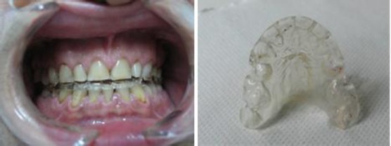
Patient with dOVD and with all accompanying symptoms. OS incorporated in patient’s mouth (left) and outside view of OS for correction of dOVD
Wassell et al. [15] described the success of 80% in patients with temporomandibular disorders treated with OS. The authors got the efficiency in treatment after five months uses of OS. In our study, the percent of efficiency was 75%, and that means the therapy in six patients was positive, while in two other patients there was no reduction of symptoms of temporomandibular problems.
After complete reduction of the signs and symptoms which were present at the beginning of the treatment from which suffer our patients the definitive prosthetic treatment was done. The kind of the prosthetic treatment was in relation with the indication of prosthetic therapy of every single patient. The need of the use of OS in dOVD treatment was also stated by Cutbirth [16]. In five patients the prosthetic treatment was done with fixed constructions, while in three cases the treatment was done with corresponding removable dentures. It’s important to notice that prosthetic constructions, fixed or removable, must be fabricated after previously corrected OVD with the use of OS.
Corrected OVD with OS as an interphase in treatment which gives positive effects on masticatory muscle activity, state Abekura et al. [17] and Stumbaun et al. [18]. The use of OS minimises the uncontrolled habit of clenching and bruxing with teeth, and there is a complete reduction of the symptomatology which is in relation with this habit.
Authors as Millet, Chandu [19] and Torii [20] suggest that in cases where the dOVD has to be changed, it has to be done with the use of OS. This approach gives us the chances of additional corrections in increasing of OVD and following the positive effects in uses of OS. This approach reduced most of the signs and symptoms of temporomandibular disorders. It’s that the definitive prosthetic treatment can be done after complete reduction of all signs and symptoms of dOVD.
In conclusion, after a clinical investigation of patients with dOVD and after the treatment following facts can be concluded: 1. dOVD is one of the reasons for the development of temporomandibular disorders; and 2. Definitive prosthetic treatment can be done after correction of the dOVD with the use of OS.
Footnotes
Funding: This research did not receive any financial support.
Competing Interests: The authors have declared that no competing interests exist.
References
- 1.Ekberg EC, Vallon D, Nilner M. Occlusal appliance therapy in patients with temporomandibular disorders. A double-blind controlled study in a short-term perspective. Acta Odontol Scand. 1998;56(2):122–8. doi: 10.1080/00016359850136102. https://doi.org/10.1080/00016359850136102 PMid:9669465. [DOI] [PubMed] [Google Scholar]
- 2.Hagag G, Yoshida K, Miura H. Occlusion, prosthodontic treatment, and temporomandibular disorders:a review. J Med Dent Sci. 2000;47(1):61–6. PMid:12162528. [PubMed] [Google Scholar]
- 3.Unger F. The management of temporomandibular joint disorders. The role of occlusal splints. Rev Stomatol Chir Maxillofac. 2001;102(1):47–54. PMid:11345625. [PubMed] [Google Scholar]
- 4.Guguvčevski LJ. Conservative Approach to the Temporomandibular Joint Hypermobility Treatment. (Plenary thesis) 11th Congress of BaSS. Sarajevo: Book of Abstracts; 2006. p. 27. [Google Scholar]
- 5.Wassell RW, Adams N, Kelly PJ. The treatment of temporomandibular disorders with stabilising splints in general dental practice:one-year follow-up. J Am Dent Assoc. 2006;137(8):1089–98. doi: 10.14219/jada.archive.2006.0347. https://doi.org/10.14219/jada.archive.2006.0347 PMid:16873324. [DOI] [PubMed] [Google Scholar]
- 6.Savabi O, Nejatidanesh F, Khosravi S. Effect of occlusal splints on the electromyographic activities of masseter and temporal muscles during maximum clenching. Quintessence Int. 2007;38(2):e129–32. PMid:17510715. [PubMed] [Google Scholar]
- 7.Barão VA, Gallo AK, Zuim PR, Garcia AR, Assunção WG. Effect of occlusal splint treatment on the temperature of different muscles in patients with TMD. J Prosthodont Res. 2011;55(1):19–23. doi: 10.1016/j.jpor.2010.06.001. https://doi.org/10.1016/j.jpor.2010.06.001 PMid:20591761. [DOI] [PubMed] [Google Scholar]
- 8.Millet C, Leterme A, Jeannin C, Jaudoin P. Vertical dimension in the treatment of the edentulous patient. Rev Stomatol Chir Maxillofac. 2010;111(5-6):315–30. doi: 10.1016/j.stomax.2010.09.001. PMid:21192343. [DOI] [PubMed] [Google Scholar]
- 9.Guguvčevski LJ. Derangements of the TMJ Disc. Apolonia. 2010;12(24):47–54. [Google Scholar]
- 10.Li Y, Zhang Z, Wu S, Qiao Y. A novel experimental design model for increasing occlusal vertical dimension. J Craniofac Surg. 2010;21(2):450–7. doi: 10.1097/SCS.0b013e3181cfe986. https://doi.org/10.1097/SCS.0b013e3181cf.e986 PMid:20489449. [DOI] [PubMed] [Google Scholar]
- 11.Botelho AL, Silva BC, Gentil FH, Sforza C, da Silva MA. The immediate effect of the resilient splint evaluated using surface electromyography in patients with TMD. Cranio. 2010;28(4):266–73. doi: 10.1179/crn.2010.034. https://doi.org/10.1179/crn.2010.034 PMid:21032981. [DOI] [PubMed] [Google Scholar]
- 12.Nilsson H. Resilient appliance therapy of temporomandibular disorders. Subdiagnoses, sense of coherence and treatment outcome. Swed Dent J Suppl. 2010;206:9–88. [PubMed] [Google Scholar]
- 13.Chang SW, Chuang CY, Li JR, Lin CY, Chiu CT. Treatment effects of maxillary flat occlusal splints for the painful clicking of the temporomandibular joint. Kaohsiung J Med Sci. 2010;26(6):299–307. doi: 10.1016/S1607-551X(10)70043-7. https://doi.org/10.1016/S1607-551X(10)70043-7. [DOI] [PMC free article] [PubMed] [Google Scholar]
- 14.Naito S, Ishida T, Kokai S, Fujita K, Shibata M, Yabushita T, Ono T. Functional adaptability of temporomandibular joint mechanoreceptors after an increase in the occlusal vertical dimension in rats. The Angle Orthodontist. 2011;81(3):453–9. doi: 10.2319/082010-489.1. https://doi.org/10.2319/082010-489.1 PMid:21261493. [DOI] [PMC free article] [PubMed] [Google Scholar]
- 15.Wassell RW, Adams N, Kelly PJ. Treatment of temporomandibular disorders by stabilising splints in general dental practice:results after initial treatment. Br Dent J. 2004;197(1):35–41. doi: 10.1038/sj.bdj.4811420. https://doi.org/10.1038/sj.bdj.4811420 PMid:15243608. [DOI] [PubMed] [Google Scholar]
- 16.Cutbirth ST. Increasing vertical dimension:considerations and steps in the reconstruction of the severely worn dentition. Pract Proced Aesthet Dent. 2008;20(10):619–26. PMid:19274959. [PubMed] [Google Scholar]
- 17.Abekura H, Yokomura M, Sadamori S, Hamada T. The initial effects of occlusal splint vertical thickness on the nocturnal EMG activities of masticatory muscles in subjects with a bruxism habit. Int J Prosthodont. 2008;21(2):116–20. PMid:18546763. [PubMed] [Google Scholar]
- 18.Stumbaum M, Konec D, Schweiger J, Gernet W. Reconstruction of the vertical jaw relation using CAD/CAM. Int J Comput Dent. 2010;13(1):9–25. PMid:20481288. [PubMed] [Google Scholar]
- 19.Chandu A, Suvinen TI, Reade PC, Borromeo GL. The effect of an interocclusal appliance on bite force and masseter electromyography in asymptomatic subjects and patients with temporomandibular pain and dysfunction. J Oral Rehabil. 2004;31(6):530–7. doi: 10.1111/j.1365-2842.2004.01377.x. https://doi.org/10.1111/j.1365-2842.2004.01377.x PMid:15189309. [DOI] [PubMed] [Google Scholar]
- 20.Torii K, Chiwata I. A case report of the symptom-relieving action of an anterior flat plane bite plate for temporomandibular disorder. Open Dent J. 2010;4:218–22. doi: 10.2174/1874210601004010218. https://doi.org/10.2174/1874210601004010218 PMid:21243070 PMCid:PMC3020490. [DOI] [PMC free article] [PubMed] [Google Scholar]