Abstract
Background
Data are limited on the various orthotic devices available for patients with Stage II posterior tibial tendon dysfunction (PTTD). Foot kinematics observed while walking with an orthotic device are hypothesized to be associated with clinical outcomes and could be used to refine future device designs.
Methods
Fifteen subjects (age, 63.6 ± 6.8 years) with Stage II PTTD walked in the lab under four conditions: (1) shoe only (control condition), (2) shoe with a custom solid AFO (Arizona Co, Mesa, AZ), (3) shoe with a custom articulated AFO (Arizona Co, Mesa, AZ), and (4) shoe with an off-the-shelf AFO (AirLift, DJ Orthopedics). Kinematic data were collected to determine the degree of hindfoot inversion, forefoot plantarflexion (reflective of raising the MLA), and forefoot adduction associated with each condition.
Results
The custom articulated device was associated with greater hindfoot inversion compared to the shoe only condition at loading response (p = 0.002), mid-stance (p < 0.001), and terminal stance (p = 0.02). The custom articulated device, custom solid device, and off-the-shelf device were associated with greater forefoot plantarflexion compared to the shoe only condition across all four phases of stance. There were no differences between any of the devices and the shoe condition associated with forefoot adduction.
Conclusion
The custom devices were associated with greater hindfoot inversion and forefoot plantarflexion compared to walking with only a shoe, while the off-the-shelf device was associated with forefoot plantarflexion but no change in hindfoot motion. None of the devices corrected forefoot abduction compared to the shoe only condition.
Clinical Relevance
The current biomechanical data may aid in understanding the clinical outcomes seen using these devices as well as provide data to support new designs.
Keywords: Biomechanics, PTTD, Posterior Tibial Tendon Dysfunction, Tendinopathy
INTRODUCTION
Posterior tibial tendon dysfunction (PTTD) is a progressive disorder with hallmarks of advancing flatfoot deformity and deteriorating function. The prevalence of PTTD is reported to be around 3.3% translating to an estimate of 2.3 million cases in the United States today. Ultimately, Stage IV is identified by the presence of arthritic changes in the lateral talocrural joint.21 The time patients take to progress from the onset of medial ankle pain indicating Stage I, to Stage IV, is variable and early conservative care may slow or halt the progression.20,22 The onset of a flexible flatfoot deformity (collapsed medial arch and hindfoot eversion) is the hallmark of Stage II and may be the most amendable to conservative care due to the flexible nature of the deformity.
Clinical guidelines nearly universally recommend the use of an orthosis for the conservative management of PTTD.8,12,16,18,24,29 However, with only limited data available, deciding which device is optimal for which patient remains almost a guess. A trend, summarized with the studies described below, may suggest the use of more restrictive orthoses that cross the ankle joint to be more successful than less restrictive in-shoe orthoses for Stage II PTTD. A 2-year followup using a validated questionnaire reported 90% of patients wearing a custom Arizona ankle-foot-orthosis (AFO) had decreased pain and increased function.2 A recent 7-year followup study indicated that overall long-term success, defined as orthosis free and avoiding surgery, was achieved in 69.7% of cases wearing a custom AFO design.22 In contrast, a study using an in-shoe foot orthoses also suggested improvement in patients with PTTD with a 47.6% decrease in pain and 53% decrease in disability.3 Off-the-shelf orthoses that extend above the ankle, such as the AirLift PTTD orthosis (AirCast Inc), are also available commercially and are widely used despite limited data on effectiveness.23 Overall, data support the use of more restrictive custom orthoses that extend proximal to the ankle joint,2,7 but off-the-shelf designs offer a considerable cost savings and are clinically popular.
Recently, numerous researchers25–26,32–33 have identified specific flatfoot kinematics observed in subjects with Stage II PTTD that may be linked to damage of the tibialis posterior tendon or the spring ligament.10,26 The tibialis posterior tendon is the primary degenerative tissue in PTTD while the spring ligament has been cited as the most commonly affected ligament, with up to 87% of ligaments involved in some study samples.10 A theoretical goal for orthotic devices is to correct flatfoot kinematics linked to the tibialis posterior tendon and spring ligament. To unload the tibialis posterior tendon, correction of a patient’s flatfoot kinematics towards hindfoot inversion and forefoot adduction are proposed.26 The spring ligament prevents hindfoot eversion and plantarflexion of the talus.19 Therefore orthoses that induce inversion of the hindfoot and raise the medial longitudinal arch (MLA) are recommended to unload the spring ligament.19 It has been suggested that correction of forefoot abduction may occur by correcting hindfoot and MLA positions,1,4,13 thus eliminating the need for specific design features to target the forefoot abduction position.
Theoretically, orthotic devices that are able to correct flat-foot kinematics will be clinically more successful. Additionally, identifying correction of kinematics may help with the redesign of current devices or design of new devices if necessary. The purpose of this study was to compare the kinematic effects of three commonly used orthotic devices in subjects with Stage II PTTD while walking. The kinematic variables of interest were hindfoot inversion, forefoot adduction, and forefoot plantarflexion (raising the MLA), chosen because of the theoretical and clinical goal of unloading support structures (such as the tibialis posterior tendon and spring ligament), and improving joint alignment to limit the onset of arthritic changes in the lateral talocrural joint.
METHODS
Fifteen subjects with a diagnosis of Stage II PTTD volunteered to participate in this study (Table 1). The diagnosis of Stage II PTTD was made by a foot and ankle fellowship-trained orthopedic surgeon (F.R.L.). The inclusion criteria for classification of Stage II PTTD required subjects to have one or more signs related to tendinopathy, including (1) palpable tenderness of the tibialis posterior tendon, (2) swelling of the tibialis posterior tendon sheath, or (3) inability to complete the heel-rise test. All of the subjects had reported symptoms for less than 2 years (range, 3 to 23 months) at the time they were tested. Additionally, one or more signs of flexible flatfoot deformity were required for classification of Stage II PTTD. These included excessive non-fixed hindfoot eversion deformity during weightbearing, excessive forefoot abduction (too-many-toes sign), or demonstrated loss of height in the medial longitudinal arch. The Arch Height Index (AHI), a reliable and valid measure of MLA height,35 was used to quantify arch height. Each subject demonstrated a lower AHI on the involved side compared to the uninvolved side and the group average of 0.270 ± 0.030 indicated a lower MLA compared to normative samples (mean ± SD; 0.340 ± 0.030).6 Signs of flatfoot deformity were based on comparisons from the involved to the uninvolved side. This then required that all subjects in the PTTD group have unilateral involvement. The uninvolved side may have also demonstrated signs of flatfoot deformity in some subjects but was not painful and did not demonstrate the same severity of flatfoot deformity. Subjects were excluded if they had a history of pain or pathology in the foot or lower extremity that prevented them from ambulating greater than 15 meters. All subjects were required to have sensate feet to ensure their safety with walking. Subjects with other foot conditions, such as plantar fasciitis, were also excluded from the current study. All PTTD subjects were required to be at least 40 years of age to restrict the study to only those with the typical degenerative onset of PTTD. All subjects were informed of the experimental procedures and signed a consent form approved by Upstate Medical University Research Subject Review Board.
Table 1.
Classification Variables for the 5 Subjects With Stage II PTTD
| Subjects with Stage II Posterior Tibial Tendon Dysfunction (PTTD) | |
|---|---|
| Age (years) | 61.8 ± 10.2 |
| Height (cm) | 167.4 ± 6.9 |
| Weight (kg) | 95.3 ± 20.9 |
| BMI | 31.8 ± 6.9 |
| Sex | 10 F, 5 M |
| Arch Height Index | 0.270 ± 0.03 |
Values expressed as means ± SD.
Device design and fabrication
For each subject, three AFOs that are commonly used in clinical practice and have published data to support their use were tested. The first orthosis was an off-the-shelf AFO (AirLift PTTD, AirCast, DJ Orthopedics), the second a custom molded AFO with a non-articulating ankle (solid ankle Arizona, Arizona Inc.), and the third a custom molded AFO with an articulating ankle (articulated ankle Arizona, Arizona Inc.). These orthoses may be broadly described as utilizing two mechanisms to manage the symptoms and flat-foot deformity observed in individuals with PTTD. First, each of these orthoses provides compression to the ankle which aids in controlling swelling and provides proprioceptive support.11,23 Second, each orthosis attempts to correct foot alignment by applying forces in locations needed to return the foot to a neutral position or oppose further deformity from occurring. Limits for hindfoot eversion are provided by three points of contact (lower lateral leg, medial malleolus, lateral heel) to maintain a neutral hindfoot to leg position. To support or correct the height of the MLA, the mechanism used is different for the custom and off-the-shelf orthoses. For the custom orthoses (articulated or solid) support for the MLA comes from a foot plate that extends into the midfoot and is fit to the MLA while the foot is held in neutral hindfoot alignment and supported arch. For the off-the-shelf orthosis an airbladder component is positioned under the MLA and inflated to raise the MLA. Control of forefoot abduction is accomplished by either; relying on coupled motion in the midfoot occurring with correction of hindfoot eversion, or, three points of contact including the lateral heel, medial arch, and lateral forefoot (by the orthosis and shoe).
Each subject was casted for the custom orthoses by a certified pedorthist following the guidelines described by the manufacturer (Arizona Co., Mesa, AZ). The foot was marked for bony landmarks and wrapped with plaster. The foot was positioned in contact with a casting plate on the floor with the hindfoot positioned in subtalar neutral as palpated by the pedorthist. The resulting mold was sent to the Arizona Company for manufacturing of two orthoses for testing.
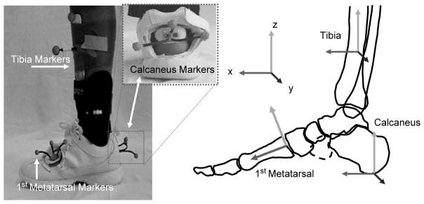
For each of the tested orthoses a “window” was needed to visualize the calcaneus and rigid body markers for gait analysis (Figure 1). The custom orthoses were constructed using a 3-mm polypropylene (plastic) ankle shell that was sewn inside a leather cover. The plastic shell covered the medial and lateral ankle (clam-shell) and continued around the foot to extend along the plantar aspect of the foot to end proximal to the metatarsal heads. The posterior portion of the heel contained no plastic support. In the solid ankle design this posterior heel was covered with leather while in the articulated ankle design the foot and shank parts of the orthosis were separated by a joint leaving the posterior heel open. In the solid ankle design the leather portion on the posterior heel was removed without altering the plastic support. In the articulated design the window was already available but was enlarged by trimming distal to the joint into the foot part of the orthosis to allow kinematic marker placement. The window locations were chosen to avoid the plastic support structure of the orthoses and qualitatively they did not appear to alter the integrity of the orthoses but rather removed the leather cover that was deemed aesthetic. Windows were also made in the testing shoe in the area of the heel marker and marker on the dorsal surface of the first metatarsal. Similar efforts to maintain the stability of the shoe were taken by adding a heel strap and replacing the shoe lacing. A previous study indicated that heel counter stability was altered less than 10% following similar shoe alterations.34 The modifications made to the shoe and orthoses were completed with input from the authors and pedorthist with efforts to maintain the integrity of the orthoses while completing the protocol. The custom orthoses were considered fit for long-term wear by the patient following the testing protocol.
Fig. 1.
Laboratory set-up for kinematic testing of the off-the-shelf orthotic device.
Each subject was seen 2 weeks prior to testing for fitting of the orthoses. Each of the custom orthoses were fit by the pedorthist to ensure there were no areas of pressure that needed to be relieved before the testing session. Three of the 15 subjects required alterations to at least one of the devices at the time of fitting, all of which were to reduce pressure on the navicular tuberosity medially. The off-the-shelf orthosis was fit according to manufacturer recommendations and was checked to ensure the same shoe could be used during the kinematic testing. Each subject was given a wearing schedule to gradually accommodate to the orthoses over the following week. Each subject was instructed to wear an orthosis for an hour each day, alternating the device worn each day. This routine continued for an average of 8 days until the testing occurred.
Motion analysis testing
The session consisted of a series of walking trials to test each of the orthoses which were tested in random order except that the off-the-shelf AFO was tested first in all subjects due to the inability to don or doff the device with the marker triads in place. Therefore, the off-the-shelf device was donned, after which the marker triads were attached to the skin through the visualization holes, finally the shoe was donned without disrupting the kinematic markers. The off-the-shelf AFO contained an airbladder along the medial side that was filled to a level of 4 PSI (27.6 kPa) of pressure in a nonweightbearing position. The 4 PSI level was chosen as a mid-level inflation comfortable to patients and previously found to achieve correction of foot kinematics.27 The subject was asked to walk down a 5-meter walkway at a comfortable walking speed. Speed was maintained within 5% using an infrared timing system for all walking trials. Following 5 successful trials where the involved foot landed completely on the force plate and the markers were in view, the shoe was removed by unlacing the front and unhooking the custom heel counter that was attached to the back of the shoe. This was done without removing the marker triads. The off-the-shelf AFO was removed by cutting the neoprene sleeve, again without disrupting the placement of the kinematic markers. Next, the custom AFO’s were tested in random order utilizing the same procedures. Each custom AFO could be slipped on or taken off by unlacing the front and holding the leather tounge aside to avoid the forefoot marker. To allow removal of each device without disrupting the position of the heel marker, a custom base was designed to allow removal of the marker wands without removing the skin-mounted base. Simply, this consisted of two “bases,” one that the marker wands attached to, and a second that was attached to a thermoplastic heel cup that was attached to the skin. These two bases were fit together and held with two small set screws. When switching between devices to be tested the triad of wands could be removed and then re-connected to the base in the same orientation. Testing of this method across five trials resulted in changes in position of each of the three markers of less than 1 mm and less than 1 degree when comparing hindfoot position relative to the shank before and after removing the marker triad.
Kinematic data were collected using a three-segment foot model including the tibia, calcaneus (hindfoot), and first metatarsal (forefoot) similar to a previously described model used to assess foot posture and motion in subjects with Stage II PTTD.33 Briefly, sets of three reflective markers were mounted on rigid thermoplastic platforms, and then attached using double-sided adhesive tape. Anatomic landmarks were digitized to establish local anatomically based coordinate systems for each segment using a static standing trial that occurred separate from the walking trials. Motion of the distal-most foot segment was then calculated relative to the adjacent proximal segment based on the Euler rotation sequence of flexion/extension, inversion/eversion, and abduction/adduction as suggested by Cole et al.9 The model used for this study consisted of the first metatarsal which was used to determine the angle of flexion/extension as well as abduction/adduction between the forefoot and hindfoot segments. A 12-camera VICON 512 motion analysis system and Workstation software (version 5.2) were used to collect marker data at a sampling rate of 60 Hz while The Motion Monitor software Version 8.52 (The Motion Monitor, Innsport Training Inc, USA) was used to develop and analyze the kinematic model. The VICON camera positions were carefully positioned to focus a field of view at an approximate 1.5 meter square centered at the force plate within the walkway. This allowed use of small 6- to 10-mm reflective spheres to be used in the kinematic model. The manufacturer reports accuracy of tracking an individual reflective marker at ± 0.1 mm with additional studies also reporting excellent precision and repeatability using the VICON motion analysis system.36 Using a 10 Newton threshold of vertical forces, collected at 1080Hz from an embedded force plate, Model 9287, (Kistler, Switzerland) initial contact and toe-off points of the gait cycle were identified. Kinematic data were smoothed using a 4th order, zero phase lag, Butterworth filter with a cut off frequency of 6 Hz.
Data analysis
The purpose of this study was to evaluate the kinematic effects of three commonly used orthotic devices in subjects with Stage II PTTD while walking. A repeated measure research design was used with each subject walking in each of the device conditions. Trials were averaged and variables of interest were interpolated to 101 points (0% to 100% stance) for comparison between the orthoses. The midpoint of each of the four stance phases of gait (10%, 35%, 65%, 90% stance) were chosen as representative of the various mechanical demands placed on the foot across the gait cycle and were used as points to compare among orthotic conditions.30 Therefore, a 4 × 4 repeated measures ANOVA model was repeated for each dependent kinematic variable (hindfoot inversion/eversion, forefoot plantarflexion/dorsiflexion, and forefoot abduction/adduction). The factors in the model included the four device conditions (shoe, custom articulated device, custom solid ankle device, and off-the-shelf) and the four phases of stance (loading response, mid-stance, terminal stance, and pre-swing). In the event of an interaction effect between the device and stance phase, the main effects were ignored and pairwise comparisons between device conditions at each phase explored. An alpha level of 0.05 was defined as a cut-off for the comparisons planned a priori.
To aid the interpretation of the data, in addition to the statistical analysis an intraclass correlation coefficient (ICC 3,1) was calculated and used to determine the standard error in the measurements (SEM) for each of the kinematic variables. This allowed an estimate of the total errors across the study to be evaluated. Two times the SEM (2SEM) was used to assess those changes that were above error and should be interpreted as meaningful differences between orthotic conditions. The 2SEM values were 1.0 degree for hindfoot eversion/inversion, 1.0 degree for forefoot plantarflexion/dorsiflexion, and 0.6 degrees for forefoot abduction/adduction.
RESULTS
Hindfoot inversion/eversion
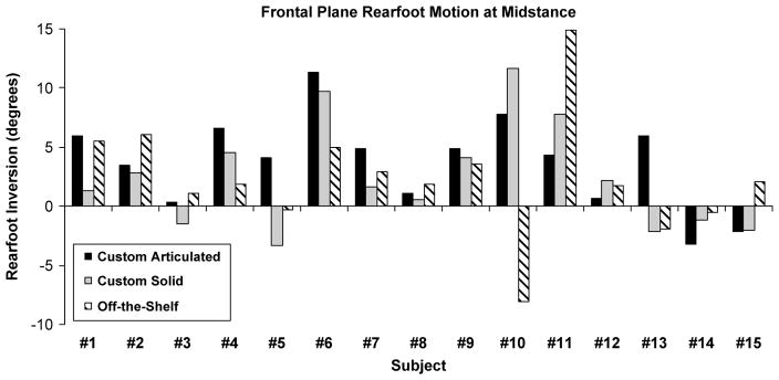
Differences between the devices was dependent on the phase of stance (significant interaction (p < 0.001)) and therefore comparisons between the devices occurred across each phase (Table 2, Figure 2). The custom articulated device was associated with greater hindfoot inversion compared to the shoe only condition at loading response (p = 0.002), mid-stance (p < 0.001), and terminal stance (p = 0.02). No other differences in hindfoot frontal plane motion were observed between the different devices or phases.
Table 2.
Descriptive Data (Mean ± SD in Degrees) for Each Kinematic Variable at the Midpoint of Each Phase Of Gait
| Shoe Only | Custom: Articulated Ankle | Custom: Solid Ankle | Off-The-Shelf | ||
|---|---|---|---|---|---|
| Hindfoot Eversion | Loading Response | −2.8 ± 5.1 | 0.5 ± 6.4α | −1.4 ± 7.1 | −1.0 ± 6.8 |
| Mid Stance | −3.2 ± 4.9 | 0.8 ± 6.4α | −1.0 ± 7.3 | −1.1 ± 7.1 | |
| Terminal Stance | −1.4 ± 5.0 | 1.4 ± 7.0α | 1.3 ± 7.4 | −0.5 ± 5.6 | |
| Pre-Swing | −0.1 ± 4.0 | 1.5 ± 6.4 | 2.1 ± 6.9 | −0.4 ± 5.2 | |
| Forefoot Plantar Flexion | Loading Response | 8.9 ± 8.3 | 13.6 ± 8.7α,β | 20.2 ± 8.9α,γ | 12.9 ± 7.7α |
| Mid Stance | 7.6 ± 9.0 | 13.4 ± 9.2α,β | 16.8 ± 10.3α,γ | 11.4 ± 8.4α | |
| Terminal Stance | 5.4 ± 10.5 | 12.8 ± 11.3α | 13.7 ± 11.7α | 9.3 ± 8.6α | |
| Pre-Swing | 6.1 ± 11.6 | 12.1 ± 11.5α | 13.2 ± 11.9α | 10.1 ± 11.0α | |
| Forefoot Abduction | −0.3 ± 2.3 | 0.9 ± 2.5γ | 1.1 ± 2.2γ | −2.9 ± 2.3 |
Positive values indicate hindfoot inversion, forefoot plantar flexion, and forefoot adduction. For the variable forefoot abduction, there were no significant differences that were dependent on phase of stance (no phase by device interaction) therefore averages across phases are presented.
Significantly different than shoe
Significantly different than custom solid
Significantly different than off-the-shelf.
Fig. 2.
Effect of each orthotic device compared to the shoe only condition (device minus shoe-only condition) at the midpoint of the midstance phase of gait. Each subject’s raw data is presented. Rearfoot Inversion is positive and indicates a hypothesized improvement in the effect of the brace compared to the shoe only condition.
Forefoot plantarflexion/dorsiflexion
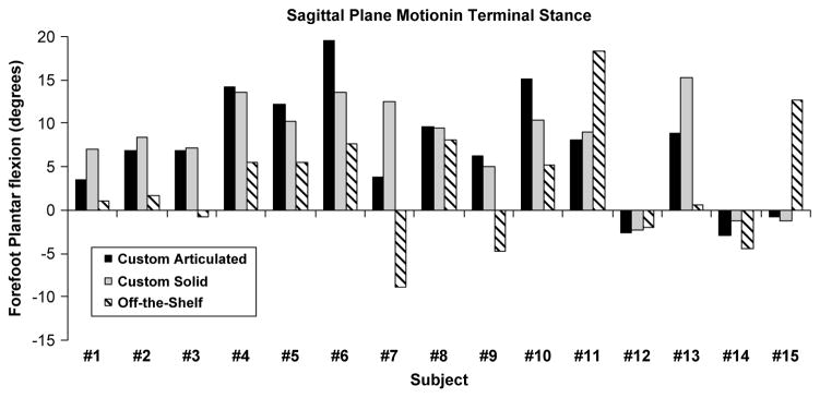
Differences between the devices was dependent on the phase of stance (significant interaction (p < 0.003)) and therefore comparisons between the devices occurred across each phase (Table 2, Figure 3). The custom articulated device, custom solid device, and off-the-shelf device were associated with greater forefoot plantarflexion compared to the shoe only condition across all four phases of stance. Additionally, the custom solid device was associated with greater forefoot plantarflexion compared to the custom articulated and off-the-shelf device during the loading response and mid-stance phases of gait.
Fig. 3.
Effect of each orthotic device compared to the shoe only condition (device minus shoe-only condition) at the midpoint of the terminal stance phase of gait. Each subject’s raw data is presented. Forefoot plantar flexion is positive and indicates a hypothesized improvement in the effect of the brace compared to the shoe only condition.
Forefoot adduction/abduction
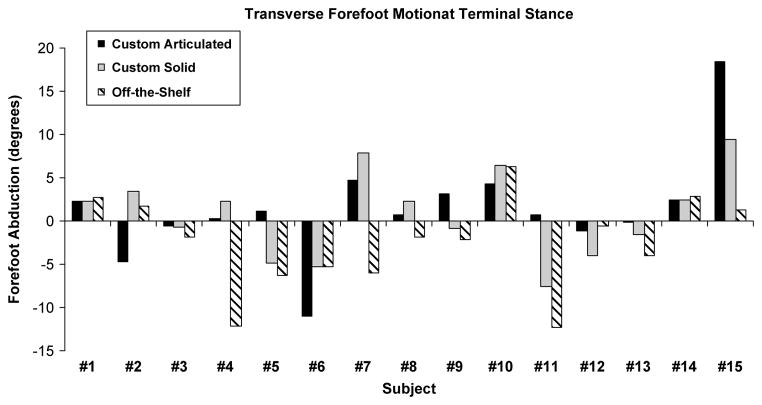
No interaction was found between the device and phase conditions but a main effect for device (p = 0.016) was observed. For this reason, the data were averaged across phases to represent the average effect of each device for all phases (Table 2, Figure 4). The custom articulated and custom solid devices were associated with significantly (p = 0.03 and p = 0.006, respectively) greater forefoot adduction compared to the off-the-shelf device. No differences between any of the devices and the shoe condition were observed.
Fig. 4.
Effect of each orthotic device compared to the shoe only condition (device minus shoe-only condition) at the midpoint of the terminal stance phase of gait. Each subject’s raw data is presented. Forefoot adduction is positive and indicates a hypothesized improvement in the effect of the brace compared to the shoe only condition.
DISCUSSION
Kinematic changes toward a foot posture that would support the tibialis posterior tendon and ligaments of the foot (e.g., spring ligament) were seen with each of the three devices tested. No previous studies provide head-to-head comparisons between available devices in patients with PTTD. New to this study are data on the kinematic effectiveness of different devices, and device designs, to inform current clinical practice and future device development. The custom devices were associated with greater hindfoot inversion and forefoot plantarflexion compared to walking with only a shoe, while the off-the-shelf device was associated with forefoot plantarflexion but no change in hindfoot motion. None of the devices corrected forefoot abduction compared to the shoe only condition. These kinematic changes may explain the positive clinical outcomes that have been reported in patients wearing each of the devices. However, new devices may be indicated to improve correction of foot posture, especially targeting forefoot abduction deformity.
Overall, for hindfoot inversion, the 3 points of pressure used by each of the tested orthoses to correct alignment produced small changes that were above the error of the measurement system. (Figure 2) Based on in vitro testing, limiting peak eversion may unload the TP muscle-tendon unit.14 Averaged across all phases, limiting peak eversion was greater when using the custom articulated device (3.3 degrees - effect of the articulated device minus the effect of the shoe alone) (Table 2). This effect was 2.3 degrees and 2.2 degrees for the custom solid ankle device and off-the-shelf device, respectively. This outcome is consistent with a previous case study where the articulated device was the most successful in limiting hindfoot eversion.28 The design of the articulated device has a close fitting heel cup which may improve inversion control. Another hypothesis is that greater muscle function may accompany allowing ankle motion to aid with greater inversion motion. These hypotheses are consistent with comparisons between the device designs but require further study to inform future device design as we had a relatively small number of subjects. It remains unknown if greater hindfoot control will translate to improved clinical outcomes over the custom solid ankle design or off-the-shelf design. However, positive outcomes in previous studies using the custom solid ankle device may be consistent with the greater-than-2-degree change observed in the current study.2 It has been argued that small changes of 2 degrees may be clinically meaningful if these changes are able to unload support structures during repetitive tasks such as walking.17
Each of the devices tested were associated with greater amounts of forefoot plantarflexion (raising the MLA) compared to the shoe only condition and these effects were the largest of the kinematic effects recorded (Table 2, Figure 3). Across all phases, the average change in plantarflexion compared to the shoe-only condition was 6.0 degrees for the articulated device, 9.0 degrees for the solid ankle device, and 3.9 degrees for the off-the-shelf device. These changes suggest the mechanisms used to support the MLA (airbladder or custom molded foot plate) are successful in subjects with PTTD and may achieve their theoretical purpose of unloading ligaments such as the spring ligament. The custom solid ankle device, when tested in a cadaver flatfoot model, demonstrated effects of restoring measures of arch height but had less effect on hindfoot inversion movement, consistent with the in-vivo data from this study.18 Although the articulated ankle design was associated with the greatest change in hind-foot inversion motion, the solid ankle design was associated with the greatest forefoot plantarflexion (raising the MLA) motion. One reasonable suggestion for the greater forefoot plantarflexion in the solid ankle design may be that allowing ankle dorsiflexion in the articulated design allows a smooth transfer of body weight across the midfoot and may load the MLA. In contrast, the solid ankle design limits ankle dorsiflexion, therefore encouraging transfer of body weight straight to the metatarsal heads, bypassing the midfoot and consistent with the current data improving arch support.
New designs may need to be considered to improve control of forefoot abduction deformity in subjects with Stage II PTTD. None of the devices tested in the current study produced changes in forefoot abduction deformity compared to the control condition of walking in a shoe alone (Table 2, Figure 4). The custom designs, both solid ankle and articulated ankle, were associated with small non-significant changes towards forefoot adduction while the off-the-shelf device was associated with a small non-significant change towards forefoot abduction. It should be noted that although none of the devices were successful in producing forefoot adduction relative to the shoe condition, the difference between the two custom devices and the off-the-shelf device was significant. This suggests that new designs may be needed to address this specific foot kinematic and in future designs custom foot plates that are fitted to the foot may be explored as better alternatives than an air bladder component. It is also apparent that with the devices tested there is large subject variability. The largest standard deviations for the smallest movements are observed with the forefoot abduction variable with some subjects demonstrating forefoot adduction (improvement) while others demonstrate forefoot abduction (worsening) testing the same device (Table 2). Previous studies have repeatedly shown that individual responses to orthotic devices are common17 and the individual foot posture or degree of deformity may be a factor specific to the success in correcting forefoot abduction using these devices.
The kinematic foot model used for this study has been shown to be successful in capturing the flatfoot posture associated with subjects with Stage II PTTD but the findings from this study are likely sensitive to the model used and alternative models have been proposed.25, 31–32 The subjects in this study were classified as having Stage II PTTD but classification schemes for PTTD continue to be refined with proposed sub-divisions within Stage II.5 These sub-divisions may help to explain the individual variability observed across the devices tested in this study and could be considered in future studies of subjects with Stage II PTTD. Future study samples may include refined classification schemes with expanded numbers within the selected groups to further elucidate the effects of orthotic design on foot kinematics. Numerous orthotic devices including in-shoe foot orthoses as well as ankle-foot-orthoses have been recommended for the conservative management of PTTD and the current study was not meant to be a comprehensive evaluation of all of the possible devices. Rather, the current study was meant to evaluate specific types of designs that are commonly used. Conclusions may be able to be transferred to other similar designs or future designs where the features or components used are similar, but further study on the kinematic as well as clinical outcomes from the numerous devices used in PTTD are needed. It should also be noted that the control condition for this study was walking while wearing a shoe. The foot kinematics of subjects with PTTD walking barefoot have been described in other studies and it is likely that the shoe included as a control condition in this study had an effect, by itself, on the foot kinematics described. This study focused on the isolated effect of the devices but further studies may include comparisons between orthotic devices and shoe designs as each in isolation, and in combination, likely have an impact on foot kinematics.
The primary foot kinematics deemed important to control for patients with PTTD included hindfoot eversion, forefoot abduction, and a low MLA. The three orthoses compared in this study were all designed to target these foot kinematics and have demonstrated positive clinical outcomes in previous studies.2,7,22 In summary, each of the devices tested were associated with raising the MLA while the articulated custom AFO was associated with greater hindfoot inversion. In general, the custom fit devices were associated with greater changes than the off-the-shelf device. None of the devices tested were associated with forefoot adduction. The current biomechanical data may aid in understanding the clinical outcomes seen using these devices as well as provide data to support new designs. Additionally, experimentally controlled studies are needed to provide more information on the long-term clinical outcomes expected from different devices and device designs.
Acknowledgments
Funds were received in support of the study presented in this article from an Upstate Medical University Research Enhancement Grant.
Footnotes
For information on pricings and availability of reprints, reprints@datatrace.com or call 410-494-4994, x232.
References
- 1.Amaral De Noronha M, Borges NG., Júnior Lateral ankle sprain: isokinetic test reliability and comparison between invertors and evertors. Clin Biomech. 2004;19:868–871. doi: 10.1016/j.clinbiomech.2004.05.011. http://dx.doi.org/10.1016/j.clinbiomech.2004.05.011. [DOI] [PubMed] [Google Scholar]
- 2.Augustin JF, Lin SS, Berberian WS, Johnson JE. Nonoperative treatment of adult acquired flat foot with the Arizona brace. Foot & Ankle Clinics. 2003;8:491–502. doi: 10.1016/s1083-7515(03)00036-6. http://dx.doi.org/10.1016/S1083-7515(03)00036-6. [DOI] [PubMed] [Google Scholar]
- 3.Bek N, Oznur A, Kavlak Y, Uygur F. The effect of orthotic treatment of posterior tibial tendon insufficiency on pain and disability. The Pain Clinic. 2003;15:345–350. http://dx.doi.org/10.1163/156856903767650907. [Google Scholar]
- 4.Blackwood CB, Yuen TJ, Sangeorzan BJ, Ledoux WR. The midtarsal joint locking mechanism. Foot Ankle International. 2005;26:1074–1080. doi: 10.1177/107110070502601213. [DOI] [PubMed] [Google Scholar]
- 5.Bluman E, Title CI, Myerson MS. Posterior Tibial Tendon Rupture: A Refined Classification System. Foot and ankle clinics. 2007;12:233. doi: 10.1016/j.fcl.2007.03.003. http://dx.doi.org/10.1016/j.fcl.2007.03.003. [DOI] [PubMed] [Google Scholar]
- 6.Butler RJ, Hillstrom H, Song J, et al. Arch height index measurement system: establishment of reliability and normative values. J Am Podiatr Med Assoc. 2008;98:102–106. doi: 10.7547/0980102. [DOI] [PubMed] [Google Scholar]
- 7.Chao W, Wapner KL, Lee TH, Adams J, Hecht PJ. Nonoperative management of posterior tibial tendon dysfunction.[see comment] Foot Ankle Int. 1996;17:736–741. doi: 10.1177/107110079601701204. [DOI] [PubMed] [Google Scholar]
- 8.Churchill RS, Sferra JJ. Posterior tibial tendon insufficiency. Its Diagnosis Management and Treatment. American Journal of Orthopedics (Chatham Nj) 1998;27:339–347. [PubMed] [Google Scholar]
- 9.Cole GK, Nigg BM, Ronsky JL, Yeadon MR. Application of the joint coordinate system to three-dimensional joint attitude and movement representation: a standardization proposal. J Biomech Eng. 1993;115:344–349. doi: 10.1115/1.2895496. http://dx.doi.org/10.1115/1.2895496. [DOI] [PubMed] [Google Scholar]
- 10.Deland JT, de Asla RJ, Sung IH, Ernberg LA, Potter HG. Posterior tibial tendon insufficiency: which ligaments are involved? Foot Ankle Int. 2005;26:427–435. doi: 10.1177/107110070502600601. [DOI] [PubMed] [Google Scholar]
- 11.Eils E, Rosenbaum D. The main function of ankle braces is to control the joint position before landing. Foot Ankle Int. 2003;24:263–268. doi: 10.1177/107110070302400312. [DOI] [PubMed] [Google Scholar]
- 12.Elftman NW. Nonsurgical treatment of adult acquired flat foot deformity. Foot & Ankle Clinics. 2003;8:473–489. doi: 10.1016/s1083-7515(03)00119-0. http://dx.doi.org/10.1016/S1083-7515(03)00119-0. [DOI] [PubMed] [Google Scholar]
- 13.Ferber R, McClay Davis I, Williams DS., III Effect of foot orthotics on rearfoot and tibia joint coupling patterns and variability. J Biomech. 2005;38:477–483. doi: 10.1016/j.jbiomech.2004.04.019. http://dx.doi.org/10.1016/j.jbiomech.2004.04.019. [DOI] [PubMed] [Google Scholar]
- 14.Flemister AS, Neville CG, Houck J. The relationship between ankle, hindfoot, and forefoot position and posterior tibial muscle excursion. Foot Ankle Int. 2007;28:448–455. doi: 10.3113/FAI.2007.0448. http://dx.doi.org/10.3113/FAI.2007.0448. [DOI] [PubMed] [Google Scholar]
- 15.Gazdag AR, Cracchiolo A., 3rd Rupture of the posterior tibial tendon. Evaluation of injury of the spring ligament and clinical assessment of tendon transfer and ligament repair. Journal of Bone Joint Surgery - American Volume. 1997;79:675–681. doi: 10.2106/00004623-199705000-00006. [DOI] [PubMed] [Google Scholar]
- 16.Geideman WM, Johnson JE. Posterior tibial tendon dysfunction. J Orthop Sports Phys Ther. 2000;30:68–77. doi: 10.2519/jospt.2000.30.2.68. [DOI] [PubMed] [Google Scholar]
- 17.Genova J, Gross MT. Effect of Foot Orthotics on Claclaneal Eversion During Standing and Treadmill Walking for Subjects with Abnormal Pronation. J Orthop Sports Phys Ther. 2000;30:664–675. doi: 10.2519/jospt.2000.30.11.664. [DOI] [PubMed] [Google Scholar]
- 18.Imhauser CW, Abidi NA, Frankel DZ, Gavin K, Siegler S. Biomechanical evaluation of the efficacy of external stabilizers in the conservative treatment of acquired flatfoot deformity. Foot Ankle Int. 2002;23:727–737. doi: 10.1177/107110070202300809. [DOI] [PubMed] [Google Scholar]
- 19.Jennings MM, Christensen JC. The effects of sectioning the spring ligament on rearfoot stability and posterior tibial tendon efficiency. J Foot Ankle Surg. 2008;47:219–224. doi: 10.1053/j.jfas.2008.02.002. http://dx.doi.org/10.1053/j.jfas.2008.02.002. [DOI] [PubMed] [Google Scholar]
- 20.Kulig K, Reischl SF, Pomrantz AB, et al. Nonsurgical management of posterior tibial tendon dysfunction with orthoses and resistive exercise: a randomized controlled trial. Phys Ther. 2009;89:26–37. doi: 10.2522/ptj.20070242. http://dx.doi.org/10.2522/ptj.20070242. [DOI] [PubMed] [Google Scholar]
- 21.Lee MS, Vanore JV, Thomas JL, et al. Diagnosis and treatment of adult flatfoot. J Foot Ankle Surg. 2005;44:78–113. doi: 10.1053/j.jfas.2004.12.001. http://dx.doi.org/10.1053/j.jfas.2004.12.001. [DOI] [PubMed] [Google Scholar]
- 22.Lin JL, Balbas J, Richardson EG. Results of non-surgical treatment of Stage II posterior tibial tendon dysfunction: a 7- to 10-year followup. Foot Ankle Int. 2008;29:781–786. doi: 10.3113/FAI.2008.0781. http://dx.doi.org/10.3113/FAI.2008.0781. [DOI] [PubMed] [Google Scholar]
- 23.Logue JD. Advances in Orthotics and Bracing. Foot and ankle clinics. 2007;12:215. doi: 10.1016/j.fcl.2007.03.012. http://dx.doi.org/10.1016/j.fcl.2007.03.012. [DOI] [PubMed] [Google Scholar]
- 24.Marzano R, Marzano R. Functional bracing of the adult acquired flatfoot. Clin Podiatr Med Surg. 2007;24:645–656. doi: 10.1016/j.cpm.2007.06.002. http://dx.doi.org/10.1016/j.cpm.2007.06.002. [DOI] [PubMed] [Google Scholar]
- 25.Ness ME, Long J, Marks R, Harris GF. Foot and Ankle Kinematics in Subjects with Posterior Tibial Tendon Dysfunction. Gait Posture. 2008;27:331–339. doi: 10.1016/j.gaitpost.2007.04.014. http://dx.doi.org/10.1016/j.gaitpost.2007.04.014. [DOI] [PubMed] [Google Scholar]
- 26.Neville C, Flemister A, Tome J, Houck J. Comparison of changes in posterior tibialis muscle length between subjects with posterior tibial tendon dysfunction and healthy controls during walking. J Orthop Sports Phys Ther. 2007;37:661–669. doi: 10.2519/jospt.2007.2539. [DOI] [PubMed] [Google Scholar]
- 27.Neville C, Flemister AS, Houck JR. Effects of the AirLift PTTD Brace on Foot Kinematics in Subjects With Stage II Posterior Tibial Tendon Dysfunction. J Orthop Sports Phys Ther. 2009;39:201–209. doi: 10.2519/jospt.2009.2908. [DOI] [PubMed] [Google Scholar]
- 28.Neville C, Houck JR. Choosing Among 3 Ankle-Foot Orthoses for a Patient With Stage II Posterior Tibial Tendon Dysfunction. J Orthop Sports Phys Ther. 2009;39:816–824. doi: 10.2519/jospt.2009.3107. [DOI] [PMC free article] [PubMed] [Google Scholar]
- 29.Noll KH. The use of orthotic devices in adult acquired flatfoot deformity. Foot & Ankle Clinics. 2001;6:25–36. doi: 10.1016/s1083-7515(03)00077-9. http://dx.doi.org/10.1016/S1083-7515(03)00077-9. [DOI] [PubMed] [Google Scholar]
- 30.Perry J. Normal and Pathologic Function, Thorofare. SLACK Incorporated; 1992. Gait Analysis. [Google Scholar]
- 31.Rattanaprasert U, Smith R, Sullivan M, Gilleard W. Three-dimensional kinematics of the forefoot, rearfoot, and leg without the function of tibialis posterior in comparison with normals during stance phase of walking. Clin Biomech. 1999;14:14–23. doi: 10.1016/s0268-0033(98)00034-5. http://dx.doi.org/10.1016/S0268-0033(98)00034-5. [DOI] [PubMed] [Google Scholar]
- 32.Ringleb SI, Kavros SJ, Kotajarvi BR, et al. Changes in gait associated with acute Stage II posterior tibial tendon dysfunction. Gait Posture. 2007;25:555–564. doi: 10.1016/j.gaitpost.2006.06.008. http://dx.doi.org/10.1016/j.gaitpost.2006.06.008. [DOI] [PubMed] [Google Scholar]
- 33.Tome J, Nawoczenski DA, Flemister A, Houck J. Comparison of foot kinematics between subjects with posterior tibialis tendon dysfunction and healthy controls. J Orthop Sports Phys Ther. 2006;36:635–644. doi: 10.2519/jospt.2006.2293. [DOI] [PubMed] [Google Scholar]
- 34.Williams DS, 3rd, McClay Davis I, Baitch SP. Effect of inverted orthoses on lower-extremity mechanics in runners. Med Sci Sports Exerc. 2003;35:2060–2068. doi: 10.1249/01.MSS.0000098988.17182.8A. http://dx.doi.org/10.1249/01.MSS.0000098988.17182.8A. [DOI] [PubMed] [Google Scholar]
- 35.Williams DS, McClay IS. Measurements used to characterize the foot and the medial longitudinal arch: reliability and validity. Phys Ther. 2000;80:864–871. [PubMed] [Google Scholar]
- 36.Windolf M, Götzen N, Morlock M. Systematic accuracy and precision analysis of video motion capturing systems–exemplified on the Vicon-460 system. J Biomech. 2008;41:2776–2780. doi: 10.1016/j.jbiomech.2008.06.024. http://dx.doi.org/10.1016/j.jbiomech.2008.06.024. [DOI] [PubMed] [Google Scholar]


