Abstract
The secondary cysts of the fish pathogen oomycete Saprolegnia parasitica possess bundles of long hooked hairs that are characteristic to this economically important pathogenic species. Few studies have been carried out on elucidating their specific role in the S. parasitica life cycle and the role they may have in the infection process. We show here their function by employing several strategies that focus on descriptive, developmental and predictive approaches. The strength of attachment of the secondary cysts of this pathogen was compared to other closely related species where bundles of long hooked hairs are absent. We found that the attachment of the S. parasitica cysts was around three times stronger than that of other species. The time sequence and influence of selected factors on morphology and the number of the bundles of long hooked hairs conducted by scanning electron microscopy study revealed that these are dynamic structures. They are deployed early after encystment, i.e., within 30 sec of zoospore encystment, and the length, but not the number, of the bundles steadily increased over the encystment period. We also observed that the number and length of the bundles was influenced by the type of substrate and encystment treatment applied, suggesting that these structures can adapt to different substrates (glass or fish scales) and can be modulated by different signals (i.e., protein media, 50 mM CaCl2 concentrations, carbon particles). Immunolocalization studies evidenced the presence of an adhesive extracellular matrix. The bioinformatic analyses of the S. parasitica secreted proteins showed that there is a high expression of genes encoding domains of putative proteins related to the attachment process and cell adhesion (fibronectin and thrombospondin) coinciding with the deployment stage of the bundles of long hooked hairs formation. This suggests that the bundles are structures that might contribute to the adhesion of the cysts to the host because they are composed of these adhesive proteins and/or by increasing the surface of attachment of this extracellular matrix.
Introduction
Oomycetes are successful and widespread groups of parasites [1]. In order to colonize and parasitize, they rely on asexual motile zoospores, i.e., secondary zoospores [2–4] that form secondary cysts that attach to their host prior the infection [5–8]. The ability of the oomycetes to invade a wide range of hosts such as fungi, algae, plants, invertebrates, or vertebrates, indicates that they might have adaptable mechanisms for substrate and host colonization [9–13]. Recent genome and secretome studies have shown that these mechanisms involve diverse molecules including translocated host-targeting proteins [14], degrading enzymes, such as glycosyl-hydrolases and proteases [15], or adhesive biopolymers [16, 17]. These adhesive compounds allow cell-substratum attachment, prevent the pathogen from removal, facilitates host-pathogen interaction and host colonization [18].
Biological attachment is based on adhesive polymers that can vary in structure and capabilities involving interactions and components with different functions [19]. In the plant pathogenic oomycetous species, Phytophthora cinnamomi, the adhesion of cysts to plants roots involved the secretion of high molecular weight glycoproteins by rapid exocytosis [11]. In 2005, Robold and Hardham [10], identified a potential adhesive molecule of 220-kDa protein with thrombospondin repeats [10]. In Saprolegnia parasitica and Saprolegnia diclina, the presence of an extracellular matrix was evidenced by Burr and Beakes [20]. The identification of these components represents a biochemical and genetic challenge since little results have been obtained because of the complexity of the methods and approach [12, 16, 17, 20].
In fungi, similar molecules have been described in Colletotrichum graminicola [21], Erysiphe graminis [22], Nectria haematococca [23]. In the attachment process, some plant pathogenic oomycetes develop specialized attachment structures named appressoria that help the host penetration and subsequent infection [24]. Apart from appressoria, the only structures that have been related to attachment in the oomycetes are the so-called hooked “hairs” or “spines” on the secondary cysts of the fish pathogen S. parasitica [25]. Theses hooked hairs seem to be unique to the genus Saprolegnia, and were described for the first time by Manton et al., [26] using transmission electron microscopy (TEM) and a few years later Meier and Webster [27] reported that S. parasitica had long bundles of these hooked hairs. All studies on hooked hairs were focused on their morphology and structure and cyst coat ornamentation in Saprolegnia spp. [28–35]. Thus, these studies showed that the hooked hairs of S. parasitica are specifically longer, over 2 μm in length than those of other Saprolegnia spp. and are organized in bundles [29, 31]. These hairs appear to be rapidly synthesized during the secondary cyst stage [29]. Beakes [29] presented the overall ontogeny of encystment vesicles in several S. parasitica isolates. In these vesicles, the hairs seem to be deposited filled with matrix material that seems to be composed of glucose and mannose since it binds to lectin Concavalin A [20]. Later, Beakes et al. [31] suggested that the bundles of long hooked hairs might play a role as a potential virulent factor by increasing the attachment efficiency. Their function is unclear and has been extensively debated in the literature [25]. Thus, it has been suggested that these structures assist the attachment to a substrate [27, 28].
The synchronization of Saprolegnia developmental stages in vitro [35, 36] as well as the recent sequencing of the whole S. parasitica genome makes it now possible to investigate secreted protein families with potential roles in virulence at different developmental stages [15]. Therefore, the main objective of this work was to elucidate the function of the bundles of long hooked hairs by: (i) comparing the strength of attachment of the secondary cysts of this pathogen with those of other closely related species that have no long hairs, (ii) understanding the time sequence of external development of these structures, and the influence of the selected factors on the morphology and the number of bundles of long hooked hairs developed, and (iii) finally, by investigating proteins that might be produced during their formation.
Material and methods
Strain cultivation, sporulation, and production of cysts
The strain of S. parasitica (SAP0206) [37] was used in all the experiments. For microscopic studies and assessment of the strength of adhesion of the secondary cysts, the strains SAP0655 of Saprolegnia delica [35], and SAP1148 of Saprolegnia anisospora [38], from the culture collection of Real Jardín Botánico at CSIC were used. Cultures were maintained on a peptone-glucose agar (PGA) medium [39], and sporulation was performed following the protocol described by Diéguez-Uribeondo et al., [36]. Briefly, mycelia colonies were grown in 0.5 mL peptone glucose liquid media (PG-l) for 24–48 hours at 20°C. The sporulation was induced by washing the mycelia with autoclaved tap water three times and then incubated for 15 hours at 20°C to allow release of the zoospores. The secondary zoospores were collected according to Cerenius and Söderhäll [40] followed by the secondary cysts production through vigorous agitation for 30 sec at 1400 rpm, except of those experiments designed to evaluate the effect of the encystment triggers on the secondary cysts morphology.
Scanning and transmission electron microscopy
The secondary zoospores were encysted as described above and volumes of 0.5 mL of secondary cysts suspensions were transferred onto glass cover slips (8 mm in diameter). Samples were incubated at 20°C for 70 min and fixed for 1 hour in 2% glutaraldehyde at room temperature. Fixed samples were dehydrated in alcohol series from 30 to 100% and critical point dried. Then, the secondary cysts on selected surfaces were sputter coated in a vacuum with an electrically conductive layer of gold to a thickness of approximately 80 nm. Samples were observed under a Hitachi s3000N scanning electron microscope, SEM, (Real Jardín Botánico, CSIC, Madrid, Spain) at a beam specimen angle of 45°. Accelerating voltage was 20 kV; final aperture was 200 μm.
For transmission electron microscopy, TEM, the samples of secondary cysts obtained as above were processed by using a slight modification of the method described by Beakes [29]. Thus, formvar-coated copper grids (30 mm, 200 mesh) were kept in autoclaved tap water in order to avoid drying, and then fixed in uranyl acetate 2%. The specimens were observed in a JEM-1010, Jeol transmission electron microscope (Centro Nacional de Microscopía Electronica, University of Complutense, Madrid, Spain). Accelerating voltage was 100 kV; resolution 0.3 nm.
Evaluation of the adhesion strength of the secondary cysts
The adhesion strength of the secondary cysts was tested on selected strains of the species S. anisospora, S. delica, and S. parasitica. Volumes of 0.5 mL of a concentration of 104 secondary cysts/mL were placed onto glass cover slips immediately after encystment. A number of 50 independent suspensions for each species were generated for the experiment. Measurements of the strength of adhesion were performed 70 min after encystment. This allowed secondary cysts to extend their hooked hairs and to attach firmly to the surface. In order to measure the strength required to remove the secondary cysts, we followed the protocol of Letourneau et al., [41]. Briefly, coverslips (8 mm in diameter) with a monolayer of attached secondary cysts were individually vortexed in 1.5 mL Eppendorf tubes for 60 sec at 1400 rpm. The total number of secondary cysts that remained attached was counted before and after the treatment by selecting three arbitrary fields of view and using a light microscope 20x (Olympus BX51, Olympus Optical, Japan). The adhesion strength of the secondary cysts was calculated by first obtaining the average numbers of cysts attached for each replicate (each having measurements of three arbitrary fields) before and after the treatment, and then obtaining the ratio of these averages after and before the treatment.
Time sequence study of extracellular deployment of bundles of long hooked hairs
The length and number of bundles of long hooked hairs (from now on will be named bundles) produced by the secondary cysts of S. parasitica were studied in a time sequence series on fixed samples using SEM. Thus, suspensions of secondary cysts obtained as described above were transferred onto glass cover slips (8 mm in diameter). The time series were prepared by vortexing the zoospores for 30 s followed by incubations for 5, 15, 70 and 115 min. After incubations, samples were fixed and processed as described above. For measurements of the length and number of bundles, 25 randomly selected secondary cysts were taken at each treatment tested. The length and number of the bundles was measured for each cyst and the whole number and length of the 25 samples were analyzed for each selected time point. Measurements were performed using ImageJ v.1.51a software [42].
Effect of substrate, area of contact, and encystment triggers on the length and number of bundles of long hooked hairs
To evaluate the effect of area of contact, and encystment triggers on the number and length of bundles of S. parasitica, the secondary cysts were obtained as described above and were transferred onto the fish scales (Salmon salar). In order to check the effect of an additional surface exposure of the cysts, a suspension of carbon particles was added when the secondary cysts were vortexing. For the experiment, a carbon particle solution was prepared as described in Diéguez-Uribeondo et al., [43]. In the encystment trigger experiments, the secondary cysts were obtained by adding an equal volume of peptone (PG-l), or 100 mM calcium solution (CaCl2) to the zoospore suspension with the concentration of 104 zoospore/ml as described by Diéguez-Uribeondo et al., [44].
In all experiments, the suspensions of secondary cysts were incubated for 70 min at 20° C and fixed for SEM as described above. The encystment induced by vortexing followed by sample incubation on glass coverslips was used as control. To count and measure the number and the length of bundles, 25 random SEM micrographs were selected. All counts and measurements were done using ImageJ v.1.51a software [42].
Enzyme digestions and immunolocalization of bundles of long hooked hairs and extracellular matrix
Enzymatic treatments
The following enzyme preparations were tried for digestion of cyst spines: (i) Peptide:N-Glycosidase F (PNGase F, from Elizabethkingia miricola, Sigma-Aldrich, Co. LLC, St. Louis, IL) and O-glycosidase (from Streptococcus pneumoniae, Sigma-Aldrich, Co. LLC, St. Louis, IL) mix for 2 hours at room temperature with a final enzyme concentration 1.25μ/mL for enzymatic deglycolysation surface proteins; and (ii) Trypsin (from porcine pancreas, Life technologies) for spines dissociation. Trypsin digestion was performed at room temperature for 48 hours with a final enzyme concentration 25mg/mL. Suspensions of cysts circa 0,5mL were allowed to lay on glass slides, and were placed onto Petri dishes to prevent evaporation, and incubated for 2 hours followed by fixing step for 1 hour in 4% paraformaldehyde in phosphate buffer 25mM at pH 6. All samples were prepared in two variants on fixed and not fixed cells.
Immunolocalization
The cysts were treated with (1) PNGase F only or (2) mix solution of PNGase F and O-glycosidase as described above. Later anti-β-tubulin antibodies, produced in mouse, were applied as described in [45]. Briefly, after enzymatic treatment, samples were permeabilized with 0.1% Triton-X 100 for 15 min and incubated in the presence of monoclonal anti-β-tubulin antibodies (diluted 1: 5000) (Sigma-Aldrich, Co. LLC, St. Louis, IL), produced in mouse for 2 hours followed by a fixing step for 1 hour in 4% paraformaldehyde in PBS. Secondary cysts monolayers were washed carefully three times with PBS, before fixation in 4% paraformaldehyde/PBS for 2 hours at room temperature. The bind of primary antibody was detected using fluorescein isothiocyanate (FITC)-conjugated conjugated goat anti-mouse IgG (H+L). Secondary antibodies were used as controls under identical conditions. Subsequently, the samples were washed three times with phosphate buffer solution (PBS) and incubated with the secondary antibody (goat-anti-rabbit Alexa Fluor 488 conjugate; Invitrogen, No. A31627) according to the manufacturer’s protocol. Samples were washed with PBS three times, and viewed using a Zeiss LSM 510 Meta confocal microscope with a Plan Apochromat x 63/1.0 water-dipping objective lens.
Bioinformatic analyses
In order to provide the basis for the bioinformatic identification of the potential bundles components, we analyzed the S. parasitica strain CBS223.65 secretome generated by Broad Institute of MIT and Harvard (https://www.broadinstitute.org) on different life stages, i.e., growing mycelium, sporulating mycelium, cysts, and germinating cysts(Jiang et al. 2013) To verify this, the predicted secretome proteins of S. parasitica were analyzed for the presence of signal peptide using SignalP tool (http://www.cbs.dtu.dk/services/SignalP/). Potential secreted proteins were selected based on presence of glycosylation sites by NetNGlyc 4.0 [46] and/or search for conserved catalytic protein domains using ScanProsite [47] database. For functional annotation of the secreted proteins, BLAST tools were used to compare the protein sequences to the NCBI (http://www.ncbi.nlm.nih.gov/).
Statistical analysis
The data sets were analyzed using a Shapiro-Wilk test to assess the normal distribution of the results. One-way ANOVA analysis was used to compare: (i) the ratio of adhesion strength of secondary cysts among the Saprolegnia spp (ii) the length and the number of the bundles of S. parasitica produced at each time sequence, (iii) and the length and the number of bundles of S. parasitica produced when secondary cysts were exposed to additional surface and encystment triggers. ANOVA was followed by a Tukey post-hoc test to determinate significant differences among the groups. The ANOVA analyses were carried out in R v. 3.3.0 software [48].
The length of the bundles of the S. parasitica produced at each time point was evaluated to determinate the corresponding biological development model using four non-linear models, i.e., Brody, Logistic, Von Bertalanffy, and Gompertz [49]. The best biological development model was found using the easynls R package [50] by comparing multiple non-linear models. To evaluate if the length of bundles is related to the length of anchor points (carbon particle) a linear model was used. The analysis was done using lineal model function of R v. 3.3.0 software [48]. P-values <0.05 were considered statistically significant.
Results
Scanning and transmission electron microscopy
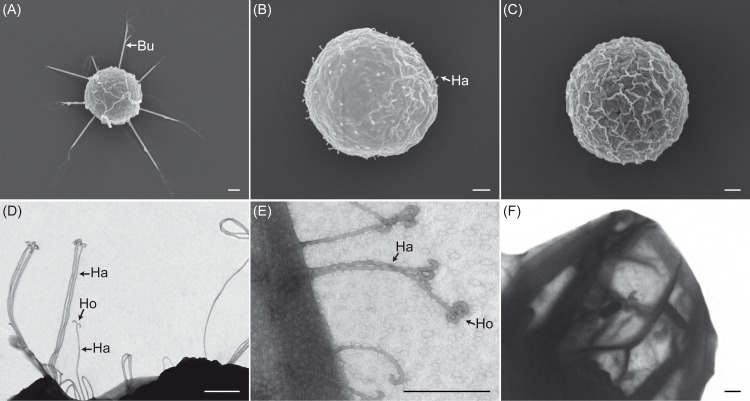
Scanning and transmission electron microscopy studies on secondary cysts and cell walls of secondary cysts obtained from selected Saprolegnia species confirmed that the cysts of S. parasitica possess bundles of long hooked hairs (10.14±1.40 μm) (Fig 1A and 1D), while S. delica has single short hooked hairs (0.67±0.09 μm) (Fig 1B and 1E), and S. anisospora has undecorated cysts (Fig 1C and 1F). Transmission electron micrographs of the hairs of both S. parasitica and S. delica showed presence of hooks at their tips (Fig 1D and 1E). In S. parasitica, these hairs were long and grouped in bundles while in S. delica the hairs were short and single (Fig 1D and 1E).
Fig 1. Electron micrographs of secondary cysts and their cell walls from selected Saprolegnia species.
Scanning electron micrographs of secondary cysts of S. parasitica (A), S. delica (B), and S. anisospora (C) showing presence or absence of bundles (Bu) or single hairs (Ha). Transmission electron micrographs of both S. parasitica (D), and S. delica (E) confirmed the presence of hooks (Ho) at their tips and that cysts of S. parasitica have longer hairs (Ha) than those of S. delica. Cysts of S. anisospora do not form any hairs (F). Scale bar 1μm.
Evaluation of the adhesion strength of the secondary cysts
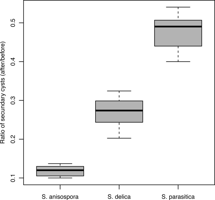
The adhesion strength of the secondary cysts was significantly different for S. parasitica, S. delica and S. anisospora (F2 = 1776, p <2e-16) (Fig 2). The adhesion strength of the secondary cysts was 0.48±0.04 for S. parasitica, 0.27±0.03 for S. delica, and 0.12±0.01 for S. anisospora (Fig 2).
Fig 2. Adhesion strength of the secondary cysts of selected Saprolegnia species.
The figure shows the ratio secondary cysts attached to glass coverslips after and before treatment in S. parasitica, S. delica, and S. anisospora.
Time sequence study on extracellular deployment of bundles of long hooked hairs in S. parasitica
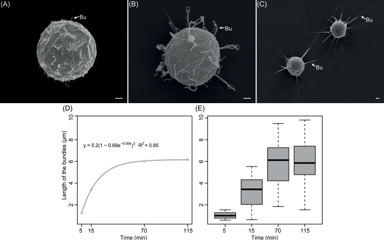
The statistical analyses of SEM micrographs of bundles of S. parasitica revealed that their length varies and increases during the time laps study (Fig 3). Thus, the length was different at time points 5, 15, and 70–115 min (F3 = 128.90, p < 2e-16) (Fig 3D and 3E). At time points 70 min and 115 min, however, the length of the bundles was not significantly different (Fig 3D and 3E). The results indicated that the length of bundles increased immediately after initiation of the encystment, e.g., 5 min, until about 70 min after encystment following a Brody model (y = a*(1–b*e(–c*t)), a = 5.97, b = 1.10, c = 0.06; AIC = 263,30, R2 = 0.85) as for the number of bundles at selected time points during encystment revealed no significant differences (12.56±1.59, F3 = 1.18, p = 0.32).
Fig 3. Time sequence of the extracellular deployment of bundles of long hooked hairs on secondary cysts of Saprolegnia parasitica.
Scanning electron micrographs of the time sequence of the bundles (Bu) of long hooked hairs deployment at 5 (A), 15 (B), and 70 min (C) after encystment (scale bar 1μm). The increase in average length of the bundles of a total of 25 cysts analyzed followed a Brody a non-linear model (D) and was significantly different between selected periods except for periods 70 and 115 min after encystment (E).
The secondary cysts fixed at 5 min after encystment seemed to have initiated the unfurling of the bundles (Fig 3A). At this time after encystment, the length of bundles was on average of 1.03±0.29 μm (Fig 3E). At time point 15 min after encystment, the bundles increased in length (Fig 3B), and were on average 3.22±1.42 μm (Fig 3E), and in some cases adopted a loop shape (Fig 3B). At 70 and 115 min (Fig 3C), all secondary cysts showed well-extended bundles, and on average, they measured 5.78±2.00 μm, and 5.96±1.98 μm, respectively (Fig 3C and 3E).
Effect of substrate, area of contact, and encystment triggers on the length and number of bundles of long hooked hairs
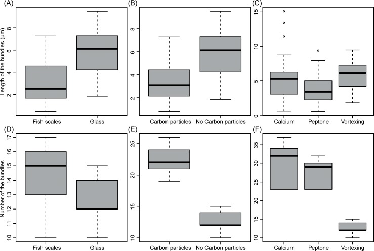
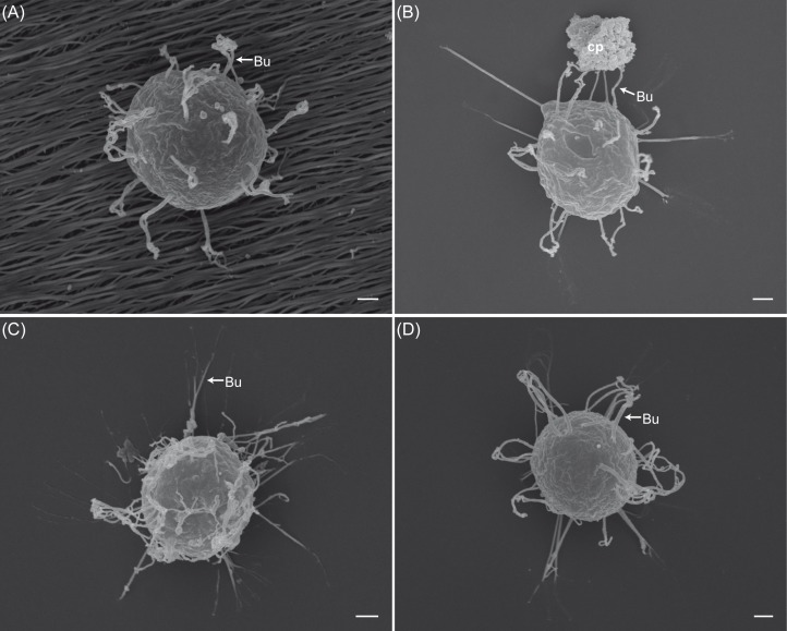
The length and number of the bundles on cysts of S. parasitica obtained by vortexing and incubated on fish scales were significantly different from those incubated on glass cover slips (Figs 4A and 4D and 6A). The length of the bundles produced on fish scales were on average 3.15±1.87 μm and were shorter than those produced on glass slides (5.78±2.00 μm) (F1 = 77.39, p < 1.8e-15) (Fig 4A). In contrast, the number of bundles significantly increased on the fish scales (14.36±2.22) compared with those incubated on glass slides (12.64±1.47, F1 = 10.47, p = 0.002) (Fig 4D).
Fig 4. Effect of substrate, area of contact and encystment triggers on the length and number of bundles of long hooked hairs of Saprolegnia parasitica.
The length of bundles on cysts produced by vortexing and incubated on fish scales was significantly shorter than those incubated on glass slides (A), while the number of bundles was significantly different and higher on fish scales (D). The exposure of secondary cysts to carbon particles resulted in reduction of the length (B) and an increase of the number of bundles (E). The bundles produced by either vortexing or addition of 50 mM CaCl2 were significantly longer than those produced by using peptone (3g/l) (C). The number of bundles produced by using calcium or peptone treatments was higher than bundles produced by vortexing (F).
Fig 6. Scanning electron micrographs of secondary cysts of Saprolegnia parasitica encysted with selected triggers.
Samples of secondary cysts were obtained by: vortexing and incubated on fish scales (A), vortexing with carbon particles (B), incubating in a 50 mM CaCl2 on glass cover slips (C), or by incubating in a peptone (3g/l) on glass cover slips (D). The bundles (Bu) of long hooked hairs were shorter on fish scales (A) than those produced by using other treatments (B, C, and D). The number of bundles increased with calcium and peptone treatments (C, D). Carbon particles (cp) seem to represent anchor points for the bundles (Bu). Scale bar 1μm.
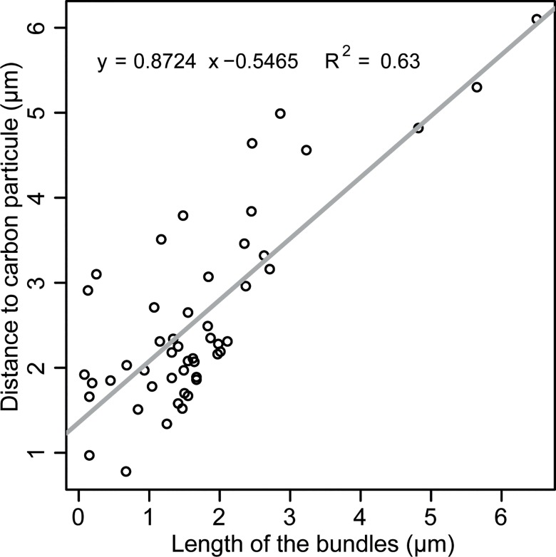
We found that the exposure of secondary cysts suspensions to an additional surface area, i.e., carbon particles, resulted in reduction of the length of bundles induced if compared to those produced with no addition of carbon particles (3.35±1.54 μm versus 5.78±2.00 μm, F1 = 82.8, p <2e-16) (Figs 4B and 6B), while the number of bundles, however, increased (22.68±2.25 versus 12.64±1.47, F1 = 349.2, p <2e-1677.39) (Fig 4E). The bundles of cysts produced in this manner, i.e., carbon particles, were distributed heterogeneously on the surface of the secondary cysts, and we found that there is a correlation between the length of the bundles to the distance from the cyst surface to the carbon particles, i.e., anchor points (F1,48 = 81.5, R2 = 0.63, p = 6.58e-12) (Fig 5).
Fig 5. Regression of the length of the bundles of long hooked hairs of Saprolegnia parasitica on the distance to anchor points (carbon particles).
The figure shows a significant regression of the length (μm) of the bundles produced by the cysts on the distance to carbon particles that were used as anchors points.
Regarding the effect of encystment triggers on length of the bundles, the analysis showed that there are significant differences among encystment triggers (F2 = 24.75, p = 1.09e-10). For example, the average length of the bundles produced by either vortexing (5.78±2.00 μm) or calcium (5.03±2.64 μm) was longer than that produced by using peptone as encystment trigger (3.71±1.87 μm) (Tukey test: p = 0.063; Tukey test: p = 2.30e-5, respectively) (Figs 4C and 6C and 6D). However, the average length of the bundles obtained by vortexing or using calcium was not significantly different (Tukey test: p = 0.063) (Fig 4C).
The analysis of the number of bundles also showed significant differences among encystment triggers (12.64±1.47, F2 = 141.80, p < 2e-16). The number of bundles produced by using calcium (29.48±5.40) or peptone (27.36±3.63) as encystment triggers were significantly different from that produced by vortexing (12.64±1.47) (Tukey test: p = 0.00; Tukey test: p = 0.00, respectively) (Fig 4F). However, the number of bundles on cysts of S. parasitica produced by calcium or peptone was not significantly different (Tukey test: p = 0.13) (Fig 4F).
Enzyme digestions and immunolocalization
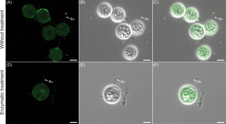
The use of histochemical staining was not entirely informative for assessing spines composition due to the presence of Saprolegnia adhesive protein complex. This was evidenced by the unspecific binding of the antibodies (Fig 7). Therefore, the digestion with an enzymatic mix consisting of Trypsin, PNGase F, O-glycosidases was applied prior the incubation with antibodies (Fig 7). Digestions with trypsin did not resulted in any rupture of the hairs (Fig 7) and the anti-β-tubulin antibody tested did not bind to the hairs.
Fig 7. Protein immunolocalization of bundles of hooked hairs of Saprolegnia parasitica.
The figure shows that treatment with PNGase F prevents the unspecific binding of the anti-β-tubulin antibodies. Differential interference contrast (A and D), confocal light fluorescent (B and E) and merged micrographs (C and F) of bundles (Bu) of hooked hairs of secondary cysts. The cysts were enzymatically treated (D, E, and F) or not (A, B and C) with PNGase F, and incubated in the presence of monoclonal anti-β-tubulin antibodies (1:5000) and goat-anti-rabbit Alexa Fluor 488 conjugate as secondary antibodies (shown in green color). Scale bar 1μm.
Bioinformatics
The data from the secretome generated by Broad Institute of MIT and Harvard (S1 Table) shows the abundance of approximately 1000 putative secreted proteins from the developmental stages: growing mycelium, sporulating mycelium, cysts, and germinating cysts. We found five putative proteins encoded by the genes SPRG_18636, SPRG_05384, SPRG_19863, SPRG_03974 and SPRG_19320 that might be potential bundle components (Table 1). However, no functional characterization of these or their orthologues have yet been presented. According to the S. parasitica secretome these five putative proteins are more highly expressed in cysts than in the mycelia (Table 1). SPRG_18636 encodes a 562 amino acid protein, SPRG_05384 encodes a 6547 amino acid protein, SPRG_19863 encodes a 9440 amino acid protein, SPRG_03974 encodes a 5297 amino acid protein and SPRG_19320 encodes a 936 amino acid protein. The predicted catalytic domain of the SPRG_18636, SPRG_05384, SPRG_19863 and SPRG_03974 contain one or more ScanProcite fibronectin III domains, and the SPRG_19320 contains thrombospondin domain (Table 1). The NetNGlyc tool reported positive results for the amino acid sequences of the SPRG_18636, SPRG_05384, SPRG_19863, SPRG_03974 and SPRG_19320 proteins, with 4 to 20 predicted N-glycosylated sites (Table 1).
Table 1. List of selected secreted proteins of Saprolegnia parasitica with their description and expression levels in different life stages.
| Code | PFAM Description | Mycelium | Sporulating mycelium | Cysts | Germinated cysts |
|---|---|---|---|---|---|
| SPRG_18636 | Fibronectin type III domain | 0 | 0.35 | 1.22 | 0.30 |
| SPRG_05384 | Fibronectin type III domain | 0.02 | 0.09 | 8.48 | 4.68 |
| SPRG_19863 | Fibronectin type III domain | 0.04 | 0.30 | 4.60 | 1.84 |
| SPRG_03974 | Fibronectin type III domain | 0.03 | 6.66 | 16.44 | 8.30 |
| SPRG_19320 | Thrombospondin type 1 domain | 0.50 | 0.42 | 227.72 | 88.59 |
Discussion
The process of attachment is a key host-pathogen interaction mediator and accumulating evidence suggests that pathogenic oomycetes can release specific substances to attach to the host [17, 51, 52]. The involvement in the attachment process of specialized structures is less known. In this study, several approaches have been used to evaluate the attachment strategy of the fish pathogenic oomycete, S. parasitica. Thus, it has been demonstrated that the bundles of long hooked hairs of S. parasitica cysts are dynamic structures involved in pathogen attachment.
The strength of cyst attachment of selected Saprolegnia spp. appears to be correlated with the length of bundles. Thus, the strength of attachment of S. parasitica cysts was around three times stronger than that of cysts of two selected Saprolegnia spp. that have shorter or lack of hooked hairs. This might indicate that the bundles of long hooked hairs might allow a stronger attachment either by themselves, or by enlarging the area of stickiness [20]. However, it cannot yet be excluded that the more efficient attachment of S. parasitica cysts might also be the result of different stickiness properties (i.e., composition) of the cyst extracellular matrix between these three species.
In addition, the evidences found here indicate that these bundles are not passive structures for hooking to the host as proposed by Wood et al., [25]. Instead, they appear to be dynamic attaching structures that are influenced by the characteristics and nature of the attaching surface. We also showed that the deployment of these structures is modulated by both physical and chemical stimuli mimicking those occurring at the area of contact with the host. Thus, the bundles of hooked hairs start being arranged extracellularly as soon as 5 min after encystment, suggesting that their components are pre-synthesized before encystment as previously described by Beakes [29]. The bundles increase length but their number remains constant after undergoing either germination or the release of a new zoospore. These two developmental stages are of key importance for the Saprolegnia pathogenic species since they determine the successful detection, attachment and colonization, or the formation of a new zoospore [53].
In the plant pathogen Phytophthora signals such as Ca2+ uptake or mechanical stimulation have been reported to be responsible to induce full and transient fusion of selective secretion of vesicle proteins (which likely to be an important aspect of host infection [13]. In this study, we investigated the effect of both chemical and mechanical stimuli on the length and number of bundles. Thus, it was found that these vary depending on substrate, area contact, and encystment triggers. Both the addition of nutrients (e.g., peptone) or of mechanical stimuli (e.g., carbon particles, rough surfaces) influenced the length and number of the bundles produced. The external formation of the bundles, therefore, might be receptor-mediated and calcium-dependent receptors could be involved. This is evidenced by the fact that addition of high concentration of Ca2+ to zoospores increased the number and affected their length throughout the surface of the cyst if compared with mechanical encystment triggers. Burr and Beakes [20], already showed that Ca2+ was the only ion that specifically enhanced adhesion of S. parasitica cyst over S. delica. Calcium is a non-specific signaling ion that also plays a central role in ion signaling pathways (for review see [54]) and in several important development processes of oomycetes such as encystment, adhesion and germination [55–57]. A variety of specialized structures such as those aiding ingresses of the fungal pathogen into its host, or adsorption of nutrients (appressoria, hyphodamia, haustoria, etc.) are triggered by the presence of certain ions [13, 16, 58, 59]. Furthermore, the dynamic nature of the bundles was evidenced by the fact that their length is also affected by the nature of the surface. Thus, zoospores encysted by agitation and exposed to rough surfaces, such as fish scales, or addition of carbon particles to a surface, formed twisted and curly bundles compared to those on flat surfaces such as glass slides. This might indicate that bundles of hooked hairs external formation could be also the response to mechanical stimuli, such as inductive surface cues, or by local accumulation of glue polymerizing agents as it occurs for other oomycetes and also fungi, i.e., transglutaminases [60].
The immunolocalization studies evidenced the presence of an adhesive extracellular matrix and suggested that β-tubulin is not a component of the bundles. The presence of adhesive matrix on cysts of S. parasitica was confirmed by enzymatic digestion conducted with both N-glycosidases and O-glycosidases and is an indication of the presence of N-glycosylated glycoproteins covering the surface of the bundles. This finding suggests that this extracellular matrix is composed of N-acetyl-b-glucosaminyl and/or sialic residues as well as N-acetyl-b-d-glucosamineoligomers, which have been shown to be involved in cell attachment of other oomycetes [61].
According to RNA sequence data of the S. parasitica genome, genes encoding putative secreted proteins [15] are highly expressed in cysts. To verify this, a bioinformatic analysis conducted on secreted proteins specifically expressed at cyst stage [15] has revealed a high expression of putative N-glycosilated proteins with either thrombospondin or fibronectin domains in S. parasitica. Proteins with these domains are known to be involved in attachment [60]. A similar study conducted on the adhesion of zoospores and cysts to biological and non-biological surfaces by P. cinnamomi showed importance of glycoproteins in cell binding [12]. The plant pathogen P. cinnamomi is known to release a glycoprotein cyst coat containing N-acetil galactosamine polysaccharides [17, 62]. Our analyses are unlikely to be sufficient to infer the components of the bundles of hooked hairs but evidence the relationship between the hairs and extracellular matrix, probably, in connection to cell attachment.
Overall, this research provides a number of microscopic, physiological, and bioinformatic evidences supporting the function of the bundles of long hooked hairs as attachment structures of the fish pathogen oomycete, S. parasitica. These results suggest that these structures are either composed by N-glycosilated proteins or may assist in dispersing the cyst extracellular matrix on the host surface. Further studies should focus on identifying the actual composition of the hairs by for example, selecting secreted proteins by gene disruption or gene silencing [16, 63]. This advance in genomics has opened unprecedented opportunities for Saprolegnia investigations. A complete identification of bundles of long hooked hairs compounds and their characterization remains a major research challenge that will help us understand composition of these attachment structures and the manner, in which the adhesion process occurs.
Supporting information
(TXT)
Acknowledgments
The funding or sources of support were grants of: (1) European Union (ITN-SAPRO- 238550), (2) Ministerio de Economía y Competitividad, Spain (CGL2016-80526). Moreover, Svetlana Rezinciuc and José V. Sandoval-Sierra were supported by fellowships from ITN-SAPRO-238550 and Ministerio de Economía y Competitividad, Spain (CGL2016-80526) partially supported lab-bench expenses and experiments.
Data Availability
All relevant data are within the paper.
Funding Statement
The funding or sources of support were grants of: (1) European Union (ITN-SAPRO-238550), (2) Ministerio de Economía y Competitividad, Spain (CGL2016-80526), which supported lab-bench expenses and experiments. Moreover, Svetlana Rezinciuc and José V. Sandoval-Sierra were supported by fellowships from ITN-SAPRO-238550.
References
- 1.Carris LM, Little CR, Stiles CM. Introduction to Fungi The Plant Health Instructor. Washington State University, Kansas State University, and Georgia Military College; 2012. [Google Scholar]
- 2.Kim KS, Judelson HS. Sporangium-Specific Gene Expression in the Oomycete Phytopathogen Phytophthora infestans. Eukaryot Cell. 2003;2(6):1376–85. 10.1128/EC.2.6.1376-1385.2003 [DOI] [PMC free article] [PubMed] [Google Scholar]
- 3.Walker CA, van West P. Zoospore development in the oomycetes. Fungal Biol Rev. 2007;21(1):10–8. 10.1016/j.fbr.2007.02.001. [DOI] [Google Scholar]
- 4.Xiang Q, Kim KS, Roy S, Judelson HS. A motif within a complex promoter from the oomycete Phytophthora infestans determines transcription during an intermediate stage of sporulation. Fungal Genet Biol. 2009;46(5):400–9. 10.1016/j.fgb.2009.02.006. 10.1016/j.fgb.2009.02.006 [DOI] [PubMed] [Google Scholar]
- 5.Cerenius L, Söderhäll K. Repeated zoospore emergence as a possible adaptation to parasitism in Aphanomyces. Exp Mycol. 1985;9(3):9–63. 10.1016/0147-5975(85)90022-2. [DOI] [Google Scholar]
- 6.Rand TG, Munden D. Chemotaxis of Zoospores of Two Fish-Egg-Pathogenic Strains of Saprolegnia diclina (Oomycotina: Saprolegniaceae) toward Salmonid Egg Chorion Extracts and Selected Amino Acids and Sugars. Journal of Aquatic Animal Health. 1993;5(4):240–5. [DOI] [Google Scholar]
- 7.Raftoyannis Y, Dick MW. Zoospore encystment and pathogenicity of Phytophthora and Pythium species on plant roots. Microbiol Res. 2006;161(1):1–8. 10.1016/j.micres.2005.04.003. 10.1016/j.micres.2005.04.003 [DOI] [PubMed] [Google Scholar]
- 8.Deacon JW, Saxena G. Germination triggers of zoospore cysts of Aphanomyces euteiches and Phytophthora parasitica. Mycol Res. 1998;102(1):33–41. 10.1017/S0953756297004358. [DOI] [Google Scholar]
- 9.Donaldson SP, Deacon JW. Role of calcium in adhesion and germination of zoospore cysts of Pythium: a model to explain infection of host plants. Microbiology. 1992;138(10):2051–9. 10.1099/00221287-138-10-2051 [DOI] [Google Scholar]
- 10.Robold AV, Hardham AR. During attachment Phytophthora spores secrete proteins containing thrombospondin type 1 repeats. Curr Genet. 2005;47(5):307–15. 10.1007/s00294-004-0559-8 [DOI] [PubMed] [Google Scholar]
- 11.Gubler F, Hardham AR. Secretion of adhesive material during encystment of Phytophthora cinnamomi zoospores, characterized by immunogold labelling with monoclonal antibodies to components of peripheral vesicles. J Cell Sci. 1988;90(2):225. [Google Scholar]
- 12.Gubler F, Hardham AR, Duniec J. Characterising adhesiveness of Phytophthora cinnamomi zoospores during encystment. Protoplasma. 1989;149(1):24–30. 10.1007/bf01623979 [DOI] [Google Scholar]
- 13.Zhang W, Blackman LM, Hardham AR. Transient fusion and selective secretion of vesicle proteins in Phytophthora nicotianae zoospores. PeerJ. 2013;1:e221 10.7717/peerj.221 [DOI] [PMC free article] [PubMed] [Google Scholar]
- 14.Wawra S, Bain J, Durward E, de Bruijn I, Minor KL, Matena A, et al. Host-targeting protein 1 (SpHtp1) from the oomycete Saprolegnia parasitica translocates specifically into fish cells in a tyrosine-O-sulphate–dependent manner. Proceedings of the National Academy of Sciences. 2012;109(6):2096–101. 10.1073/pnas.1113775109 [DOI] [PMC free article] [PubMed] [Google Scholar]
- 15.Jiang RHY, de Bruijn I, Haas BJ, Belmonte R, Löbach L, Christie J, et al. Distinctive expansion of potential virulence genes in the genome of the oomycete fish pathogen Saprolegnia parasitica. PLoS Genetics. 2013;9(6):e1003272 10.1371/journal.pgen.1003272 [DOI] [PMC free article] [PubMed] [Google Scholar]
- 16.Epstein L, Nicholson R. Adhesion and adhesives of Fungi and Oomycetes In: Smith A, Callow J, editors. Biological Adhesives: Springer Berlin Heidelberg; 2006. p. 41–62. [Google Scholar]
- 17.Hardham AR. Cell Biology of Fungal Infection of Plants In: Howard RJ, Gow NAR, editors. Biology of the Fungal Cell. Berlin, Heidelberg: Springer Berlin Heidelberg; 2001. p. 91–123. [Google Scholar]
- 18.Bechinger C, Giebel K-F, Schnell M, Leiderer P, Deising HB, Bastmeyer M. Optical Measurements of Invasive Forces Exerted by Appressoria of a Plant Pathogenic Fungus. Science. 1999;285(5435):1896–9. 10.1126/science.285.5435.1896 [DOI] [PubMed] [Google Scholar]
- 19.Fabritius A-L, Judelson HS. A Mating-Induced Protein of Phytophthora infestans Is a Member of a Family of Elicitors with Divergent Structures and Stage-Specific Patterns of Expression. Molecular Plant-Microbe Interactions. 2003;16(10):926–35. 10.1094/MPMI.2003.16.10.926 [DOI] [PubMed] [Google Scholar]
- 20.Burr AW, Beakes GW. Characterization of zoospore and cyst surface structure in saprophytic and fish pathogenic Saprolegnia species (Oomycete fungal protists). Protoplasma. 1994;181(1–4):142–63. 10.1007/BF01666393 [DOI] [Google Scholar]
- 21.Mercure EW, Leite B, Nicholson RL. Adhesion of ungerminated conidia of Colletotrichum graminicola to artificial hydrophobic surfaces. Physiological and Molecular Plant Pathology. 1994;45(6):421–40. 10.1016/S0885-5765(05)80040-2. [DOI] [Google Scholar]
- 22.Kunoh H, Yamaoka N, Yoshioka H, Nicholson RL. Preparation of the infection court by Erysiphe graminis. Exp Mycol. 1988;12(4):325–35. 10.1016/0147-5975(88)90024-2. [DOI] [Google Scholar]
- 23.Jones MJ, Epstein L. Adhesion of Macroconidia to the Plant Surface and Virulence of Nectria haematococca. Appl Environ Microbiol. 1990;56(12):3772–8. [DOI] [PMC free article] [PubMed] [Google Scholar]
- 24.Grenville-Briggs LJ, Anderson VL, Fugelstad J, Avrova AO, Bouzenzana J, Williams A, et al. Cellulose synthesis in Phytophthora infestans is required for normal appressorium formation and successful infection of potato. The Plant Cell Online. 2008;20(3):720–38. 10.1105/tpc.107.052043 [DOI] [PMC free article] [PubMed] [Google Scholar]
- 25.Wood SE, Willoughby LG, Beakes GW. Experimental studies on uptake and interaction of spores of the Saprolegnia diclina-parasitica complex with external mucus of brown trout (Salmo trutta). Transactions of the British Mycological Society. 1988;90(1):63–73. 10.1016/S0007-1536(88)80181-5. [DOI] [Google Scholar]
- 26.Manton I, Clarke B, Greenwood AD. Observations with the electron microscope on a species of Saprolegnia. Journal of Experimental Botany. 1951;2(3):321–31. 10.1093/jxb/2.3.321 [DOI] [Google Scholar]
- 27.Meier H, Webster J. An electron microscope study of cysts in the Saprolegniaceae. Journal of Experimental Botany. 1954;5(3):401–9. 10.1093/jxb/5.3.401 [DOI] [Google Scholar]
- 28.Pickering AD, Willoughby LG, McGrory CB. Fine structure of secondary zoospore cyst cases of Saprolegnia isolates from infected fish. Transactions of the British Mycological Society. 1979;72(3):427–36. 10.1016/S0007-1536(79)80150-3. [DOI] [Google Scholar]
- 29.Beakes GW. A comparative account of cyst coat ontogeny in saprophytic and fish-lesion (pathogenic) isolates of the Saprolegnia diclina-parasitica complex. Canadian Journal of Botany. 1983;61(2):603–25. 10.1139/b83-068 [DOI] [Google Scholar]
- 30.Beakes GW, Ford H. Esterase isoenzyme variation in the genus Saprolegnia, with particular reference to the fish-pathogenic S. diclina-parasitica complex. Journal of General Microbiology. 1983;129(8):2605–19. 10.1099/00221287-129-8-2605 [DOI] [PubMed] [Google Scholar]
- 31.Beakes GW, Wood SE, Burr AW. Features which characterize Saprolegnia isolates from salmon fish lesions–a review. In: Mueller GJ, editor. Salmon Saprolegniasis. Portland, Oregon: Report to Bonneville Power Administration. Environment, Fish and Wildlife Division; 1994. p. 33–66.
- 32.Stueland S, Hatai K, Skaar I. Morphological and physiological characteristics of Saprolegnia spp. strains pathogenic to Atlantic salmon, Salmo salar L. J Fish Dis. 2005;28(8):445–53. 10.1111/j.1365-2761.2005.00635.x [DOI] [PubMed] [Google Scholar]
- 33.Mueller GJ, Whisler HC. Fungal parasites of Salmon from the Columbia River Watershed. In: Mueller GJ, editor. Salmon Saprolegniasis. Portland, Oregon: Report to Bonneville Power Administration. Environment, Fish and Wildlife Division; 1994. p. 163–88.
- 34.Fregeneda-Grandes JM, Rodríguez-Cadenas F, Aller-Gancedo JM. Fungi isolated from cultured eggs, alevins and broodfish of brown trout in a hatchery affected by saprolegniosis. Journal of Fish Biology. 2007;71(2):510–8. 10.1111/j.1095-8649.2007.01510.x [DOI] [Google Scholar]
- 35.Diéguez-Uribeondo J, Fregeneda-Grandes JM, Cerenius L, Pérez-Iniesta E, Aller-Gancedo JM, Tellería MT, et al. Re-evaluation of the enigmatic species complex Saprolegnia diclina-Saprolegnia parasitica based on morphological, physiological and molecular data. Fungal Genet Biol. 2007;44:585–601. 10.1016/j.fgb.2007.02.010 [DOI] [PubMed] [Google Scholar]
- 36.Diéguez-Uribeondo J, Cerenius L, Söderhäll K. Repeated zoospore emergence in Saprolegnia parasitica. Mycol Res. 1994;98(7):810–5. [Google Scholar]
- 37.Söderhäll K, Dick MW, Clark G, Fürst M, Constantinescu O. Isolation of Saprolegnia parasitica from the crayfish Astacus leptodactylus. Aquaculture. 1991;92(0):121–5. 10.1016/0044-8486(91)90014-X. [DOI] [Google Scholar]
- 38.Sandoval-Sierra JV, Martín MP, Diéguez-Uribeondo J. Species identification in the genus Saprolegnia (Oomycetes): defining DNA-based molecular operational taxonomic units. Fungal Biol. 2014;118:559–78. 10.1016/j.funbio.2013.10.005 [DOI] [PubMed] [Google Scholar]
- 39.Unestam T. Studies on the crayfish plague fungus Aphanomyces astaci. Some factors affecting growth in vitro. Physiologia Plantarum. 1965;18(2):483–505. 10.1111/j.1399-3054.1965.tb06911.x [DOI] [Google Scholar]
- 40.Cerenius L, Söderhäll K. Chemotaxis in Aphanomyces astaci, an arthropod-parasitic fungus. J Invertebr Pathol. 1984;43(2):278–81. 10.1016/0022-2011(84)90150-2. [DOI] [Google Scholar]
- 41.Letourneau J, Levesque C, Berthiaume F, Jacques M, Mourez M. In vitro assay of bacterial adhesion onto mammalian epithelial cells. Journal of Visualized Experiments. 2011;51:1–4. [DOI] [PMC free article] [PubMed] [Google Scholar]
- 42.Schneider CA, Rasband WS, Eliceiri KW. NIH Image to ImageJ: 25 years of image analysis. Nat Methods. 2012;9:671–5. 10.1038/nmeth.2089 [DOI] [PMC free article] [PubMed] [Google Scholar]
- 43.Diéguez-Uribeondo J, Gierz G, Bartnicki-García S. Image analysis of hyphal morphogenesis in Saprolegniaceae (Oomycetes). Fungal Genet Biol. 2004;41(3):293–307. 10.1016/j.fgb.2003.10.012. 10.1016/j.fgb.2003.10.012 [DOI] [PubMed] [Google Scholar]
- 44.Diéguez-Uribeondo J, Cerenius L, Söderhäll K. Saprolegnia parasitica and its virulence on three different species of freshwater crayfish. Aquaculture. 1994;120(3):219–28. 10.1016/0044-8486(94)90080-9 [DOI] [Google Scholar]
- 45.Van West P, De Bruijn I, Minor KL, Phillips AJ, Robertson EJ, Wawra S, et al. The putative RxLR effector protein SpHtp1 from the fish pathogenic oomycete Saprolegnia parasitica is translocated into fish cells. FEMS Microbiol Lett. 2010;310(2):127–37. 10.1111/j.1574-6968.2010.02055.x [DOI] [PubMed] [Google Scholar]
- 46.Steentoft C, Vakhrushev SY, Joshi HJ, Kong Y, Vester-Christensen MB, Schjoldager KTBG, et al. Precision mapping of the human O-GalNAc glycoproteome through SimpleCell technology. The EMBO Journal. 2013;32(10):1478–88. 10.1038/emboj.2013.79. [DOI] [PMC free article] [PubMed] [Google Scholar]
- 47.Sigrist CJA, de Castro E, Cerutti L, Cuche BA, Hulo N, Bridge A, et al. New and continuing developments at PROSITE. Nucleic Acids Res. 2013;41(D1):D344–D7. 10.1093/nar/gks1067 [DOI] [PMC free article] [PubMed] [Google Scholar]
- 48.R Development Core Team. R: A language and environment for statistical computing Vienna, Austria: R Foundation for Statistical Computing; 2017. [Google Scholar]
- 49.Koya PR, Goshu AT. Generalized Mathematical Model for Biological Growths. Open Journal of Modelling and Simulation. 2013;Vol.01No.04:12 10.4236/ojmsi.2013.14008 [DOI] [Google Scholar]
- 50.Arnhold E. easynls: Easy nonlinear model. R package version 4.0; 2015.
- 51.Mendgen K, Hahn M, Deising H. Morphogenesis and Mechanisms of Penetration by Plant Pathogenic Fungi. Annu Rev Phytopathol. 1996;34(1):367–86. 10.1146/annurev.phyto.34.1.367 [DOI] [PubMed] [Google Scholar]
- 52.Epstein L, Nicholson RL. Adhesion of Spores and Hyphae to Plant Surfaces In: Carroll GC, Tudzynski P, editors. Plant Relationships: Part A. Berlin, Heidelberg: Springer Berlin Heidelberg; 1997. p. 11–25. [Google Scholar]
- 53.Andersson MG, Cerenius L. Pumilio homologue from Saprolegnia parasitica specifically expressed in undifferentiated spore cysts. Eukaryot Cell. 2002;1(1):105–11. 10.1128/EC.1.1.105-111.2002 [DOI] [PMC free article] [PubMed] [Google Scholar]
- 54.Shaw BD, Hoch HC. Ions Regulate Spore Attachment, Germination, and Fungal Growth In: Howard RJ, Gow NAR, editors. Biology of the Fungal Cell. Berlin, Heidelberg: Springer Berlin Heidelberg; 2007. p. 219–36. [Google Scholar]
- 55.Deacon JW, Donaldson SP. Molecular recognition in the homing responses of zoosporic fungi, with special reference to Pythium and Phytophthora. Mycol Res. 1993;97(10):1153–71. 10.1016/S0953-7562(09)81278-1. [DOI] [Google Scholar]
- 56.Reid B, Morris BM, Gow NAR. Calcium-Dependent, Genus-Specific, Autoaggregation of Zoospores of Phytopathogenic Fungi. Exp Mycol. 1995;19(3):202–13. 10.1006/emyc.1995.1025. [DOI] [Google Scholar]
- 57.Brazee NJ, Lindner DL, Fraver S, D'Amato AW, Milo AM. Wood-inhabiting, polyporoid fungi in aspen-dominated forests managed for biomass in the U.S. Lake States. Fungal Ecol. 2012;5(5):600–9. 10.1016/j.funeco.2012.03.002. [DOI] [Google Scholar]
- 58.Takano Y, Kikuchi T, Kubo Y, Hamer JE, Mise K, Furusawa I. The Colletotrichum lagenarium MAP Kinase Gene CMK1 Regulates Diverse Aspects of Fungal Pathogenesis. Molecular Plant-Microbe Interactions. 2000;13(4):374–83. 10.1094/MPMI.2000.13.4.374 [DOI] [PubMed] [Google Scholar]
- 59.Yamaoka N, Takeuchi Y. Morphogenesis of the powdery mildew fungus in water (4) The significance of conidium adhesion to the substratum for normal appressorium development in water. Physiological and Molecular Plant Pathology. 1999;54(5):145–54. 10.1006/pmpp.1998.0194. [DOI] [Google Scholar]
- 60.Smith AM, Callow JA, editors. Biological Adhesives. Berlin: Springer Berlin Heidelberg; 2006. [Google Scholar]
- 61.Prieto AL, Andersson-Fisone C, Crossin KL. Characterization of multiple adhesive and counteradhesive domains in the extracellular matrix protein cytotactin. The Journal of Cell Biology. 1992;119(3):663 [DOI] [PMC free article] [PubMed] [Google Scholar]
- 62.Hardham AR, Suzaki E. Glycoconjugates on the surface of spores of the pathogenic fungus Phytophthora cinnamomi studied using fluorescence and electron microscopy and flow cytometry. Can J Microbiol. 1990;36(3):183–92. 10.1139/m90-032 [DOI] [Google Scholar]
- 63.Saraiva M, de Bruijn I, Grenville-Briggs L, McLaggan D, Willems A, Bulone V, et al. Functional characterization of a tyrosinase gene from the oomycete Saprolegnia parasitica by RNAi silencing. Fungal Biol. 2014;118(7):621–9. 10.1016/j.funbio.2014.01.011. 10.1016/j.funbio.2014.01.011 [DOI] [PMC free article] [PubMed] [Google Scholar]
Associated Data
This section collects any data citations, data availability statements, or supplementary materials included in this article.
Supplementary Materials
(TXT)
Data Availability Statement
All relevant data are within the paper.


