Abstract
The incidence of multidrug-resistant (MDR) organisms, including methicillin-resistant Staphylococcus aureus (MRSA), is a serious threat to public health. Progress in developing new therapeutics is being outpaced by antibiotic resistance development, and alternative agents that rapidly permeabilize bacteria hold tremendous potential for treating MDR infections. A new class of glycopolymers includes polycationic poly-N (acetyl, arginyl) glucosamine (PAAG) is under development as an alternative to traditional antibiotic strategies to treat MRSA infections. This study demonstrates the antibacterial activity of PAAG against clinical isolates of methicillin and mupirocin-resistant Staphylococcus aureus. Multidrug-resistant S. aureus was rapidly killed by PAAG, which completely eradicated 88% (15/17) of all tested strains (6-log reduction in CFU) in ≤ 12-hours at doses that are non-toxic to mammalian cells. PAAG also sensitized all the clinical MRSA strains (17/17) to oxacillin as demonstrated by the observed reduction in the oxacillin MIC to below the antibiotic resistance breakpoint. The effect of PAAG and standard antibiotics including vancomycin, oxacillin, mupirocin and bacitracin on MRSA permeability was studied by measuring propidium iodide (PI) uptake by bacterial cells. Antimicrobial resistance studies showed that S. aureus developed resistance to PAAG at a rate slower than to mupirocin but similar to bacitracin. PAAG was observed to resensitize drug-resistant S. aureus strains sampled from passage 13 and 20 of the multi-passage resistance study, reducing MICs of mupirocin and bacitracin below their clinical sensitivity breakpoints. This class of bacterial permeabilizing glycopolymers may provide a new tool in the battle against multidrug-resistant bacteria.
Introduction
Staphylococcus aureus is the most frequently isolated human bacterial pathogen associated with several severe clinical infections including soft-tissue and endovascular infections, pneumonia, and sepsis in patients frequenting hospitals or other healthcare facilities [1]. Over 2 million people every year in the US acquire serious infections with strains of S. aureus that are resistant to one or more of the antibiotics designed to treat those infections. At least 23,000 people in the US die each year as a direct result of these antibiotic-resistant infections. Misuse and overuse of antimicrobials has accelerated the prevalence of antimicrobial resistance and tolerance to the first-line drugs used to treat infections caused by S. aureus.
Methicillin resistant S. aureus (MRSA) is a well-known nosocomial pathogen often associated with health care-associated and community acquired infections [2] and is also a significant contributor to pulmonary decline in patients with cystic fibrosis [3, 4]. First described in 1960’s, MRSA continues to spread worldwide despite efforts of novel therapeutic development. MRSA isolates are resistant to available penicillin’s and other β-lactam antimicrobial drugs, limiting the potential treatment options with standard antibiotic therapy [5–8]. Patients affected by MRSA strains have an increased risk of poorer clinical outcomes, and 64% of them are more likely to die than patients infected with non-resistant strains of the same bacteria [9]. In 2013, MRSA was classified as a serious threat by the Centers for Disease Control and Prevention (CDC) [10]. Current research continues to provide evidence showing increasing incidence and virulence among resistant strains of S. aureus, despite the development of alternative strategies to combat MRSA infections [6, 11–14].
Standard of care treatments for multidrug-resistant S. aureus infections include vancomycin, linezolid, daptomycin and β-lactams. Vancomycin has been a mainstay of therapy for MRSA infections, but its effectiveness has been challenged by its extensive use, generating a selective pressure that favors the outgrowth of rare VRSA strains. Glycopeptides, like vancomycin and teicoplanin, have been effective bactericidal agents against MRSA infections but are often associated with adverse effects including renal failure and nephrotoxicity [15, 16]. The combination effect of vancomycin and β-lactam against S. aureus clinical isolates, including additive and/or synergistic effects [17–18] and antagonistic effects [19–22] have been contradictory.
Mupirocin, oxacillin, and bacitracin have been used as topical agents or intranasal treatments to prevent onset and spread of primary and secondary MRSA infections for over 10 years [23–27]. However, extensive use of mupirocin, bacitracin and oxacillin has also culminated in increased resistance in MRSA strains [28, 29], which is also consistently reported in community-associated MRSA infections [30]. Studies and clinical trials have also focused on antimicrobial peptides (AMP’s) for their wide-spectrum of antibacterial activity. However, many antimicrobial peptides are polycationic and have some cytotoxicity, limiting their use [31]. Recent studies suggest that a combination of vancomycin and ceftaroline possess greater bactericidal activity [21, 22], however, clinical experience with these therapies is limited. Ceftaroline is commonly associated with adverse effects such as skin rash, nausea, vomiting, and diarrhea neutropenia and agranulocytosis, particularly with high doses or prolonged exposure [32–35]. Development and successful implementation of novel, non-toxic strategies for treating emerging resistant pathogens is critical to limiting the potential increased lethality of S. aureus, minimizing the side effects of last-resort therapies, and reducing the spread of acquired nosocomial infections.
Poly-N (acetyl, arginyl) glucosamine (PAAG) represents a novel class of glycopolymers with antibacterial activity against a wide range of pathogenic Gram- negative and Gram- positive bacteria including MRSA, Pseudomonas aeruginosa, and non-tuberculous Mycobacteria (NTM) [36, 37]. Polyglucosamines are not commonly associated with toxicity [38–42]. PAAG has been shown to be versatile, surface-active glycopolymer with broad antimicrobial activity and excellent biocompatibility.
The goal of this study is to complete a preliminary assessment of the antibacterial efficacy of PAAG against clinical isolates of multidrug-resistant S. aureus, to evaluate PAAG’s capability to kill MRSA at its stationary phase of growth, to permeabilize bacteria, to re-sensitize MRSA isolates to conventional antibiotics and to study antimicrobial resistance development to PAAG, using the antibiotics mupirocin and bacitracin as comparators.
Materials and methods
Bacterial strains and culture conditions
Seventeen opportunistic pathogens of methicillin resistant S. aureus isolated from respiratory, skin and blood infections were used in the study. The MRSA strains designated SA01-SA12 were hospital acquired and obtained from California State University, San Bernardino (Paul Orwin). The mupirocin resistant MRSA strains (MMRSA) were obtained from Washington University, Minneapolis (Mike Dunn). S. aureus ATCC 6538 was used as a control in the microdilution test procedure for antibiotics. The experimental preparation of the cultures was adapted from CLSI [43]. Bacterial cultures were maintained in -80°C for up to 6 months. Luria-Bertani Broth (LB, BD) and Mueller Hinton broth (MHB, BD) media were used to grow the bacteria.
Antimicrobial agents
Three antibiotics, oxacillin (TCI), bacitracin (Alfa Aesar) and mupirocin (USP) were used in the study. Stock solutions were freshly prepared from dry powders according to the manufacturer’s instructions. Synedgen's polycationic proprietary glycopolymer, PAAG, is an arginine derivative of a natural polysaccharide poly-N (acetyl, arginyl) glucosamine. It is polycationic and soluble at physiologic pH.
In vitro susceptibility testing
Minimum inhibitory concentrations (MICs) of antibiotics and PAAG were determined with the broth microdilution technique described by the Clinical and Laboratory Standards Institute (CLSI). Each of the clinical isolates was tested against oxacillin (0.125–512 μg/mL), mupirocin (0.125–2048 μg/mL), and PAAG (8–2048 μg/mL). Serial two-fold dilutions of oxacillin, mupirocin, and PAAG were prepared in supplemented MH broth and 100 μl was aliquoted into each well of 96-well flat bottom microtiter plates. The clinical isolates of MRSA and S. aureus ATCC 6538 (control strain) were grown overnight in MH broth were diluted to a 0.5 McFarland turbidity standard in MH broth. A final inoculum of 1.5 X 10⁶ was added to the wells being tested. Bacteria without the addition of antibiotics or PAAG were used as controls. The plates were incubated at 37°C for 24h. The isolates were categorized as sensitive, intermediate, or resistant according to the CLSI guidelines.
Checkerboard assay
All of the seventeen-drug resistant clinical isolates of methicillin resistant S. aureus were used for the study, based on their resistance to oxacillin determined by the MIC microdilution assay. The MICs of oxacillin and PAAG in combination were determined using two-dimensional checkerboard microdilution assay with MHB and a final inoculum of 1.5 X 10⁶ CFU/mL. The 96-well plate contained increasing concentrations of oxacillin ranging from 1–128 μg/mL on the x-axis and increasing concentrations of PAAG ranging from 2–256 μg/mL on the y-axis. MICs and fractional inhibitory concentrations (FIC’s) were determined after 24 h growth at 37°C. The FIC of each drug A or B is determined by dividing the MIC of each drug when used in combination by the MIC of each drug when used alone. The FICA/B of the combination is the sum of each individual FIC: FICA/B = FICA + FICB, where FICA = MIC of A (combination)/ MIC of A alone and FICB = MIC of B (combination)/ MIC of B alone. The effect of in vitro antibiotic combinations is interpreted based on the fractional inhibitory concentration index (FICI) which is described as FICI < 0.5 synergy, 0.5 < FICI < 4 additive effects or indifference, and FICI ≥ 4 antagonism [44–47].
Time-kill analysis
Seventeen 24-hour experiments including a growth control were used to study the bactericidal effect of PAAG on each of the clinical isolates of MRSA. Bacteria were grown for 24 h at 37°C in MHB. The bacterial culture was further diluted to 0.5 by McFarland standard turbidity. The diluted bacterial culture was serially diluted, and spot plated onto LB agar plates. The initial inoculum was found to be 1.5 X 10⁶ CFU/mL. The bacterial inoculums were treated with PAAG at a concentration of 100 μg/mL and incubated at 37°C for 1, 4, 12 and 24 h. Culture aliquots were removed at 0 h, 1 h, 4 h, 12 h, and 24 h and then serially diluted in sterile water, plated in duplicates onto LB agar plates and incubated at 37°C for 24 h. Cell viability was determined by enumerating the number of visible colonies in the LB plates. Effective bactericidal activity was defined as a 3-log reduction in the CFU/mL from the baseline. Time-kill curves were generated using GraphPad Prism 6 software, by plotting the mean colony counts (log10 CFU/mL) versus time.
PI permeability study
The effect of the PAAG on MRSA permeability was studied by measuring propidium iodide (PI) uptake by bacterial cells. Five MRSA strains (SA10, 2- 4C, 2- 1A, 2-9A and 2- 5A) used in the study demonstrated high resistance to mupirocin and oxacillin (Table 1). Overnight cultures were centrifuged at 13,000 rpm for 1 min. The supernatant was discarded. Bacterial pellets were resuspended in water and diluted to obtain an inoculum of 1 × 10⁸ cells/mL. All assays were performed at room temperature. For each assay, 3 ml of bacterial suspension was mixed with PI (2.5 mg/mL in water) to make a final concentration of 17μg/mL [36]. The mixture was added to the wells of a 96 well plate and fluorescence was measured via SpectraMax Gemini XPS (Molecular Devices). PAAG (50–200 μg/mL), vancomycin, oxacillin, mupirocin, or bacitracin at concentrations of 100 μg/mL or no antibiotic control was added to the wells containing the mixture. The mixture was mixed thoroughly before fluorescence measurements were taken again. Fluorescence was measured at excitation and emission wavelengths of 535 and 625 nm, respectively. Fluorescence intensity was taken every ten minutes for up to 4 hours.
Table 1. In vitro minimum inhibitory concentration (MIC) of PAAG, oxacillin and mupirocin against S. aureus and clinical MRSA isolates.
| S. aureus Strains | MIC (μg/mL) | ||
|---|---|---|---|
| PAAG | Oxacillin | Mupirocin | |
| ATCC 6538* | 32 | <0.125 (S) | <0.125 (S) |
| MW-2 | 32 | 16 (R) | <0.125 (S) |
| SA01 | 32 | 16 (R) | <0.125 (S) |
| SA02 | 32 | 32 (R) | 16 (LR) |
| SA03 | 16 | 16 (R) | <0.125 (S) |
| SA04 | 32 | 8 (R) | <0.125 (S) |
| SA05 | 32 | 4 (R) | >64 (R) |
| SA06 | 32 | 8 (R) | 16 (LR) |
| SA07 | 32 | 16 (R) | <0.125 (S) |
| SA08 | 32 | 128 (R) | 8 (LR) |
| SA09 | 32 | 16 (R) | <0.125 (S) |
| SA10 | 32 | 64 (R) | <0.125 (S) |
| SA11 | 32 | 16 (R) | <0.125 (S) |
| SA12 | 16 | 64 (R) | <0.125 (S) |
| 2-1A | 128 | 256 (R) | >512 (HR) |
| 2-4C | 64 | 64 (R) | >512 (HR) |
| 2-5A | 64 | 128 (R) | >512 (HR) |
| 2-9A | 64 | 128 (R) | >512 (HR) |
Clinical resistance breakpoints according to CLSI: mupirocin > 512 μg/mL (high resistance -HR); 8–64 μg/mL (low resistance-LR); oxacillin ≥ 4 μg/mL (resistant–R); oxacillin < 4μg/mL (sensitive-S).
*MIC bacitracin was found to be 200 μg/mL.
In vitro resistance development study
Serial-passage experiments were completed in 96-well plates as a series of individual MIC experiments with a broad range of PAAG, bacitracin and mupirocin concentrations. S. aureus 6538 was the parental strain selected because it is susceptible to bacitracin and mupirocin. A single colony of S. aureus ATCC 6538 was inoculated in MHB and grown for 16–24 hours at 37°C. On day 1, each plate (in duplicate) containing serial dilutions of PAAG, bacitracin or mupirocin was seeded with 1.5 X 10⁶ CFU/mL of exponentially growing S. aureus ATCC 6538. After overnight growth, the MIC was determined, and the highest drug concentration that allowed growth was diluted 1 in 10−3 in MHB. The diluted bacteria were used as an inoculum for the next day’s MIC assay following which, plates were incubated overnight as before, and the process was repeated for 20 days. Aliquots of bacteria from each serial-passage experiment were stored at -80°C in MHB supplemented with 15% glycerol [48, 3].
Re-sensitization of antimicrobial resistant strains
Additional checkerboard assays were performed on mupirocin and bacitracin resistant S. aureus 6538 samples collected from the 13th and 20th passages respectively. The synergistic effects of PAAG in combination with the antibiotics (bacitracin and mupirocin) were assessed by the checkerboard assay. Two-fold serial dilutions of bacitracin (100–6400 μg/mL) and mupirocin (4–512 μg/mL) were tested individually in combinations with PAAG ranging from 2–256 μg/mL. The FIC’s were calculated as described above.
Results
In vitro antimicrobial activity
Seventeen clinical isolates of MRSA were acquired from patients with opportunistic respiratory, skin and blood infections. The antimicrobial activity of PAAG, oxacillin and mupirocin against the MRSA clinical isolates was measured using standard planktonic measurements of minimum inhibitory concentrations (MICs). The measured MICs were used to characterize the multidrug resistance in known MRSA strains with respect to their resistance to oxacillin and mupirocin. A minimum inhibitory concentration (MIC) of ≥ 4 μg/mL for oxacillin defines MRSA [43]. All seventeen clinical isolates of MRSA exhibited significant resistance to oxacillin, the clinical resistance breakpoint of oxacillin being ≥ 4 μg/mL (CLSI). Six strains out of the 17 MRSA clinical isolates tested showed resistance as well to mupirocin (MMRSA), the clinical resistance breakpoint for mupirocin being > 512 μg/mL. PAAG MICs against multidrug-resistant (mupirocin and oxacillin) clinical isolates of S. aureus ranged between 16–128 μg/mL as shown in Table 1.
Checkerboard microdilution assay
A standard checkerboard microdilution assay was used to test combination treatments of oxacillin and PAAG against planktonic cells of a wide range of clinical isolates of MRSA characterized in Table 1. The fractional inhibitory concentrations (FIC’s) were calculated from the highest dilution of antibiotic combination permitting no visible growth. The FICI values < 0.5, 1.0, and > 4 were defined as synergistic, additive or indifferent, and antagonistic respectively, according to the previously published methods [46, 47]. Table 2 reports the MICs and fractional inhibitory concentration (FICPAAG/OXA) values calculated for combination of PAAG and oxacillin, for each strain tested. PAAG, when used in combination with oxacillin, demonstrated a strong reduction in MIC of oxacillin (upto 128-fold), decreasing the MICOXA below the clinical sensitivity break points for all the MRSA strains tested (Table 2). The addition of PAAG at concentrations ≤ 8 μg/mL (2 μg/mL being the lowest concentration of PAAG that exhibited synergy) was sufficient to re-sensitize all of the antibiotic resistant clinical isolates of MRSA to oxacillin.
Table 2. In vitro activities of PAAG in combination with oxacillin against clinical isolates of multidrug-resistant S. aureus.
| S. aureus Strains | MIC (μg/mL) | FIC* PAAG/OXA | Relationship | |
|---|---|---|---|---|
| PAAG [with OXA] | OXA [with PAAG)] | |||
| MW-2 | 2 | 1 | 0.2 | Synergistic |
| SA01 | 2 | 2 | 0.2 | Synergistic |
| SA02 | 4 | 8 | 0.4 | Synergistic |
| SA03 | 4 | 4 | 0.5 | Synergistic |
| SA04 | 4 | 2 | 0.4 | Synergistic |
| SA05 | 4 | 1 | 0.4 | Synergistic |
| SA06 | 4 | 1 | 0.4 | Synergistic |
| SA07 | 4 | 1 | 0.3 | Synergistic |
| SA08 | 4 | 2 | 0.1 | Synergistic |
| SA09 | 8 | 4 | 0.5 | Synergistic |
| SA10 | 16 | 1 | 0.5 | Synergistic |
| SA11 | 2 | 4 | 0.3 | Synergistic |
| SA12 | 2 | 1 | 0.1 | Synergistic |
| 2-1A | 4 | 2 | 0.2 | Synergistic |
| 2-4C | 8 | 1 | 0.2 | Synergistic |
| 2-5A | 4 | 1 | 0.3 | Synergistic |
| 2-9A | 16 | 1 | 0.3 | Synergistic |
*FICA/B = FICA + FICB, where FICA = MIC of A (combination)/ (MIC of A alone) and FICB = MIC of B(combination)/ (MIC of B alone). The FICI interpretation used was FICI < 0.5 synergy, 0.5 < FICI < 4 additive effects or indifference, and FICI ≥ 4 antagonism.
Time-kill assay
The MIC and the checkerboard assays confirmed PAAG’s ability to inhibit bacterial growth of MRSA clinical isolates for the standard 24-hour assay period. The time-kill study further characterized PAAG kinetics of bactericidal activity. PAAG was used at a concentration of 100 μg/mL and compared to untreated controls for all seventeen MRSA strains tested. Samples were plated to determine colony forming units (CFU) in triplicate (n = 3) for the MRSA strains at time points 0, prior to the introduction of PAAG, and at 1, 4, 12, and 24 h after the addition of PAAG.
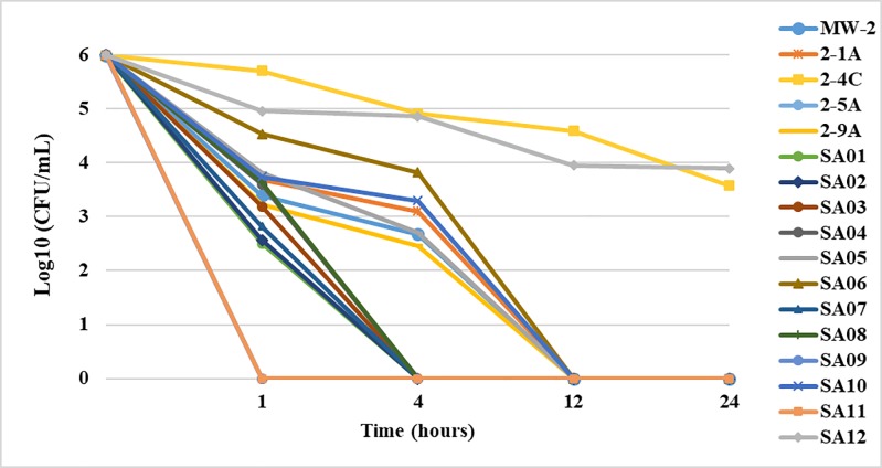
Fig 1 shows the time-dependent killing of the clinical MRSA strains with the addition of PAAG. PAAG (100 μg/mL) demonstrated rapid bactericidal activity against 58% of the MRSA isolates tested, eliminating a starting inoculum of 1.5 x 10⁶ CFU/mL within 1- 4h. A 2-log reduction in the CFU/mL was observed upon 1 h treatment of PAAG for 15 of the 17 clinical isolates tested. PAAG completely cleared 88% of the MRSA isolates with 6- log reduction in CFU/mL upon 12 h of treatment with PAAG. PAAG exhibited slower bactericidal activity against strains SA10 and 2-4C with < 2 log reduction (CFU/mL) in 24 hours. Complete elimination of these two strains was not possible even after 24 hours at PAAG concentrations of 100 μg/mL.
Fig 1. Bactericidal activity of PAAG against seventeen MRSA clinical isolates.
PAAG at a concentration of 100 μg/mL was added at timepoint 0 and monitored until 24h. Six log reductions in CFU/mL were observed in 58% of the MRSA isolates tested in 1-4h of treatment. 88% of the MRSA strains was observed within 12h of PAAG treatment. Data is presented as mean ± SD (n = 3).
Permeabilization of MRSA by PI uptake assay
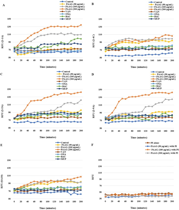
To assess the mechanism of antibacterial activity of PAAG, the PI uptake assay was used to measure bacterial permeabilization by PAAG. PI shifts emission upon intercalation into the bacterial DNA. Untreated cells did not display any PI fluorescence intensity. (Fig 2A–2E) shows the relative fluorescence unit (RFU) of emission showing PI fluorescence on exposure of 50–200 μg/mL of PAAG and 100μg/mL of oxacillin, bacitracin, mupirocin and control. A steady increase in fluorescence intensity was observed with increasing PAAG concentrations for isolates 2-1A, 2-5A and 2-9A. The fluorescence intensity increased dramatically within 30 minutes of addition of PAAG (200 μg/mL) when compared to the antibiotics (Fig 2). The increase in fluorescence in treated samples clearly indicates the permeabilization of bacterial cells as a result of PAAG treatment.
Fig 2.
(A-F). The relative fluorescence units (RFU) measured reflects PI intercalation into bacterial DNA by 2-1A (A), 2-4C (B), 2-5A (C), 2-9A (D), SA10 (E), PI alone and PI with PAAG (F) over 200 minutes, with the addition of control, 50–200 μg/mL PAAG, 100 μg/mL of vancomycin, oxacillin, mupirocin or bacitracin respectively.
Strains SA10 and 2-4C were observed in the time kill assay to be less affected by PAAG treatment. Treatment with PAAG resulted in minimal increase in PI fluorescence intensity for strains SA10 and 2-4C compared to the other strains. The lack of significant increase in intensity upon PAAG treatment on these two strains explains the slower bactericidal action of PAAG observed in the time kill curves. These strains will be further investigated in future studies.
Vancomycin, oxacillin, bacitracin and mupirocin showed minimal increase in fluorescent intensity even after 4h of treatment, as shown in Fig 2. While bacitracin, oxacillin and vancomycin are known to disrupt cell wall synthesis, mupirocin reversibly inhibits bacterial protein and RNA synthesis. None of these traditional antibiotics act upon contact to permeabilize bacteria, and thus the potential bactericidal effects may take longer than the duration of this study. Control experiments were carried out by adding PI onto the bacteria, in the absence of PAAG. No change was observed for the fluorescence intensity of PI alone and PI with PAAG (Fig 2F).
Antimicrobial resistance study
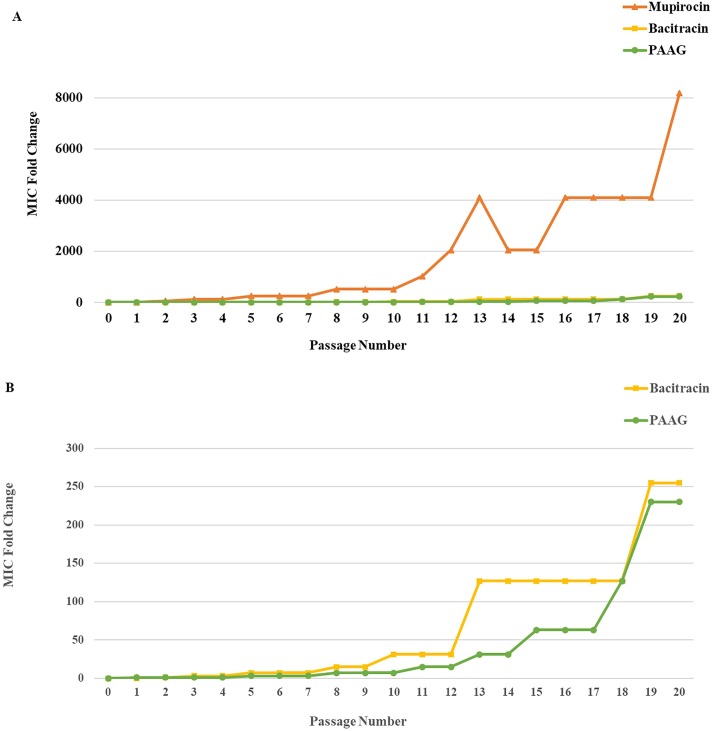
The results of the serial incubation experiments are presented in Fig 3 as the fold changes in MIC plotted against number of incubations. All experiments demonstrated emergence of resistance, allowing S. aureus to grow in successively higher concentrations of mupirocin, bacitracin and PAAG over time. After 20 days of serial passage, the MICs were found to be 1024 μg/mL for mupirocin (MIC > 512 μg/mL classified as high resistance (CLSI)), 51 mg/mL for bacitracin, and 8194 μg/mL for PAAG. The general trend toward bacterial growth in higher concentrations of PAAG and bacitracin resulted from a series of small incremental changes in MIC over the course of 20 days, as opposed to mupirocin, where an early dramatic change in MIC was followed by similar increases upon subsequent passages.
Fig 3. In vitro resistance development study.
Development of in vitro resistance to PAAG was compared to that of two other topical antibiotics, mupirocin and bacitracin, plotted as MIC fold change vs passage number (A). S. aureus ATCC# 6538 developed resistance to PAAG and bacitracin at a slower rate compared to the rapid onset of resistance to mupirocin. Fig 3B is an expansion of Fig 3A showing the MIC fold change to PAAG and Bacitracin.
Resensitization of the antimicrobial resistant strains
Additional checkerboard assays were performed on mupirocin and bacitracin resistant strains of Staphylococcus aureus 6538 collected at 13th and 20th serial MIC passage. PAAG in combination with bacitracin re-sensitized the bacitracin resistant Staphylococcus aureus ATCC# 6538 displaying a reduction up to 256 -fold in the MIC. PAAG concentrations as low as 4 μg/mL, significantly reduced MICs of bacitracin from both 25.6 mg/mL and 51.2 mg/mL to 0.2 μg/mL for both passage 13 and 20, respectively. PAAG (4 μg/mL) also re-sensitized highly mupirocin-resistant isolates of S. aureus 6538, facilitating substantial reduction in MICs from 1024 μg/mL (passage 20) and 512 μg/mL (passage 13) to 0.125 μg/mL, resulting in a 4 order of magnitude change in MIC. MICs of mupirocin and bacitracin for these resistant isolates of S. aureus were reduced below their clinical susceptibility breakpoints (≤ 2 μg/mL) [43]. The assay demonstrated the ability of PAAG to potentiate the activity of antibiotics in a strain with recent resistance development, there by re-sensitizing S. aureus to bacitracin and mupirocin.
Discussion
The panel of isolates tested represent a collection of a wide range of clinical isolates, associated with respiratory, skin and blood infections. All the isolates tested were found to be resistant to oxacillin and 35% of the clinical isolates of MRSA tested were found to be highly resistant to mupirocin (Table 1), according to the CLSI standards [43]. PAAG demonstrated potent antibacterial activity against all the multidrug resistant clinical isolates of MRSA tested, with MICs ranging from 16–128 μg/mL (Table 1). The antimicrobial activity exhibited by PAAG is hypothesized to be a result of its polycationic nature, and is a result of permeabilization of the bacterial membrane, as shown in Fig 2 and demonstrated previously for its interaction with the Gram-negative E. coli [36].
Oxacillin has been used in combination with vancomycin in clinical use, though has not been proven to be a therapeutically effective combination in treating MRSA infections. PAAG when combined with oxacillin exhibited strong synergy against all seventeen clinical isolates of MRSA tested with FIC ranging from 0.1 to 0.5. The MIC for oxacillin was reduced up to 128-fold in the presence of PAAG. No antagonistic interactions were observed between PAAG and oxacillin for all strains tested. PAAG was able to decrease the MIC of oxacillin below clinical susceptibility breakpoints of <4 μg/mL for 94% of the MRSA strains tested (16/17). The direct antimicrobial and synergistic activities of PAAG suggest its potential to be used alone or in combination with oxacillin to treat infections that would otherwise be resistant to oxacillin. These findings are supported by similar observations for PAAG in potentiating the activity of antibiotics against multidrug-resistant Burkholderia isolates [37]. The fact that synergistic, and not additive, effects were observed is hypothesized to reflect that these drug resistant S. aureus strains are likely less fit than sensitive strains. Multidrug resistance in bacteria is often linked to a fitness cost associated with utilizing a less efficient system or the energy cost of antibiotic pumps and clearance efforts, which explains their reduced growth rates and virulence [49, 50]. If a strain is less fit, the ability to respond to a pore-forming agent could be less robust, resulting in a synergistic rather than additive effect of the combination therapy.
PAAG’s ability to eliminate bacteria through direct bactericidal activity, in addition to inhibition of bacterial growth, was assessed though time-kill assays. The time dependent reduction in CFU/mL of all the 17 clinical isolates over a 24-hour period post treatment with PAAG (100 μg/mL) has been shown in Fig 1. Treatment with PAAG at a concentration of 100 μg/mL resulted in eradication of 88% (15/17) of the multidrug resistant strains followed by a 6-log reduction in CFU/mL. Strains highly resistant to mupirocin (MIC >512 μg/mL) were eradicated (> 5 log reduction) within 4 hours of treatment with PAAG (100 μg/mL). Most commonly isolated opportunistic respiratory pathogens such as MW-2 and 2-1A was found to be eradicated (6- log reduction) with in 1 hour of treatment with PAAG (100 μg/mL). It was noted that PAAG at concentrations 3x MIC (100 μg/mL) exhibited slower bactericidal activity against SA10 and 2-4C with < 2 log reduction in CFU/mL in 24 hours. In the clinical setting, bacteria exposed to sublethal doses of antibiotics may accumulate point mutations resulting in amino acid substitutions that result in increased MICs. Specifically, mutations in mprF, yycG, and rpoB have demonstrated treatment associated increases in MICs and suggest that genetic changes in these genes can influence antimicrobial susceptibility [51]. Studies have also shown that bacteria can resist killing by peptides, such as AMP’s, by membrane modifications, active efflux and reduced uptake [52]. These resistant strains might have a phenotype defined by increased cell wall thickness and increased membrane fluidity [53]. Therefore, it is hypothesized that these altered membrane arrangements may be limiting efficient PAAG insertion into the membrane [54–56]. Slower bactericidal activity of PAAG against SA10 and 2-4C was further analyzed and investigated using a PI uptake study.
Bacterial permeabilization upon treatment with PAAG and standard antibiotics were assessed using a fluorescent probe, propidium iodide (PI). PI cannot enter the bacteria unless the outer layer of the cell is permeabilized. If PI enters the bacteria, DNA-bound PI fluoresces with excitation and emission at 544 nm and 620 nm, respectively. The results of the study confirm that bacteria treated with PAAG are leaky, allowing increased uptake of PI. PAAG rapidly permeabilized five of the MRSA isolates tested in a dose dependent manner as indicated by an increase in PI fluorescence (Fig 2). The antibiotics tested showed minimal increase in PI intensity, even after 4h of treatment, which helps to explains bacterial persistence. One of the major reasons for emergence of antibiotic resistance is poor permeability of the outer cell wall to antimicrobial agents [57]. Vancomycin resistance arises from thickening of the cell wall followed by the segregation of vancomycin at non-active cell wall targets [3, 57]. Permeabilization of the outer cell wall and membrane might play a role in the mechanism of PAAG mediated killing of the clinical MRSA isolates and is likely a contributor to PAAG’s synergistic activity. Similar to antimicrobial peptides (AMP’s), polycationic PAAG most likely first interacts with the net negative charge of the bacterial cell surface, followed by disruption of membrane integrity.
Treatment with PAAG did not lead to as much PI uptake in strains SA10 and 2-4C compared to the other three strains. A number of known cell wall and membrane resistance mechanisms [54–56] could account for the reduced permeabilization of these two strains. Further studies on the changes in cell wall and membrane composition of these resistant strains will be conducted to better understand the slower killing exhibited by PAAG on SA10 and 2-4C.
The investigation of the development of resistance of S. aureus 6538 to PAAG, mupirocin and bacitracin is shown in Fig 3, where the fold change in MIC is shown over the course of 20 passages. Mupirocin and bacitracin were selected as comparators as they have very different mechanisms of action. Bacitracin disrupts Gram-positive bacteria by interfering with cell wall synthesis and is used topically. Mupirocin selectively binds to bacterial isoleucyl-tRNA synthetase, which stops or slows bacterial protein synthesis. Also, mupirocin is known to have genetic transfer of resistance [58, 59] and bacitracin is known to develop resistance slowly, compared to other conventional antibiotics [60].
Mupirocin was observed to express nearly a 63-fold increase in MIC following the first passage, suggesting that the genes for the resistance were available. A rapid increase in resistance continued, leading to >8000-fold increase in MIC, over the next 20 passages. Conversely, both PAAG and bacitracin exhibited slower development of resistance, with incremental changes upon passages up to slightly greater than 200-fold increase in MIC by 20 passages. While studies are ongoing to unravel the mechanism of PAAG resistance, these results shed light to the point that both PAAG and bacitracin interactions with the cell walls result in similar time lines for developing resistance in this ATCC strain. However, this observation may not be generalizable to all strains.
PAAG displayed substantial potential to re-sensitize mupirocin and bacitracin resistant S. aureus isolated from passages 13 and 20 below clinical sensitivity breakpoints (≤ 2 μg/mL) [43]. Synergistic combinations of PAAG at concentrations as low as 4 μg/mL with mupirocin/ bacitracin resulted in a 4-order magnitude reduction in the MICs of mupirocin and bacitracin respectively.
Combination therapies have been reported to minimize the likelihood of resistance development, to reduce drug toxicity by lowering the efficacious dose and to broaden the range of pathogenic bacteria that can be targeted [3, 61]. While the MICs of antibiotics are used to quantify the in vitro antimicrobial activities of antibiotics against infectious microorganisms, consideration of bactericidal effects of the drug is also a key factor to consider when determining antibiotic dosing regimens. The findings of the current study point to the potential of PAAG as a means to treat MRSA infections through a direct bactericidal activity or through combination therapies. PAAG was observed to enhance the antibacterial activity of oxacillin, potentially increasing its clinical efficacy. PAAG was also shown to re-sensitize a resistant strain of S. aureus to both mupirocin and bacitracin, suggesting that PAAG could potentially expand the range of multidrug-resistant bacteria that can be treated, should these observations continue for a broader range of bacteria [4].
Data Availability
All relevant data are within the paper.
Funding Statement
The funders, through Synedgen Inc., provided support in the form of salaries for authors SMT, VPN, SG, SMB, and WPW, but did not have any additional role in the study design, data collection and analysis, decision to publish, or preparation of the manuscript. The specific roles of these authors are articulated in the 'author contributions' section.
References
- 1.David MZ, Daum RS. Community-associated methicillin-resistant Staphylococcus aureus: epidemiology and clinical consequences of an emerging epidemic. Clinical microbiology reviews. 2010; 1;23(3):616 doi: 10.1128/CMR.00081-09 [DOI] [PMC free article] [PubMed] [Google Scholar]
- 2.Reddy CM, Thati V, Shivannavar CT, Gaddad SM. Vancomycin resistance among methicillin resistant Staphylococcus aureus isolates in Rayalaseema region Andhra Pradesh, South India. World J Sci Tech. 2012; 2:6–8. [Google Scholar]
- 3.Dasenbrook EC, Checkley W, Merlo CA, Konstan MW, Lechtzin N, Boyle MP. Association between respiratory tract methicillin-resistant Staphylococcus aureus and survival in cystic fibrosis. Jama. 2010. June 16;303(23):2386–92. doi: 10.1001/jama.2010.791 [DOI] [PubMed] [Google Scholar]
- 4.Döring G, Flume P, Heijerman H, Elborn JS, Consensus Study Group. Treatment of lung infection in patients with cystic fibrosis: current and future strategies. Journal of Cystic Fibrosis. 2012. December 31;11(6):461–79. doi: 10.1016/j.jcf.2012.10.004 [DOI] [PubMed] [Google Scholar]
- 5.Mohamed MF, Abdelkhalek A, Seleem MN. Evaluation of short synthetic antimicrobial peptides for treatment of drug-resistant and intracellular Staphylococcus aureus. Scientific Reports. 2016;6. [DOI] [PMC free article] [PubMed] [Google Scholar]
- 6.deLeo FR, Chambers HF. Reemergence of antibiotic-resistant Staphylococcus aureus in the genomics era. J Clin Invest. 2009; 119(9):2464–2474. doi: 10.1172/JCI38226 [DOI] [PMC free article] [PubMed] [Google Scholar]
- 7.Frank PT, Christopher WC, John S. Antibiotic Therapy of Methicillin-Resistant Staphylococcus Aureus in Critical Care Critical Care Clinics. Volume 24 Issue 2 249–260 doi: 10.1016/j.ccc.2007.12.013 [DOI] [PubMed] [Google Scholar]
- 8.Pulido MR, García-Quintanilla M, Martín-Peña R, Cisneros JM, McConnell MJ. Progress on the development of rapid methods for antimicrobial susceptibility testing. Journal of Antimicrobial Chemotherapy. 2013; 68(12):2710–7. doi: 10.1093/jac/dkt253 [DOI] [PubMed] [Google Scholar]
- 9.World health organization [Internet]. Antimicrobial resistance; c2016 [cited 2016 Sep]. Available from: http://www.who.int/mediacentre/factsheets/fs194/en/.
- 10.Center for disease control and prevention [Internet]. Biggest threats; [cited 2013]: Antibiotic/ antimicrobial resistance; Available from: https://www.cdc.gov/drugresistance/biggest_threats.html.
- 11.Klevens RM. Changes in the epidemiology of methicillin-resistant Staphylococcus aureus in intensive care units in US hospitals, 1992–2003. Clin. Infect. Dis. 2006; 42:389–391. doi: 10.1086/499367 [DOI] [PubMed] [Google Scholar]
- 12.Klein E, Smith DL, Laxminarayan R. Hospitalizations and deaths caused by methicillin-resistant Staphylococcus aureus, United States, 1999–2005. Emerg. Infect. Dis. 13:1840–1846. doi: 10.3201/eid1312.070629 [DOI] [PMC free article] [PubMed] [Google Scholar]
- 13.Liu C. A population-based study of the incidence and molecular epidemiology of methicillin-resistant Staphylococcus aureus disease in San Francisco, 2004–2005. Clin. Infect. Dis. 2008; 46:1637–1646. doi: 10.1086/587893 [DOI] [PubMed] [Google Scholar]
- 14.Andersson DI, Hughes D. 2011. Persistence of antibiotic resistance in bacterial populations. FEMS Microbiol. Rev. 35:901–911. doi: 10.1111/j.1574-6976.2011.00289.x [DOI] [PubMed] [Google Scholar]
- 15.Drago L, De Vecchi E, Nicola L, Gismondo MR. In vitro evaluation of antibiotics’ combinations for empirical therapy of suspected methicillin resistant Staphylococcus aureus severe respiratory infections. BMC Infect Dis. 2007; 7, 111 doi: 10.1186/1471-2334-7-111 [DOI] [PMC free article] [PubMed] [Google Scholar]
- 16.Svetitsky S, Leibovici L, Paul M. Comparative efficacy and safety of vancomycin versus teicoplanin: systematic review and meta-analysis. Antimicrobial agents and chemotherapy. 2009; 1;53(10):4069–79. doi: 10.1128/AAC.00341-09 [DOI] [PMC free article] [PubMed] [Google Scholar]
- 17.Barr JG, Smyth ET, Hogg GM. In vitro antimicrobial activity of imipenem in combination with vancomycin or teicoplanin against Staphylococcus aureus and Staphylococcus epidermidis. Eur. J. Clin. Microbiol. Infect. Dis. 1990; 9:804–809 [DOI] [PubMed] [Google Scholar]
- 18.Domaracki BE, Evans, Preston KE, Fraimov H, Venezia RA. Increased oxacillin activity associated with glycopeptides in coagulasenegative staphylococci. Eur. J. Clin. Microbiol. Infect. Dis. 1998; 17:143–150. [DOI] [PubMed] [Google Scholar]
- 19.Aritaka N, Hanaki H, Cui L, Hiramatsu K. Combination effect of vancomycin and β-lactams against a Staphylococcus aureus strain, Mu3, with heterogeneous resistance to vancomycin. Antimicrobial agents and chemotherapy. 2001;45(4):1292–4. doi: 10.1128/AAC.45.4.1292-1294.2001 [DOI] [PMC free article] [PubMed] [Google Scholar]
- 20.Tabuchi F, Matsumoto Y, Ishii M, Tatsuno K, Okazaki M, Sato T, et al. "Synergistic effects of vancomycin and β-lactams against vancomycin highly resistant Staphylococcus aureus." The Journal of Antibiotics 70.6 (2017): 771–774). [DOI] [PubMed] [Google Scholar]
- 21.EClimo MW, Patron RL, Archer GL. Combinations of vancomycin and beta-lactams are synergistic against staphylococci with reduced susceptibilities to vancomycin. Antimicrob Agents Chemother. 1999; 43: 1747–1753 [DOI] [PMC free article] [PubMed] [Google Scholar]
- 22.Barber KE, Rybak MJ, Sakoulas G. Vancomycin plus ceftaroline shows potent in vitro synergy and was successfully utilized to clear persistent daptomycin-non-susceptible MRSA bacteraemia. J Antimicrob Chemother. 2015; 70: 311–313 doi: 10.1093/jac/dku322 [DOI] [PubMed] [Google Scholar]
- 23.Wertheim HFL, Vos MC, Ott A, Voss A, Kluytmans JAJW, Vandenbroucke-Grauls CMJE. Mupirocin prophylaxis against nosocomial Staphylococcus aureus infections in nonsurgical patients. A randomized study. Ann. Intern. Med. 2004; 40:419–425. [DOI] [PubMed] [Google Scholar]
- 24.Leyden JJ. Studies on the safety of Bactroban ointment: potential for contact allergy, contact irritation, phototoxicity and photo-allergy. Excerpta Medica Curr Clin Pract Ser. 1985; 16:68–71. [Google Scholar]
- 25.Maddin S, Larochelle S, Wilkinson RD, Carey WD, Gratton D, Haydey RP. Bactroban: efficacy and tolerance of a novel, new topical antibiotic vs. conventional systemic and topical treatment of primary and secondary skin infections. Contemp. Dermatol. 1987; 32–39. [Google Scholar]
- 26.McRipley RJ, Whitney RR. Characterization and quantitation of experimental wound infections used to evaluate topical antibacterial agents. Antimicrob Agents Chemother. 1976; 10:38–44. [DOI] [PMC free article] [PubMed] [Google Scholar]
- 27.Gisby J, Bryant J. Efficacy of a New Cream Formulation of Mupirocin: Comparison with Oral and Topical Agents in Experimental Skin Infections. Antimicrobial Agents and Chemotherapy. 2000; 44(2):255–260. [DOI] [PMC free article] [PubMed] [Google Scholar]
- 28.Dos Santos KRN, Fonseca DSL, Filho PPG. Emergence of high-level mupirocin resistance in methicillin-resistant Staphylococcus aureus isolated from Brazilian university hospitals. Infect. Control Hosp. Epidemiol. 1996; 17:813–816. [PubMed] [Google Scholar]
- 29.Vasquez JE, Walker ES, Franzus BW, Overbay BK, Reagan DR, Sarubbi FA. The epidemiology of mupirocin resistance among methicillin-resistant Staphylococcus aureus at a Veterans' Affairs hospital. Infect. Control Hosp. Epidemiol. 2000, 21:459–464. doi: 10.1086/501788 [DOI] [PubMed] [Google Scholar]
- 30.Han LL, McDouga LK, Gorwitz RJ, Mayer KH, Patel JB, Sennott JM et al. High frequencies of clindamycin and tetracycline resistance in methicillin-resistant Staphylococcus aureus pulsed-field type USA300 isolates collected at a Boston ambulatory health center. J. Clin. Microbiol. 2007; 45:1350–1352. doi: 10.1128/JCM.02274-06 [DOI] [PMC free article] [PubMed] [Google Scholar]
- 31.Dawson RM, Liu CQ. Analogues of peptide SMAP-29 with comparable antimicrobial potency and reduced cytotoxicity. Int. J. Antimicrob. Agents. 2011; 37, 432–437. doi: 10.1016/j.ijantimicag.2011.01.007 [DOI] [PubMed] [Google Scholar]
- 32.Ho TT, Cadena J, Childs LK. et al. Methicillin-resistant Staphylococcus aureus bacteremia and endocarditis treated with ceftaroline salvage therapy. J Antimicrob Chemother. 2012; 67: 1267–1270 doi: 10.1093/jac/dks006 [DOI] [PubMed] [Google Scholar]
- 33.Jain R, Chan JD, Rogers L et al. High incidence of discontinuations due to adverse events in patients treated with ceftaroline. Pharmacotherapy. 2014; 34: 758–763 doi: 10.1002/phar.1435 [DOI] [PubMed] [Google Scholar]
- 34.Yam FK, Kwan BK. A case of profound neutropenia and agranulocytosis associated with off-label use of ceftaroline. Am J Health Syst Pharm. 2014; 71: 1457–1461 doi: 10.2146/ajhp130474 [DOI] [PubMed] [Google Scholar]
- 35.Rimawi RH, Frenkel A, Cook PP. Ceftaroline—a cause for neutropenia. J Clin Pharm Ther. 2013; 38: 330–332 doi: 10.1111/jcpt.12062 [DOI] [PubMed] [Google Scholar]
- 36.Tang H, Zhang P, Kieft TL, Ryan SJ, Baker SM, Wiesmann WP, et al. Antibacterial action of a novel functionalized chitosan-arginine against Gram-negative bacteria. Acta Biomaterialia. 2010; 6(7):2562–71. doi: 10.1016/j.actbio.2010.01.002 [DOI] [PMC free article] [PubMed] [Google Scholar]
- 37.Narayanaswamy VP, Giatpaiboon S, Baker SM, Wiesmann WP, LiPuma JJ, Towsend SM. Novel glycopolymer sensitizes Burkholderia cepacia complex isolates from cystic fibrosis patients to tobramycin and meropenem. PLoS ONE 2017; 12(6): e0179776 https://doi.org/10.1371/journal.pone.0179776 doi: 10.1371/journal.pone.0179776 [DOI] [PMC free article] [PubMed] [Google Scholar]
- 38.Baxter RM, Dai T, Kimball J, Wang E, Hamblin MR, Wiesmann WP, et al. Chitosan dressing promotes healing in third degree burns in mice: gene expression analysis shows biphasic effects for rapid tissue regeneration and decreased fibrotic signaling. J Biomed Mat Res. 2013; Part A, 101(2), 340–348. [DOI] [PMC free article] [PubMed] [Google Scholar]
- 39.Burkatovskaya M, Tegos GP, Swietlik E, Demidova TN, P Castano A, Hamblin MR. Use of chitosan bandage to prevent fatal infections developing from highly contaminated wounds in mice. Biomaterials. 2006; 27(22): p. 4157–64. doi: 10.1016/j.biomaterials.2006.03.028 [DOI] [PMC free article] [PubMed] [Google Scholar]
- 40.Fischer TH, Arthur PB, Marina D, John NV. Hemostatic properties of glucosamine-based materials. J Biomed Mater Res A, 2007; 80(1): p. 167–74. doi: 10.1002/jbm.a.30877 [DOI] [PubMed] [Google Scholar]
- 41.Mi FL, Wu YB, Shyu SS, Schoung JY, Huang YB, Tsai YH et al. Control of wound infections using a bilayer chitosan wound dressing with sustainable antibiotic delivery. J Biomed Mater Res. 2002; 59(3): p. 438–49. 214. [DOI] [PubMed] [Google Scholar]
- 42.Noble L, Gray AI, Sadiq L, Uchegbu IF. A non-covalently cross-linked chitosan based hydrogel. Int J Pharm. 1999; 192(2): p. 173–82. [DOI] [PubMed] [Google Scholar]
- 43.National Committee for Clinical Laboratory Standards. Performance standards for antimicrobial susceptibility testing. 2013. CLSI approved standard M100-S23. Clinical and Laboratory Standards Institute, Wayne, PA
- 44.Claudia S, Oliwia M, Jurgen AB, Yvonne P, Miriam K, Stefen H, et al. Three-dimensional Checkerboard Synergy analysis of colistin, Meropenem, Tigecycline against Multidrug-Resistant Clinical Klebsiella pneumonia Isolates, PLoSONE. 2015; 10(6): e0126479. [DOI] [PMC free article] [PubMed] [Google Scholar]
- 45.Eliopoulos GM, Moellering RC. In Lorian V. (ed.), Antibiotics in laboratory medicine, 3rd ed. Antibiotic combinations; 1991; p. 432–492. [Google Scholar]
- 46.Meletiadis J, Pournaras S, Roilides E, Walsh TJ. Defining Fractional Inhibitory Concentration Index Cutoffs for Additive Interactions Based on Self-Drug Additive Combinations, Monte Carlo Simulation Analysis, and In Vitro-In Vivo Correlation Data for Antifungal Drug Combinations against Aspergillus fumigatus. Antimicrob Agents Chemother. 2010; 54(2):602–609. doi: 10.1128/AAC.00999-09 [DOI] [PMC free article] [PubMed] [Google Scholar]
- 47.Odds FC. Synergy, antagonism, and what the checkerboard puts between them. J Antimicrob Chemother. 2003; 52:1 doi: 10.1093/jac/dkg301 [DOI] [PubMed] [Google Scholar]
- 48.Farrell DJ, Robbins M, Rhys-Williams W, Love WG. Investigation of the potential for mutational resistance to XF-73, retapamulin, mupirocin, fusidic acid, daptomycin, and vancomycin in methicillin-resistant Staphylococcus aureus isolates during a 55-passage study Antimicrob Agents Chemother. 2011; 55(3):1177–81. doi: 10.1128/AAC.01285-10 [DOI] [PMC free article] [PubMed] [Google Scholar]
- 49.Nilsson AI, Zorzet A, Kanth A, Dahlstrom S, Berg OG, Andersson DI. Reducing the fitness cost of antibiotic resistance by amplification of initiator tRNA genes. Proc Natl Acad Sci USA. 2006; 103: 6976–6981. doi: 10.1073/pnas.0602171103 [DOI] [PMC free article] [PubMed] [Google Scholar]
- 50.Besier S, Ludwig A, Brade V, Wichelhaus TA. Compensatory adaptation to the loss of biological fitness associated with acquisition of fusidic acid resistance in Staphylococcus aureus. Antimicrob Agents Chemother. 2005; 49: 1426–1431. doi: 10.1128/AAC.49.4.1426-1431.2005 [DOI] [PMC free article] [PubMed] [Google Scholar]
- 51.Friedman L, Alder JD, Silverman JA. Genetic changes that correlate with reduced susceptibility to daptomycin in Staphylococcus aureus. Antimicrobial agents and chemotherapy. 2006. June 1;50(6):2137–45. doi: 10.1128/AAC.00039-06 [DOI] [PMC free article] [PubMed] [Google Scholar]
- 52.Kubicek-Sutherland JZ, Lofton H, Vestergaard M, Hjort K, Ingmer H, Andersson DI. Antimicrobial peptide exposure selects for Staphylococcus aureus resistance to human defence peptides. Journal of Antimicrobial Chemotherapy, 2017. 72(1), 115–127. http://doi.org/10.1093/jac/dkw381 doi: 10.1093/jac/dkw381 [DOI] [PMC free article] [PubMed] [Google Scholar]
- 53.Mishra NN, McKinnell J, Yeaman MR, Rubio A, Nast CC, Chen L, et al. In vitro cross-resistance to daptomycin and host defense cationic antimicrobial peptides in clinical methicillin-resistant Staphylococcus aureus isolates. Antimicrob. Agents Chemother. 2011; 55:4012–4018. doi: 10.1128/AAC.00223-11 [DOI] [PMC free article] [PubMed] [Google Scholar]
- 54.Bayer AS, Prasad R, Chandra J, Koul A, Smriti M, Varma A, et al. In vitro resistance of Staphylococcus aureus to thrombin-induced platelet microbicidal protein is associated with alterations in cytoplasmic membrane fluidity. Infect. Immun. 2000; 68:3548–3553. [DOI] [PMC free article] [PubMed] [Google Scholar]
- 55.Van Blitterswijk WJ, van der Meer BW, Hilkmann H. Quantitative contributions of cholesterol and the individual classes of phospholipids and their degree of fatty acyl (un)saturation to membrane fluidity measured by fluorescence polarization. Biochemistry. 1987; 26:1746–1756. [DOI] [PubMed] [Google Scholar]
- 56.Nawrocki KL, Crispell EK, McBride SM. Antimicrobial peptide resistance mechanisms of gram-positive bacteria. Antibiotics. 2014. October 13;3(4):461–92. doi: 10.3390/antibiotics3040461 [DOI] [PMC free article] [PubMed] [Google Scholar]
- 57.Kollef MH. Limitations of vancomycin in the management of resistant staphylococcal infections. Clinical infectious diseases: an official publication of the Infectious Diseases Society of America. 2007; 45 Suppl 3, S191–195. [DOI] [PubMed] [Google Scholar]
- 58.Seah C, Alexander DC, Louie L, Simor A, Low DE, Longtin J, et al. MupB, a new high-level mupirocin resistance mechanism in Staphylococcus aureus. Antimicrobial agents and chemotherapy. 2012; 56(4):1916–20. doi: 10.1128/AAC.05325-11 [DOI] [PMC free article] [PubMed] [Google Scholar]
- 59.Hurdle JG, O'neill AJ, Ingham E, Fishwick C, Chopra I. Analysis of mupirocin resistance and fitness in Staphylococcus aureus by molecular genetic and structural modeling techniques. Antimicrobial Agents and Chemotherapy. 2004. November 1;48(11):4366–76. doi: 10.1128/AAC.48.11.4366-4376.2004 [DOI] [PMC free article] [PubMed] [Google Scholar]
- 60.Thangamani S. Antibacterial activity and mechanism of action of auranofin against multi-drug resistant bacterial pathogens. Scientific reports. 2016; 6, 22571 doi: 10.1038/srep22571 [DOI] [PMC free article] [PubMed] [Google Scholar]
- 61.Kumar A, Zarychanski R, Light B, Parrillo J, Maki D, Simon D, et al. Early combination antibiotic therapy yields improved survival compared with monotherapy in septic shock: A propensity-matched analysis. Crit. Care Med. 2010, 38, 1773–1785. doi: 10.1097/CCM.0b013e3181eb3ccd [DOI] [PubMed] [Google Scholar]
Associated Data
This section collects any data citations, data availability statements, or supplementary materials included in this article.
Data Availability Statement
All relevant data are within the paper.


