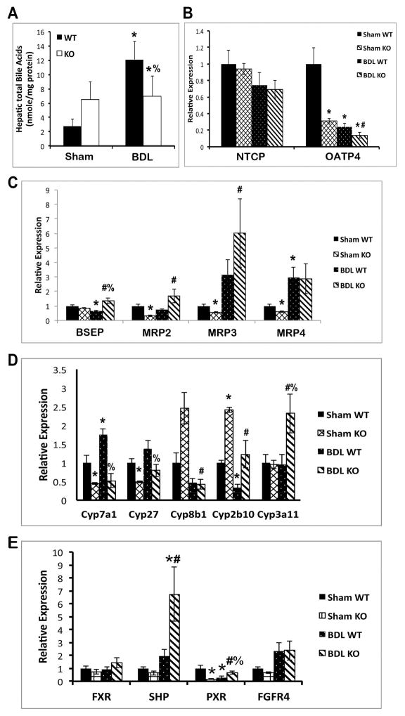
Figure 3. Decreased hepatic BA and BA synthesis in KO.
(A) Total BA remained unchanged in KO livers after BDL, while increasing significantly in WT. (B) Expression of basolateral uptake transporter OATP4 is decreased in KO after BDL. (C) Expression of apical BA exporters BSEP and MRP2 is upregulated in KO after BDL. (D) Expression of BA biosynthesis genes is suppressed, while detoxification genes are induced, in KO after BDL. (E) Expression of SHP, a known FXR/RXRα target gene, is significantly increased in KO after BDL. *p<0.05 vs. sham WT, #p<0.05 vs. sham KO, %p<0.05 vs. BDL WT.

