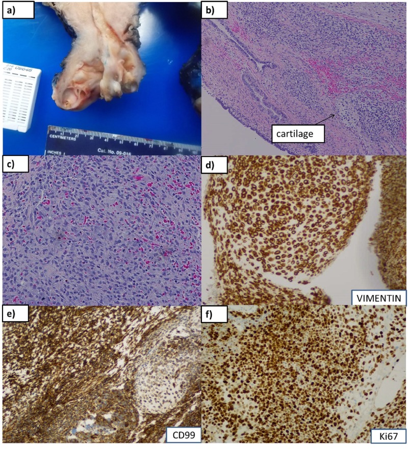
Figure 1. Gross and microscopic features.
(a): Gross polypoid mass protruding through os, 8.0 cm in greatest dimension, homogeneous, fleshy, yellow-tan to gray-pink myxoid cut surface with soft consistency. Foci of necrosis and ulceration are seen. (b), (c): Microscopic findings of primitive small cells with scant cytoplasm, islands of cartilage (arrow), alternating hypo- (with myxoid &/or edematous stroma) and hypercellular areas at 10x magnification, (b), and 20 x, (c). (d), (e), (f): Immunohistochemistry stains positive for vimentin, CD99, and Ki67, respectively.

