Description
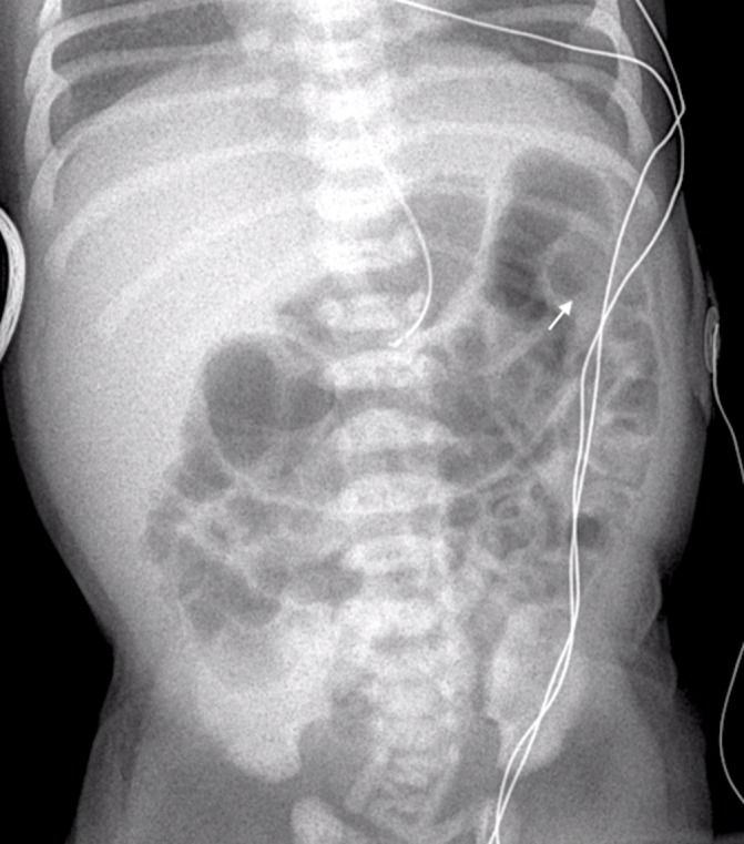
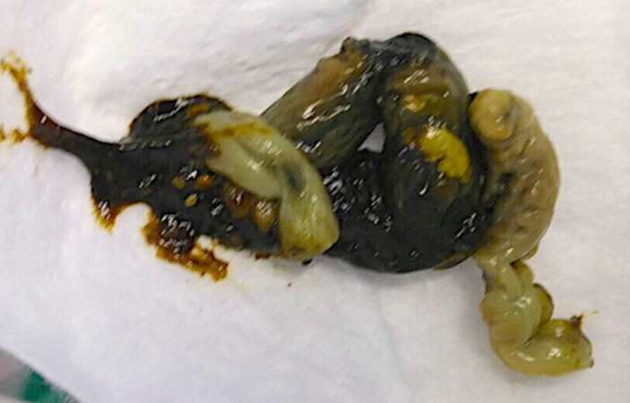
A female infant born at 35+6 weeks by caesarean section to a mother with poorly controlled type 1 diabetes was admitted to the neonatal unit due to hypoglycaemia. Birth weight was 3.2 kg (91–98th centile). On day 1, the baby required a glucose load of 10 mg/kg/min to maintain normoglycaemia. By day 2, her blood sugar levels stabilised, feeds were started and intravenous fluids were weaned. With the introduction of feeds, she had milky vomits and abdominal distension. Feeds were stopped and an abdominal X-ray showed a dilated transverse colon with an abrupt transition zone at splenic flexure (figure 1, arrow indicates transition zone). Symptoms resolved after passage of a large, thick plug of mucus and meconium that resembled an intestinal cast (figure 2).
Figure 1.

Patient’s plain abdominal X-ray. There is a dilated transverse colon and calibre change at the splenic flexure indicates a transition point (arrow) and a relatively narrow left colon.
Figure 2.
Patient’s stool. This shows a thick plug of stool consisting of mucus and meconium in the form of an intestinal cast with imprint of mucosal folds.
Feeds were introduced and tolerated without any subsequent problems. Cystic fibrosis mutation testing was negative. A diagnosis of neonatal small left colon syndrome (NSLCS) was made based on clinical and radiological findings.
NSLCS is a rare functional lower intestinal disorder, first described in 1974.1 2 The underlying pathogenesis is unknown, but there is a strong association with maternal diabetes.2 Transient dysmotility of the descending colon leads to distinctive radiological features of a narrow descending colon with an abrupt transition zone at the splenic flexure. This can be visualised on plain abdominal X-rays as well as with contrast enema. Contrast enema when performed can be therapeutic, causing the passage of a meconium plug with the contrast. Normally there are no long-term complications and there is normal intestinal motility once resolved.2 3
Patient’s perspective.
My baby had prolonged admission in the neonatal unit and I believe some of her problems were related to my diabetes. I was very worried when she was 2 days old, when doctors informed me that she has bowel obstruction and possibly might need transfer to a surgical unit. However, doctors suspected this rare association with diabetes where the tail end of the bowel is not well formed in the baby and this was picked up on X-ray. She soon passed a horrible looking poo which looked like her bowels had come out, but after that her vomiting settled and she started tolerating feeds. I think it is important to consider this complication early on in this setting so that unnecessary transfers could be avoided.
Learning points.
Infants of diabetic mothers frequently have gastrointestinal symptoms that mimic intestinal obstruction.
Neonatal small left colon syndrome is rare but an important differential in this situation and radiological findings are diagnostic.
Early suspicion of this diagnosis averts unnecessary invasive interventions and surgery.
Footnotes
Contributors: KW wrote the manuscript. AJ and SN edited the manuscript. KA provided radiology input and reported X-ray.
Competing interests: None declared.
Patient consent: Guardian consent obtained.
Provenance and peer review: Not commissioned; externally peer reviewed.
References
- 1.Davis WS, Allen RP, Favara BE, et al. Neonatal small left colon syndrome. Am J Roentgenol Radium Ther Nucl Med 1974;120:322–9. doi:10.2214/ajr.120.2.322 [DOI] [PubMed] [Google Scholar]
- 2.Ellis H, Kumar R, Kostyrka B. Neonatal small left colon syndrome in the offspring of diabetic mothers-an analysis of 105 children. J Pediatr Surg 2009;44:2343–6. doi:10.1016/j.jpedsurg.2009.07.054 [DOI] [PubMed] [Google Scholar]
- 3.Loening-Baucke V, Kimura K. Failure to pass meconium: diagnosing neonatal intestinal obstruction. Am Fam Physician 1999;60:2043–50. [PubMed] [Google Scholar]


