

Abstract
Background
Facial hypermelanosis is a significant cause of cosmetic disfigurement, social embarrassment and psychological morbidity affecting quality of life.
Objective
To study clinicoepidemlogic patterns of facial hypermelanoses among men.
Material and Methods
Medical records of all adult males presenting with facial hypermelanoses were analyzed for this retrospective cross sectional study for demographic details, duration, cosmetic usage, sun exposure, drug intake, infections, systemic or cutaneous diseases, and family history of hypermelanotic dermatosis. Laboratory investigations and skin biopsy were performed when deemed necessary.
Results
These were 300 Indian men aged 18 to 74 (mean 37.35) years with 121 (40.3%) individuals aged 31-50 years. Various patterns of melasma in 230 (76.7%) patients were the major cause of facial hypermelanosis. Periorbital hypermelanosis was observed in 32 (10.7%), freckles and lentigens in 26 (8.7%), acanthosis nigricans in 12 (4%) and lichen planus pigmentosus in 10 (3.3%), pigmented cosmetic contact dermatitis in 7, and nevus of Ota in 6 persons. The 71 (30.8%) patients with melasma had a history of frequent sun exposure, 9 (3.9%) patients had systemic comorbidities. Family history of periorbital melanosis was present in 7 (21.8%), personal or family history of atopy in 5 (15.6%) patients. Acanthosis nigricans was associated with obesity in 9 (75%) of patients and with diabetes mellitus in 4 (33.3%) cases.
Conclusions
Melasma, periorbital hypermelanosis, acanthosis nigricans and lichen planus pigmentosus remain the predominant causes for facial hypermelanosis in men.
Keywords: acanthosis nigricans, ashy dermatosis, Becker's nevus, freckles, melasma, periorbital hypermelanoses, hyperpigmentation, pigmented contact dermatitis, lentigens, lichen planus pigmentosus, nevus of Ota
Background
Facial hypermelanosis is a great cosmetic concern causing significant social embarrassment and psychological distress particularly among Asians including Indians. Facial hypermelanosis may occur from a variety of clinical entities; common ones such as melasma, lichen planus pigmentosus, Riehl’s melanosis, erythema dyschromicum perstans, and uncommonly from poikiloderma of Civatte, erythromelanosis follicularis of face and neck, nevus of Ota, periorbitalmelanosis, exogenous ochronosis, or acanthosis nigricans. Sun exposure and exposure to photosensitizing agents play a pivotal role in the pathogenesis of many of these disorders.[1] Since, most Indian populations are occupationally engaged in agrarian work or manual labor exposing them to prolonged sunshine without adequate sun protection that possibly predisposes them for facial hypermelanosis. The use of cosmetics that may contain some unlisted photosensitizing agents or have a potential for pigmented cosmetic dermatitis too has increased in recent years.[2] Systemic or topical medicaments, indigenous and over-the-counter (OTC) preparations, may have photosensitizing potential and induce facial pigmentation in some of these individuals.[3,4] The rapid urbanization leading to increased usage of computers and televisions combined with altered sleeping habits may have caused increased prevalence of periorbital hypermelanosis. Sedentary life style causing near epidemic of obesity has led to an increased prevalence of acanthosis nigricans manifesting as facial hypermelanosis sometimes. Interestingly, most studies for facial hypermelanosis report predominance of females perhaps owing to their cosmetic concerns.[5-7] However, males with facial hypermelanosis also form a sizeable number in outpatient clinics, for instance, they constitute nearly 10%-25% of patients with melasma.[8] However, causes of facial hypermeanosis in them remain under studied. We studied clinical and epidemiological features in adult Indian men presenting with facial hypermelanoses.
Materials and Methods
Records of 300 adult men with facial hypermelanosis maintained during 3 months were analyzed for this retrospective cross sectional study for demographic features, duration, occupation, cosmetics use, photo exposure, drug intake, presence of infections or systemic diseases and similar hypermelanosis in their family members. Medical examination included complete systemic examination for associated systemic disorders. The diagnosis of facial hypermelanosis was mainly clinical. Patch testing with Contact and Occupational Dermatoses Form of India approved Indian Cosmetic and Fragrance series was performed in cases suspected of pigmented contact dermatitis. Laboratory investigations, if any, included complete blood counts, blood glucose levels, and hepatorenal function tests, HBsAg and Anti-HCV serology, and lesional histopathology.
Results and Discussion
All the 300 patients were aged between 18 and 74 (mean 37.35) years and had Fitzpatrick skin type III-V. The majority, 121 (40.3%) patients were in 31-50 years of age group while 97 (32.3%) patients were above 50 years and 82 (27.3%) were between 18-30 years of age. Commonly observed facial hypermelanoses were melasma, periorbital hypermelanosis, freckles and lentigines, acanthosis nigricans, lichen planus pigmentosus (LPP), and pigmented contact dermatitis in order of frequency [Table 1].
Table 1. Types of facial hypermelanosis.
| Types of facial hypermelanosis | Number of patients (%) N=300 | Mean age (range) in years | ||
|---|---|---|---|---|
| Melasma n=230 (76.7%) | Centrofacial pattern (involving cheeks, forehead, chin, upper lip and nose) | 119 (51.7%) | 230 (76.7%) | 37.35 (18-63) |
| Malar pattern (involving cheeks and nose) | 99 (43%) | |||
| Madibular pattern ( localized over rami of mandible | 12 (5.2%) | |||
| Priorbital hyermelanosis | 32 (10.7) | 28.77 (18-45) | ||
| Freckels/Lentigens | 26 (8.7) | 26.40 (19-46) | ||
| Acanthosis nigricans | 12 (4) | 40.68 (20-64) | ||
| Lichen planus pigmentosus | 10 (3.3) | 34.46 (24-56) | ||
| Pigmented contact dermatitis | 07 (2.3) | 32.36 (29-66) | ||
| Nevus of Ota | 06 (2) | 22.78 (19-27) | ||
| Ashy dermatosis of Ramirez | 01 (0.3) | 39.0 | ||
| Becker’s nevus | 01 (0.3) | 26.0 | ||
| Erythromelanosis follicularis of face and neck | 01 (0.3) | 42.0 | ||
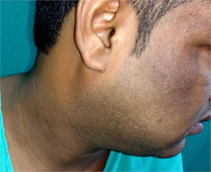
Melasma is a common disorder of facial hypermelanosis among all races particularly south-Asian and Hispanic populations affecting up to 4% of the patients attending dermatology clinics.[4,9] It affects both genders; women more than men, in their middle age with a mean age of 33.5 years in an Indian study.[8] The centrofacial pattern in 55% - 75%, the malar pattern in 24% - 43%, and the mandibular pattern in 1.5% - 2% patients are three distinct clinical patterns.[10,11] Its exact etiology is poorly understood but often imputed to genetic predisposition, pregnancy and oral contraceptives, endocrinopathies, or drugs (hormones, anticonvulsants, phenothiazines, phototoxic agents) ingestion.[3,4] Pigmented cosmetic contact dermatitis is another recognized etiologic factor.[2] However, sun exposure remains the most important exacerbating factor for 55%-100% patients.[12] Most of these features were also noted in our facial hypermelanosis patients with melasma being the commonest cause affecting 230 (76.7%) patients aged between 18-63 (mean 37.35) years [Fig. 1]. Common clinical patterns were centrofacial pattern in 119 (51.7%), malar pattern in 99 (43%) and mandibular pattern in 12 (5.2%) patients, respectively. Seventy one (30.8%) of these patients were working outdoors for long hours in the sun but only 32 (13.9%) patients were using OTC sunscreens. Commonly used cosmetics were shaving creams, cold creams, perfumes, after-shave lotions, hair color, etc. in 182 (79.1%) patients and pigmented contact dermatitis was seen in seven patients with positive patch test reactions from PPD (4 patients), gallate mix and cetrimide (1 patient), thiomersal (1 patient) and paraben mix (1 patient) correlating clinically with their cosmetic use [Fig. 2]. Forty-nine (21.3%) patients were also applying mustard oil over face while using it for hair that has photosensitizing potential because of its furocoumarin contents. However, melasma in association with chronic renal disease in 6 patients, chronic alcoholic liver disease in 2 patients and drug (levitiracetam) intake for seizure disorder in one patient in this study may be from other unidentified reasons.
Figure 1.

Malar pattern of melasma in a 28-year-old male.
Figure 2.

Pigmented cosmetic dermatitis of face predominately involving forehead, beard and moustache areas following regular use of hair color.
Periorbital hypermelanosis is frequent in women, in early adulthood (mean age 21 Years) and factors like genetic/ constitutional (in 77%), atopy (in 33%), refractive errors (in 30%), stress and inadequate/abnormal sleep pattern (in 40%), anemia, periorbital edema and skin laxity were implicated factors in a study.[13] Similarly, the periorbital hypermelanosis was second most common and noted in 32 (10.7%) patients aged between 18 and 45 (mean 28.77) years [Fig. 3]. Conformingly, family history of periorbital melanosis in 7 (21.8%) patients and personal or family history of atopy in 5 (15.6%) patients was noted. In addition, 4 (12.5%) patients had history of prolonged (>8 hours) television watching or computer work leading to eyestrain and inadequate sleep in 3 (9.4%) patients. The refractive error was noted in 7 (21.9%) patients but corrective spectacles in four of them were not benefiting in perorbital hypermelanosis. Periorbital hypermelanosis in one patient with chronic renal disease might have been from overall ill health and associated anemia. Similarly, pathogenic role of cold creams/ soaps used routinely by all these patients over face including periorbital area remains speculative.
Figure 3.

Non-familial periocular hypermelanosis in a non-atopic 25-year old male.
Freckles are common in fair skin individuals and characterized by small brown macules over the face usually appearing/increasing in summers after sun exposure. Lentigines are also hyperpigmented macules and unlike photo-distributed freckles, they may appear at any site with tendency to persist. Genetic and constitutional factors are implicated etiological factors. Freckles and lentigines are another significant cause of facial hypermlanosis and social embarrassment and were noted in our 26 (8.7%) patients with Fitzpatrick skin type III-IV aged between 19 and 46 (mean 26.4) years. Three (11.5%) patients also had another affected family member. While 20 (76.9%) patients had frequent sun exposure, only 8 (30.8%) patients were using OTC sunscreens suggesting little concern these cause among non white skin individuals.
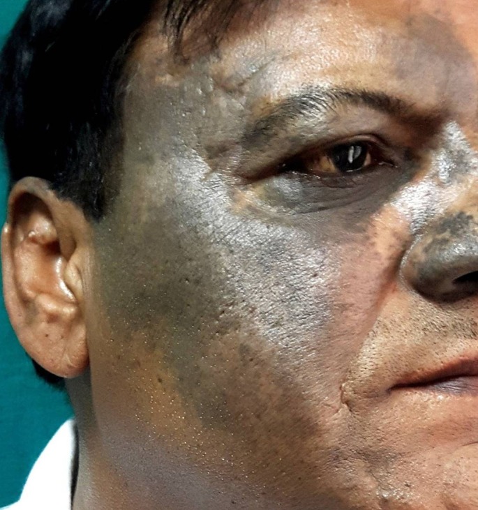
Acanthosis nigricans is characterized by hyperpigmented, velvety plaques over body folds and often involves facial skin. It may be familial or associated commonly with obesity, BMI ≥27, (in 76%), diabetes mellitus (30%), ingestion of drugs (nicotinic acid, insulin, systemic corticosteroids), or malignancy.[14] Similarly, it affected our 12 (4%) patients aged between 20 and 64 (mean 40.68) years involving neck, axillae or groins folds. Periocular involvement occurred in 7 (58.3%) patients and 5 (41.7%) patients had malar involvement [Fig. 4]. It was associated with obesity in 9 (75%) and diabetes mellitus in 4 (33.3%) patients, respectively. One patient had familial acanthosis nigricans with no obesity or other systemic disease. No patient had history of any drug intake or internal malignancy.
Figure 4.
Acanthosis nigricans: characteristic velvety hyperpigmentation involving neck fold and malar skin.
Lichen planus pigmentosus is an uncommon form of lichen planus affecting most individuals in their third or fourth decade. It is characterized by asymptomatic or mildly pruritic slate gray to brownish black, diffuse, reticular, blotchy, linear or perifollicular macular lesions of insidious onset. Face and neck are the frequent initial sites of onset followed by involvement of other body sites including body folds (LPP inversus).[15,16] Its exact etiology is poorly understood but UV light, viral infections, and topical mustard oil and amla oil have been often implicated. LPP in 10 (3.3%) patients was not associated with any systemic disease and HBsAg and Anti-HCV serology was negative [Fig. 5]. The habitual topical application of mustard oil/amla oil could not be excluded however. Nevus of Ota (6 patients, Fig. 6), ashy dermatosis (1 patient), Becker’s nevus (1 patient) and erythromelanosis follicularis of face and neck (1 patient) were other less commonly observed dermatoses.
Figure 5.
Lichen planus pigmentosus involving neck andwhole face sparing nose.
Figure 6.
Nevus of Ota (hamartoma of dermal melanocytes): characteristic bluish pigmentation in classic distribution involving first and second divisions of the trigeminal nerve.
Limitations
This was a retrospective study of data for small number of patients observed in a limited period of three months and do not represent actual number of such patients in the outdoor clinic.
Conclusion
Facial hypermelanoses are a heterogeneous group of disorders with variable etiologies. Melasma, periorbital hypermelanosis, acanthosis nigricans and lichen planus pigmentosus remain the predominant causes for facial hypermelanoses among Indian men.
References
- Khanna N, Rasool S. Facial melanoses: Indian perspective. Indian J Dermatol Venereol Leprol. 2011;77:552–564. doi: 10.4103/0378-6323.84046. [DOI] [PubMed] [Google Scholar]
- Prabha N, Mahajan VK, Mehta KS, Chauhan PS, Gupta M. Cosmetic contact sensitivity in patients with melasma: results of a pilot study. Dermatol Res Pract. 2014;2014:316219. doi: 10.1155/2014/316219. [DOI] [PMC free article] [PubMed] [Google Scholar]
- Grimes PE. Melasma. Etiologic and therapeutic considerations. Arch Dermatol. 1995;131:1453–1457. doi: 10.1001/archderm.131.12.1453. [DOI] [PubMed] [Google Scholar]
- Sivayathorn A. Melasma in Orientals. Clin Drug Investig. 1995;10 (Suppl. 10):24–40. [Google Scholar]
- Pichardo R, Vallejos Q, Feldman SR, Schulz MR, Verma A, Quandt SA, Arcury TA. The prevalence of melasma and its association with quality of life among male migrant Latino workers. Int J Dermatol. 2009;48:22–26. doi: 10.1111/j.1365-4632.2009.03778.x. [DOI] [PMC free article] [PubMed] [Google Scholar]
- Pawaskar MD, Parikh P, Markowski T, McMichael AJ, Feldman SR, Balkrishnan R. Melasma and its impact on health-related quality of life in Hispanic women. J Dermatolog Treat. 2007;18:5–9. doi: 10.1080/09546630601028778. [DOI] [PubMed] [Google Scholar]
- Vázquez M, Maldonado H, Benmamán C, Sánchez JL. Melasma in men. A clinical and histologic study. Int J Dermatol. 1988;27:25–27. doi: 10.1111/j.1365-4362.1988.tb02329.x. [DOI] [PubMed] [Google Scholar]
- Sarkar R, Puri P, Jain RK, Singh A, Desai A. Melasma in men: a clinical, aetiological and histological study. J Eur Acad Dermatol Venereol. 2010;24:768–772. doi: 10.1111/j.1468-3083.2009.03524.x. [DOI] [PubMed] [Google Scholar]
- Pasricha JS, Khaitan BK, Dash S. Pigmentary disorders in India. Dermatol Clin. 2007;25:343–352. doi: 10.1016/j.det.2007.05.004. [DOI] [PubMed] [Google Scholar]
- Achar A, Rathi SK. Melasma: a clinico-epidemiological study of 312 cases. Indian J Dermatol. 2011;56:380–382. doi: 10.4103/0019-5154.84722. [DOI] [PMC free article] [PubMed] [Google Scholar]
- Guinot C, Cheffai S, Latreille J, Dhaoui MA, Youssef S, Jaber K, Nageotte O, Doss N. Aggravating factors for melasma: a prospective study in 197 Tunisian patients. J Eur Acad Dermatol Venereol. 2010;24:1060–1069. doi: 10.1111/j.1468-3083.2010.03592.x. [DOI] [PubMed] [Google Scholar]
- KrupaShankar DS, Somani VK, Kohli M, Sharad J, Ganjoo A, Kandhari S, Mysore VR, Aurangabadkar S, Malakar S, Vedamurthy M, Kadhe G, Motlekar S, Ahirrao P. A cross-sectional, multicentric clinico-epidemiological study of melasma in India. Dermatol Ther (Heidelb) 2014;4:71–81. doi: 10.1007/s13555-014-0046-1. [DOI] [PMC free article] [PubMed] [Google Scholar]
- Sheth PB, Shah HA, Dave JN. Periorbital hyperpigmentation: a study of its prevalence, common causative factors and its association with personal habits and other disorders. Indian J Dermatol. 2014;59:151–157. doi: 10.4103/0019-5154.127675. [DOI] [PMC free article] [PubMed] [Google Scholar]
- Puri N. A study of pathogenesis of acanthosis nigricans and its clinical implications. Indian J Dermatol. 2011;56:678–683. doi: 10.4103/0019-5154.91828. [DOI] [PMC free article] [PubMed] [Google Scholar]
- Das A, Mishra V, Poddar I, Das D, Das NK. Linear lichen planus pigmentosus: a rare entity with an illusory presentation. Pigment Int. 2014;1:100–102. [Google Scholar]
- Namazi MR. Lichen planus pigmentosus presenting as diffuse facial melanosis. J Drugs Dermatol. 2004;3:436–437. [PubMed] [Google Scholar]