Abstract
Recapitulation of lung development from human pluripotent stem cells (hPSCs) in three dimensions (3D) would allow deeper insight into human development, as well as the development of innovative strategies for disease modeling, drug discovery and regenerative medicine1. We report here the generation from hPSCs of lung bud organoids (LBOs) that contain mesoderm and pulmonary endoderm and develop into branching airway and early alveolar structures after xenotransplantation and in Matrigel 3D culture. Expression analysis and structural features indicated that the branching structures reached the second trimester of human gestation. Infection in vitro with respiratory syncytial virus, which causes small airway obstruction and bronchiolitis in infants2, led to swelling, detachment and shedding of infected cells into the organoid lumens, similar to what has been observed in human lungs3. Introduction of mutation in HPS1, which causes an early-onset form of intractable pulmonary fibrosis4,5, led to accumulation of extracellular matrix and mesenchymal cells, suggesting the potential use of this model to recapitulate fibrotic lung disease in vitro. LBOs therefore recapitulate lung development and may provide a useful tool to model lung disease.
The respiratory system originates from buds that arise on the ventral aspect of the anterior foregut endoderm (AFE) and develop through a stereotyped branching process into proximal airways and distal alveolar progenitors (pseudoglandular stage). During the canalicular stage, cell cycle activity decreases, and specialization of the airway epithelium occurs in the stalks, with the emergence of basal, goblet, club, ciliated, and other cell types. This stage is followed by the saccular stage, where the canaliculi widen into distal sacculations that will give rise to primitive alveoli6,7. Organoids are in vitro generated 3D structures containing multiple cell types that are organized similar to an organ and recapitulate some specific organ function1. One group reported generation of human lung organoids8,9. However, while these small structures contained cells expressing markers of lung and airway8 and have some airway potential after subcutaneous xenografting in mice9, they do not satisfy the aforementioned criteria for organoids, as neither features of lung development, notably branching morphogenesis and proximodistal specification, nor function were observed.
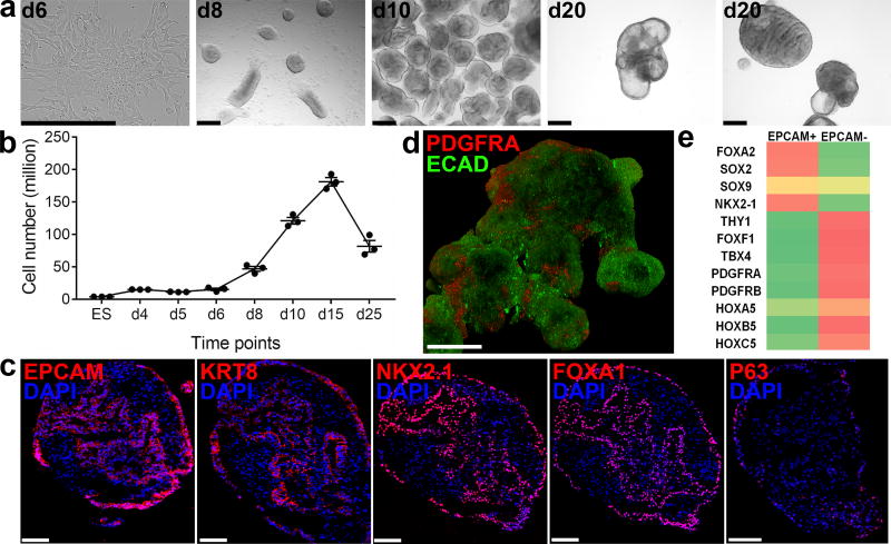
We previously reported a strategy to differentiate hPSCs (embryonic stem cells (ESCs) and induced pluripotent stem cells (iPSCs)) in 2D through sequential developmental steps from definitive endoderm (DE) to AFE, lung field progenitors, and, finally, lung and airway epithelial cells (Supplementary Fig. 1a)10–12. Early during induction of a ventral lung fate from AFE adherent structures formed that detached easily and expanded in suspension culture as clumps of cells (Fig. 1a,b) in the presence of BMP4, FGF10, FGF7, retinoic acid (RA) and the GSK3β antagonist, CHIR99201 (Supplementary Fig. 1b), factors shown previously to be required for lung development6,7. 7.5×10⁵ DE cells yielded 2490±129 clumps (n=3; RUES2 ESCs). The structures formed folding sheets of EPCAM+KRT8+ECAD+FOXA1/2+ AFE cells (FOXA2: 89.07% ± 3.36%, EPCAM+: 92.08% ± 1.88%, n=3; RUES2 ESCs) (Fig. 1c). By d25 51.26±4.37% (n=3; RUES2 ESCs) of the cells expressed the lung marker NKX2.1+ (Fig. 1c). Except for the epithelial progenitor marker, p63 (18.59% ± 1.49%, n=3; RUES2 ESCs, Fig. 1c), markers of mature lung and airway cells were absent (not shown). The cells were surrounded by mesodermal PDGFRA+ECAD− cells (Fig. 1d). RNAseq (Supplementary Fig. 1c) confirmed strong enrichment of endoderm/lung genes (FOXA2, SOX2, NKX2.1) in EPCAM+ cells (Fig. 1e). EPCAM− cells expressed mesodermal genes (Fig. 1e), some of which, such as TBX4 and HOX5 paralogs, are expressed in pulmonary mesoderm13,14. Genes expressed in mature lung and airway and in other AFE-derived lineages were nearly undetectable in the EPCAM+ fraction (Supplementary Fig. 1d). Sonic Hedgehog (SHH) was expressed in endodermal cells, and its transcriptional targets15, PTCH1, GLI1 and HHIP in mesoderm (Supplementary Fig. 1d). In situ hybridization confirmed SHH expression in the endodermal fraction at d15. At d25, SHH was expressed most strongly in the tips of budding epithelial structures (Supplementary Fig. 1e). These findings are consistent with the developing mouse lung where SHH is expressed throughout the pulmonary endoderm early but is limited to branch tips during branching morphogenesis15–17. Because they contain multiple cell types that are spatially organized similar to developing lung buds in vivo, we call these structures lung bud organoids (LBOs).
Figure 1. Generation of lung bud organoids.
(a) Development of adherent structures during ventralization of AFE between d6 and d8 (see protocol Supplementary Fig. 1b), that could be expanded in suspension culture (d10, d20). Representative of >50 independent experiments (ESCs and iPSCs). Scale bars 250 µm. (b) Cellular expansion during the generation of LBOs (mean±s.e.m, n=3 independent experiments in RUES2 ESCs). The source data can be found in Supplementary Table 4. (c) Expression of EPCAM, KRT8, NKX2.1, FOXA1, and P63 in d25 LBOs. Representative of >10 independent experiments in ESCs and iPSCs. Scale bars 100 µm. (d) Staining of d25 LBO for ECADH and PDGFRA. Representative of 3 independent experiments in RUES2 ESCs. Scale bar 250 µm. (e) Expression of endodermal and mesodermal markers in the EPCAM+ and EPCAM− fraction of d25 LBOs determined by RNAseq (3 independent biological replicates, RUES2 ESCs).
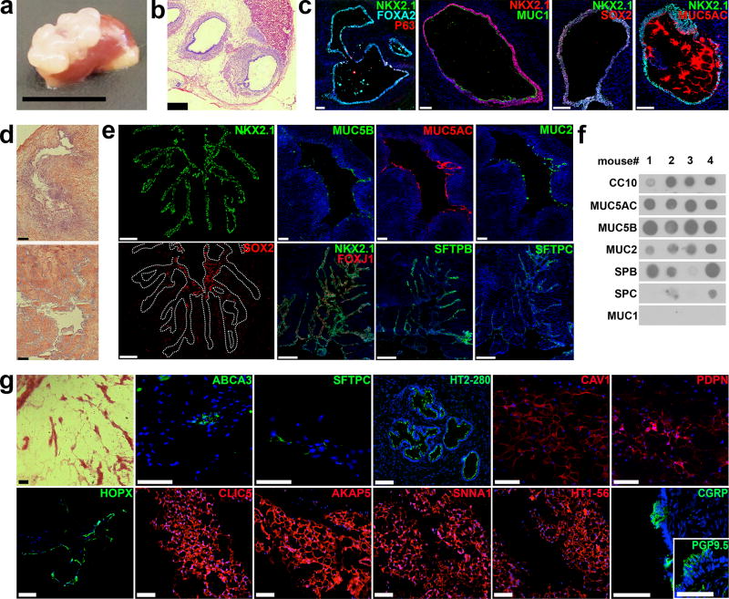
When transplanted under the kidney capsule of immunodeficient NSG mice, LBOs yielded growths (Fig. 2a) containing tubular structures surrounded by mesenchymal tissue after 1.5 months (Fig. 2b). The tubes were uniformly lined by a FOXA2+NKX2.1+SOX2+ epithelium containing MUC5AC+ (goblet) cells with p63+ cells in the basal layer (Fig. 2c), compatible with airway epithelium. All cells were human (Supplementary Fig. 2a), except for endothelial cells, which were of mouse origin (Supplementary Fig. 2b). After 5 months, branching structures (Fig. 2d, Supplementary Fig. 2c) surrounded by SMA+ mesodermal cells arose (Supplementary Fig. 2c). All epithelial cells were NKX2.1+ while SOX2, a proximal marker later in lung development18,19, was excluded from the branch tips, which expressed SFTPB and SFTPC, markers of surfactant-producing type II alveolar epithelial (ATII) cells (Fig. 2e)20. The stalks and central tubules expressed the proximal (airway) markers FOXJ1 (ciliated cells), CC10 (club cells) and mucins (goblet cells) (Fig. 2e). Hematoxylin-eosin staining showed abundant multiciliated cells (Supplementary Fig. 2d), while live imaging documented beating cilia (Supplementary Video 1). Furthermore, submucosal glands were observed near the larger tubular structures (Supplementary Fig. 2e). Overall, morphology and expression pattern within the growths are consistent with proximodistal specification during lung branching morphogenesis6,7. The fluid in the tubular structures contained all tested secretory products of lung and airway but was negative for the cell surface mucin21, MUC1, indicating detection of secreted proteins and not proteins associated with sloughed cells, and providing evidence for function (Fig. 2f). After 7 months, dome-shaped groups of CGRP+PGP9.5+ cells, compatible with neuroepithelial bodies22, were present in the airway-like structures (Fig. 2g). Furthermore, areas of the growths developed into a network of thin cell layers (Fig. 2g) containing cells expressing ATII cells markers (SFTPC, ABCA3, HT2-280)23 and cells bearing type I alveolar epithelial cell (ATI) markers (HT1-56, HOPX, PDPN, CAV1, SCNN1A, AKAP5, CLIC5)20, although other markers for mature ATI cells (RAGE, AQP5)20 were not detected (Fig. 2g). An alveolar capillary network and bronchoalveolar ducts were not observed, however. We conclude that, although full phenotypic and architectural alveolar maturation was not achieved, possibly at least in part because of the ectopic location, LBOs recapitulate many essential features of lung development, including branching morphogenesis and proximodistal specification, after xenotransplantation.
Figure 2. In vivo potential of LBOs.
(a) Macroscopic aspect of growths 1.5 months after transplantation of 106 LBO cells embedded in Matrigel under the kidney capsule of NSG mice. Scale bar 1 cm. (b) HE stain of LBO-derived growth 1.5 months after transplantation. Scale bar 500 µm. (c) Immunofluorescence for indicated markers in LBO-derived growths 1.5 months after transplantation. Scale bars 100 µm. (d) HE staining of LBO-derived growths 5 months after transplantation. Scale bars 250 µm. (e) Immunofluorescence for indicated markers in LBO-derived growth 5 months after transplantation. Scale bars 250 µm. (f) Dot blots for proteins marked on the left in aspirates from tubules in LBO-derived growth 5 months after transplantation. (g) HE staining and immunofluorescence for indicated markers in LBO-derived growths 7 months after transplantation. Scale bars 100 µm. All panels used RUES2 ESCs, representative of 4 independent experiments.
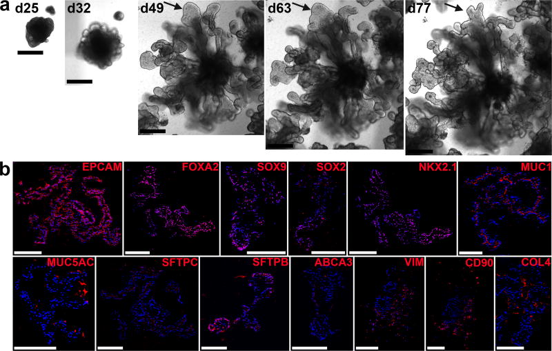
After embedding d25 LBOs in Matrigel in the presence of CHIR99021, FGF10, FGF7, BMP4 and RA (Supplementary Fig. 1b), >95% yielded rapidly expanding branching structures (Fig. 3a, Supplementary Fig. 3a for iPSCs, including C12, a line from a patient with mutations IRF7, causing acute respiratory distress syndrome after influenza infection)24 expressing markers of pulmonary endoderm (FOXA2+: 95.17%±1.54%, NKX2.1+: 74.97%± 4.37%, EPCAM+: 96.83% ± 0.62%, SOX9+: 92.42% ± 3.81% n=3 at d70; RUES2 ESCs) (Fig. 3b). Uniform luminal expression of MUC1 demonstrates polarization (Fig. 3b). Cells expressing the ATII markers SFTPC, SFTPB and ABCA3 were present in all structures (Fig. 3b). Airway goblet cells (MUC5B or MUC5AC) were rare while other airway cells (club cells (SCGB3A2), ciliated cells (FOXJ1) and basal cells (KRT5 and P63)) were not detected (not shown). While singly plated LBOs branched randomly in every direction and filled a 6.4mm well within 90 days, they formed branching trees that only occupied a section of the well when plated together in close proximity (Supplementary Fig. 3b). These findings show that branching architecture can be manipulated in vitro, and that repulsive interactions between branching structures may play a role in determining their architecture. Mesenchymal cells expressing VIMENTIN and CD90 were present surrounding the structures (Fig. 3b). Their proportion, as determined by flow cytometry for EPCAM− cells, declined during Matrigel culture to less than 2% of the total population however (Supplementary Fig. 3c). EPCAM+, but not EPCAM− cells, purified from d25 LBOs yielded branching colonies after plating in Matrigel, albeit with low cloning efficiency (0.30±0.0316%,) (Supplementary Fig. 3d). These branching colonies displayed a similar pattern of marker expression as Matrigel colonies generated from intact LBOs (Supplementary Fig. 3e). These findings indicate that rare progenitors in the LBOs are capable of generating branching colonies, and that mesenchymal cells are not required for branching in these culture conditions.
Figure 3. LBO differentiation in Matrigel at d70.
(a) Bright field images of the development of an LBO into a branching structure after plating in Matrigel. RUES2 ESCs. Representative of >50 independent experiments. Scale bars 500 µm. (b) Immunofluorescence staining for indicated markers in d70 RUES2-derived LBOs plated in Matrigel at d25. Representative of 4 independent experiments. Scale bars 250 µm.
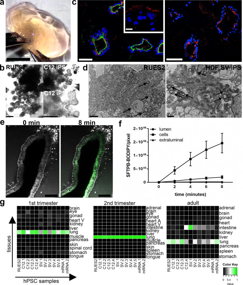
After >170 days, macroscopic tissue (Fig. 4a) consisting of branching tubules with dilated tips, reminiscent of saccules formed during the saccular stage of lung development, had developed (Fig. 4b, Supplementary Fig. 4a, Supplementary Video 2). 84.86%±5.21% cells were NKX2.1+, while most cells were SOX9+ (76.75±6.89%) and a minority (23.78±5.21%) were SOX2+ (Supplementary Fig. 4b) (n=4, one ESC and three iPS lines). Most luminal cells expressed HT2-280, MUC1, SFTPB, SFTPC and ABCA3 (RUES2 Fig. 4c, iPSCs Supplementary Fig. 4b), identifying these as ATII cells. Electron microscopy showed large numbers of lamellar bodies (LBs), the organelles where surfactant is stored25 (Fig. 4d, Supplementary Fig. 4c). To examine ATII cell function, we added SFTPB covalently linked to the fluorescent lipid, BODIPY. Within minutes, SFTPB-BODIPY was taken up by the cells and secreted in the lumens (Fig. 4e,f, Supplementary Video 3). Although HOPX, a marker of ATI cells and of putative bipotential alveolar progenitors in the mouse,20,26 was widely expressed (Supplementary Fig. 4b), other ATI markers (AQP5, CLIC5, AKAP5, CAV1, AGER) were undetectable. SOX9, a marker for the distal tips that is downregulated as alveoli mature and becomes undetectable postnatally, was mostly expressed at the tips and outer edges of the branching structures in vitro, consistent with mouse development, where SOX9 is a distal lung marker27–29 (Supplementary Fig. 4b). Expression of airway markers (MUC5AC, SCGB3A2) in the Matrigel LBO colonies was confined to structures co-expressing SOX2 and SOX9 (Supplementary Fig. 4b), and were therefore likely more proximal. While co-expression of SOX2 and SOX9 is unusual in the mouse19, numerous larger SOX2+SOX9+ structures were identified in the human second trimester fetal distal lung (Supplementary Fig. 4d), suggesting that LBOs recapitulate human lung development. The expression of SOX9, the formation of saccular structures expressing predominantly ATII markers and absence of mature ATI cells are consistent with the canalicular stage of lung development, which occurs at the end of gestation of mice, but during the late second trimester in humans. To further verify the developmental stage of d170 Matrigel LBO cultures, we performed RNAseq on 12 independent samples from RUES2 cells and from three iPSC lines. Cross-referencing with the KeyGenes database, which contains expression profiles of human organs during first and second trimesters of gestation and adulthood30, showed the best match with second trimester fetal lung, without any match with other organs (Fig. 4g, Supplementary Fig. 4e). Together, the structural, protein expression and transcriptomic data indicate that the d170 Matrigel LBO organoids reached the late second trimester of human gestation.
Figure 4. Long-term development of LBOs in vitro.
(a) Macroscopic appearance of d170 RUES2 LBOs embedded in Matrigel at d25. Representative of >50 independent experiments. Scale bar 5 mm. (b) Bright field images of d170 RUES2 and C12 LBOs embedded in Matrigel at d25. Representative of >50 independent experiments. Scale bars 500 µm. (c) Immunofluorescence for indicated markers in d170 RUES2 LBOs embedded in Matrigel at d25. Representative of 3 independent experiments. Scale bars for MUC1+SFTPB and HT2-280 100 µm. Scale bar for SFTPC 10 µm. (d) Electron microscopy of d170 LBOs embedded in Matrigel at d25 in RUES2 ESCs and HDF SV iPSCs. Arrows indicate LBs. Representative of 3 independent experiments. (e) Uptake of SFTPB-BODIPY (green) in d170 LBOs embedded in Matrigel at d25. Representative of 4 independent experiments. Scale bars 100 µm. (f) Time-course of uptake of SFTPB-BODIPY in d170 LBOs embedded in Matrigel at d25 (mean±s.e.m, n=4 independent experiments in RUES2 ESCs). The source data can be found in Supplementary Table 4. (g) Comparison of genome-wide expression in d170 LBOs derived from hESCs and hiPSCs (12 biologically independent samples) with the KeyGenes database, showing the best match with second trimester human lung.
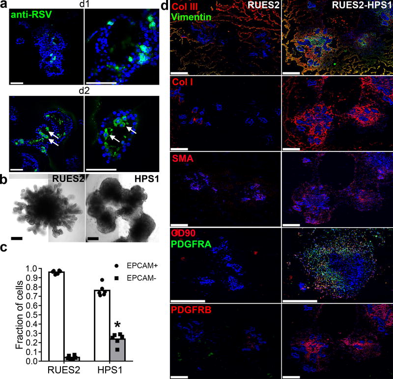
We next explored whether select infectious and fibrotic lung disease could be recapitulated. We asked whether LBOs infected with respiratory syncytial virus (RSV) display features of human lung infection. RSV is a major cause of lower respiratory tract infection in infants, and causes bronchiolitis with obstruction of small airways2,31. There is no licensed vaccine or effective antiviral drug at this time, and immunity after infection is short-lived32. RSV tropism in humans includes ciliated cells and alveolar epithelial cells2,3. Previous studies in human airway epithelial cell lines showed that cells infected with RSV swell and detach from the epithelium33, a finding consistent with obstruction of small airways by infected cells in archival pathology specimens and with the clinical syndrome of bronchiolitis3. At day 2 after infection of d170 Matrigel LBO cultures with RSV, confocal microscopy revealed shedding of swollen, infected cells into the lumen of the branching structures (Fig. 5a, arrows, Supplementary Video 4). No shedding was seen at day 1, despite evidence of viral infection. RSV infection in LBOs therefore recapitulates important features of infection in humans.
Figure 5. Potential application of LBOs in modeling human diseases.
(a) Confocal images of whole mount d170 LBOs 1 and 2 days after infection with RSV and stained using anti-RSV (all antigens) antibody. Arrows: infected cells in the lumen. Representative of 3 independent experiments. Scale bars 100 µm. (b) Bright field images of d50 LBO-derived Matrigel colonies from RUES2 and RUES2-HPS1 cells. Representative of six independent experiments. Scale bars 500 µm. (c) Fraction of EPCAM+ and EPCAM− cells in d50 LBO-derived colonies in 3D Matrigel cultures of RUES2 and RUES2-HPS1 cells. (n=6, mean±s.e.m of 3 technical replicates from two experiments; * P<0.0001; two-tailed Student’s t-test). The source data can be found in Supplementary Table 4. (d) Immunofluorescence staining for mesenchymal markers and ECM components in 3D Matrigel cultures of RUES2 and RUES2-HPS1 cells. Representative of 3 independent experiments. Scale bars 500 µm.
Next, we attempted to model pulmonary fibrosis associated with some forms of Hermansky-Pudlak Syndrome (HPS)5. HPS is characterized by pigmentation and bleeding abnormalities caused by abnormal biogenesis and trafficking of lysosome-related organelles (LROs), which include platelet dense granules and melanosomes.34 Some forms, in particular HPS1, are associated with early-onset and intractable pulmonary fibrosis (HPS interstitial pneumonia (HPSIP)) that is clinically similar Idiopathic pulmonary fibrosis (IPF)5, is characterized by fibrotic obliteration of alveoli and has a median survival of 3–4 years35. The fact that LBs of ATII cells are also LROs34 potentially explains the association of IPF with some mutations causing HPS5. Matrigel colonies derived from LBOs generation from RUES2 cells with CRISPR-CAS9-induced deletion of HPS1 (Supplementary Fig. 5a) exhibited less sharply defined branching structures in Matrigel cultures than the LBOs from parental RUES2 line (Fig. 5b), with an increased fraction of EPCAM− mesenchymal cells (Fig. 5c, Supplementary Fig. 5b), heterogeneously expressing the mesenchymal markers PDGFRA, PDGFRB, SMA, VIMENTIN and CD90 (Fig. 5d, low magnification tile scans in Supplementary Fig. 5c). The EPCAM−, but not the EPCAM+ population, showed strongly enhanced proliferation in cultures of RUES2-HPS1 cells compared to parental cells (Supplementary Fig. 5d,e), indicating that expansion of mesenchymal cells explains the increased fraction of EPCAM− cells. Surprisingly however, hyperproliferation of EPCAM− cells was already noticed in RUES2-HPS1 LBOs as early as d15 of suspension culture, prior to detection of any ATII markers. Furthermore, increased hydroxyproline content (Supplementary Fig. 5f) as well as enhanced extracellular matrix (ECM) autofluorescence (Supplementary Fig. 5c) and immunofluorescent staining for collagens 1 and 3 and fibronectin (Fig. 5d, Supplementary Fig. 5g) in RUES2-HPS1 cells indicated increased ECM deposition. Mixing experiments (Supplementary Fig. 5h–j) were consistent with notion that the accumulation of mesenchymal cells was driven by mutant epithelial cells, and not a cell intrinsic property of mutant mesenchymal cells, a finding consistent with the notion that HPSIP36 and potentially other forms of IPF4 may be caused by epithelial injury. Together, these findings suggest that it may be possible to model at least some fibrotic pulmonary disease using LBOs.
LBOs and LBO-derived branching colonies in Matrigel in vitro and growths after xenografting fulfill the definition of true organoids1. Previously reported human lung organoids did not show branching in vitro or after xenografting8,9. Furthermore, in contrast to LBOs, these were generated in the presence of serum, but in the absence of BMP4, RA and Wnt agonism, which we have shown to be essential for lung specification in vitro10. Finally these structures did not develop in vivo after grafting under the kidney capsule of immunodeficient mice, but required preculture on a bioengineered scaffold to generate airway epithelial cells after subcutaneous transplantation9.
We could reproduce the morphological features of RSV infection in the distal lung, for which there is currently no model that reproduces human infection. The LBO model also showed evidence of fibrosis in cells lacking HPS1, mutation of which is the most penetrant for a form of pulmonary fibrosis that is clinically, prognostically, radiologically and pathologically indistinguishable from IPF4,5,36. It is remarkable, however, that while HPSIP typically arises in the 3rd to 4th decade of life, a fibrotic phenotype could be reproduced in vitro within 40 days of directed differentiation. While it cannot be fully excluded that this in vitro model in fact reveals a developmental abnormality not observed in patients, it is possible that stress of in vitro culture recapitulated the changes induced by senescence and led to the very rapid appearance of the phenotype, in particular since age and telomere dysfunction are prime risk factors for IPF35,37.
The LBO model has limitations however. After 6 months of culture in Matrigel, the organoids match the second trimester of human gestation in terms of structure, marker expression and genome-wide expression signature. These findings suggest that lung development as modeled in the LBO system keeps pace with human lung development in utero. Full, terminal maturation therefore remains a challenge in the organoid field1. A second limitation is that branching appears random, a finding consistent with a, as yet unproven, ‘space-filling’ model of branching morphogenesis38. However, branching could be directed by plating several LBOs in close proximity to each other in Matrigel, in which case the organoids branch away from each other, suggesting that branching can be manipulated in vitro. A third limitation is that the exact nature and patterning of the mesenchyme present in the LBOs is unclear. In vivo xenografting revealed that LBO-associated mesodermal cells do not have the potential to generate endothelial cells, bone or skeletal muscle, suggesting that the mesenchyme is specified to some extent. The various mesenchymal lineages in the lung and their ontogeny are still poorly characterized.6 Pulmonary vasculature is likely not derived from pulmonary mesenchyme however. Proximal pulmonary vessels are derived from a common cardiopulmonary mesenchymal progenitor, while the development origin of the alveolar capillary network likely arises from VE-cadherin+ progenitors arising in preexisting trunk vessels.6,39 A fourth limitation is that the in vitro cultures are strongly biased towards distal lung, and, although some areas co-expressing SOX2 and SOX9 expressed more proximal markers for goblet cells and club cell precursors, mature club cells, ciliated cells or basal cells were not observed. We could also not achieve induction of ATI markers in vitro, although ATI potential is present after engraftment in vivo. It is possible that the cultures were not sufficiently mature to allow development of ATI cells. Alternatively, our current culture condition may drive differentiation of distal progenitor towards ATII cells at the expense of ATI cells.
Taken together, this work indicates that, despite certain limitations, LBOs will be a useful tool for the study of human lung development and possibly for lung disease modeling.
METHODS
Reagents
Reagents used are listed in Supplementary Table 1.
Human samples
The use of human fetal tissues procured by the Human Studies Core at Columbia Center for Translational Immunology was approved by the Columbia University Medical Center (CUMC) Human research review committee and the experiments were performed in accordance with the approved protocols.
Media
hPSC maintenance media consisted of DMEM/F12 (1:1) supplemented with 20% knockout serum replacement, 0.1 mM β-mercaptoethanol, Primocin, and 20 ng/ml FGF-2. Serum-free differentiation (SFD) media consisted of IMDM/Ham’s F12 (3:1) supplemented with N2, B27, 0.05% bovine serum albumin, 1% penicillin-streptomycin, 50 µg/ml ascorbic acid, 2mM Glutamax, 0.4 µM monothioglycerol and different growth factor cocktails as indicated in Supplementary Table 2.
hPSCs Maintenance
Rockefeller University Embryonic Stem Cell Line 2 (RUES2, NIH approval number NIHhESC-09-0013, Registration number 0013, passage 17–28), Sendai Virus and modified mRNA generated hiPSC lines from healthy human dermal fibroblasts7,9 (passage 16–25) and IRF7-deficient C12 hiPSC lines28 were maintained on mouse embryonic fibroblasts (MEFs) plated at 15,000–18,000 cells/cm2. Cells were cultured in hPSC maintenance media and medium was changed daily. hPSCs were passaged with Accutase/EDTA washed and replated at a dilution of 1:48. Cultures were maintained in a humidified 5% CO2 atmosphere at 37°C. Lines are karyotyped and verified for Mycoplasma contamination using PCR every 6 months.
Endoderm induction
Induction of endoderm was carried as previous described9. Briefly, MEFs were depleted by passaging onto Matrigel for 24 h supplied with hPSC maintenance media and maintained in a humidified 5% CO2 atmosphere at 37°C. After MEF depletion, primitive streak and embryoid body induction was performed in embryoid bodies/primitive streak formation media (Supplementary Table 2) in low attachment plates for 12–16 h followed by switching to endoderm induction media (Supplementary Table 2) for 36–40 h. Embryoid bodies were fed every day and maintained in a humidified 5% CO2/5% O2 atmosphere at 37°C. Endoderm yield was determined by the expression of CXCR4 and c-KIT. For iPS lines, endodermal cells were purified using human CD184 (CXCR4) MicroBead kit. Cells used in all experiments had > 90% endoderm yield.
Anterior foregut endoderm induction
Anterior foregut endoderm was induced as previous described9. On day 4, embryoid bodies were dissociated with 0.05% Trypsin/EDTA and plated on fibronectin-coated multiple well plates with a density at 80,000–105,000 cells/cm2. Cells were incubated in Anteriorization media-1 for 24 h followed by switching to Anteriorization media-2 for another 24 h.
Formation of lung bud organoids
At the end of anterior foregut endoderm induction, cells were treated with Ventralization media (Branching media) for 48 h and three-dimensional clump formation was observed. The clumps were then suspended by gently pipetting around the wells. The suspended clumps are called lung bud organoids (LBOs) hereafter. LBOs were maintained in non-tissue culture treated multiple-well plates submerged in Branching media and were fed every other day until d20–d25.
Branching morphogenesis in Matrigel
The d20-d25 LBOs were embedded in 100% Matrigel in 24-well transwell inserts and incubated in incubator until the Matrigel solidified. Branching media were added to the well, after which the transwell was inserted, branching media added into the transwell insert as well. Media were changed every other day. A step-by-step protocol describing the generation of LBOs and LBO-derived branching colonies in Matrigel can be found at Nature Protocol Exchange41.
Immunofluorescence staining
LBOs and branching Matrigel cultures were freshly embedded in Optimal Cutting Temperature (OCT). Samples were sectioned between 5–8 µm, and then air dried for 2 hours. The sections were fixed with 4% paraformaldehyde for 20 minutes at room temperature (RT) and washed with DPBS for 5 minutes. The sections were permeabilized with 0.3% Triton X-100/PBS for 30 minutes followed by blocking in 5% donkey serum for 1 hour. Primary antibodies (Supplementary Table 3) were incubated at 4°C overnight. The next day, sections were washed with DPBS 3 × 5 minutes followed by secondary antibody (Supplementary Table 3) incubation for 2 hours at RT, washed 3 × 10 minutes with DPBS then mounted with DAPI contained fluorescent mounting medium. For 3D imaging, D25 LBOs were stained as described above, but were stained as intact organoids.
Isolation of EPCAM+ and EPCAM− population from LBOs
LBOs were dissociated by 0.05% Trypsin/EDTA. The cells were stained with APC-conjugated EPCAM for 20 minutes at 4°C. EPCAM+ and EPCAM− cells were isolated by Fluorescence activated cell sorting (FACS) using a BD Influx Cell Sorter (San Jose, CA).
RNAseq
Total RNA from LBOs was purified using Direct-zol™ RNA MicroPrep kit. RNA concentration and RNA integrity number (RIN) were determined using an Agilent microfluidic RNA 6000 Nano Chip kit (Agilent Technologies, Santa Clara, CA) on the 2100 Bioanalyzer (Agilent Technologies, Santa Clara, CA). Those samples with RIN greater than 9 were used for RNAseq. Poly-A-pull-down was used to enrich mRNAs from total RNA samples. Libraries were prepared using Illumina TruSeq RNA prep kit (Illumina, San Diego, CA). Libraries were then sequenced using the Illumina HiSeq2000 (Illumina, San Diego, CA) at the Columbia Genome Center. Samples were multiplexed in each lane, yielding a targeted number of single-end/pair-end 100bp reads for each sample, as a fraction of 180 million reads for the whole lane. RTA (Illumina, San Diego, CA) was used for base calling and bc12fastq (version 1.8.4) for converting BCL to fastq format, coupled with adaptor trimming. Reads were mapped to a reference genome (NCBI/build37.2) using Tophat (version 2.0.4) with 4 mismatches and 10 maximum multiple hits. To tackle the mapping of reads that are from exon-exon junctions, Tophat infers novel exon-exon junctions ab initio, and combines them with junctions from known mRNA sequences as the reference annotation. We estimated the relative abundance of genes and splice isoforms using cufflinks (version 2.0.2) with default settings. We tested for differentially expressed genes under various conditions using DEseq, an R package based on a negative binomial distribution that models the number reads from RNAseq experiments and tests for differential expression.
In situ hybridization
In situ hybridization was performed on frozen sections (5–8 µm) using digoxigenin (DIG)-UTP-labeled SHH riboprobes. Briefly, human adult lung tissue cDNA was used as template to generate SHH PCR products containing either T7 or T3 promoter sequences (Forward: AATTAACCCTCACTAAAGGGACAGCTCGGAAGTCATCAGTT; Reverse: TAATACGACTCACTATAGGGG CCTCTGAGTGGTGGCCATCTT). The PCR products were used as templates to generate SHH riboprobes using T7 MAXIscript kit (Ambion) followed by RNeasy micro kit (Qiagen) to clean up the riboprobes. Different stages of the LBOs freshly embedded in OCT. Samples were sectioned between 5–8 µm followed by fixation with 4% paraformaldehyde for 20 minutes RT. The sections were washed with DEPC-DPBS for 3× 5 minutes and acetylated in acetylation buffer (584 µl of triethanolamine/50 ml of DEPC-H2O/125 µl acetic anhydride) for 10 minutes. Permeabilization was carried in 0.1% Triton X-100/PBS for 30 minutes at RT followed by washed with DEPC-DPBS 3 × 5 minutes. The sections were incubated with hybridization buffer (5% dextran sulfate/4× SSC/50% formamide/1× Denhardt’s/5% fish sperm DNA) for at least 2 hours at RT then overnight with 200 ng/ml of DIG-labeled SHH probe in hybridization buffer at 72°C. The next day, sections were incubated with 0.2× SSC pre-warmed to 72°C for 2 hours followed by cool down to RT for 30 minutes. The sections were washed with fresh 0.2× SSC for 5 minutes then PBS for another 5 minutes. The sections were incubated with blocking solution (2% sheep serum/TBST) for 1 hour followed by anti-DIG-AP Ig overnight at 4°C. The sections were washed with TBST 3 × 10 minutes and rinsed in color reaction buffer (100 mM Tris, pH 9.5/0.1% Tween-20/100 mM NaCl/50 mM MgCl2) for 10 minutes. Color was developed by incubating the section with BM-purple.
Mouse kidney capsule transplantation
The NOD.Cg-Prkdcscid.Il2rgtm1Wjl/SzJ (NSG) mice were housed in a specific pathogen-free mouse facility. All the mice used at 10–13 weeks of age and not selected for gender. The experiment was set up to use 5–7 mice per time point. No statistical method was used to predetermine sample size. The experiments were not randomized. Experiments and animal care were performed in accordance with the protocols approved by The Columbia University Institutional Animal Care And Use Committee. One million of d20-d25 LBO cells were mixed with 5 µl Matrigel prior to surgery and implanted under the kidney capsule. Outgrowths were excised, embedded freshly in OCT for immunofluorescence or fixed in 4% paraformaldehyde for paraffin embedding. Histology was analyzed using hematoxylin/eosin staining.
Dot blots
Three microliter of fluid aspirated from the tubular structures of 5 month grafts was deposited onto a nitrocellulose blotting membrane (GE Healthcare Life Sciences). The dot-blot membrane was air-dried for 5 minutes, and blocked in 5% milk/PBS for 1 hour and then probed with the indicated primary antibodies (Supplementary Table 3) overnight at 4°C. HRP-conjugated secondary antibodies was applied to the membranes followed by signal detection with ECL Western Blotting Detection Reagents and exposure to X-ray film.
Imaging
Samples were imaged using motorized Leica DMI6000 B (Leica Microsystems, Buffalo Grove, IL) or DMi8 (Leica Microsystems, Buffalo Grove, IL) inverted microscopes or 2-photon confocal laser scanning microscope Leica TCS SP8 (Leica Microsystems, Buffalo Grove, IL). Macroscopic images (Fig. 3a and Fig. 5a) were taken using iPhone 6 (Model: MG5A2LL/A, Apple, Cupertino, CA).
Quantification of immunofluorescence
Images for each nuclear marker were quantified using ImageJ. Briefly, images were converted to 8-bit images and the threshold was adjusted to correspond with the nuclear stain, which allows for measurement of total area. The total area was analyzed by the “Analyze Particles” function of ImageJ. Percentage of positive cells were calculated by dividing the total area of positive cells over the total area of DAPI. For extracellular matrix quantification, fluorescence intensity was quantified using Leica Application Suite X. The values were normalized to the RUES2 control for each individual experiment before statistical analysis.
Transmission Electron Microscopy
Transmission Electron Microscopy (TEM) was performed at the NYU Langone Medical Center Microscopy Core. LBOs were fixed with 2.5% glutaraldehyde in 0.1M sodium cacodylate buffer (pH7.2) for 2 hours and post-fixed with 1% osmium tetroxide for 1.5 hours at room temperature, then processed in a standard manner and embedded in EMbed 812 (Electron Microscopy Sciences, Hatfield, PA). Semi-thin sections were cut at 1 mm and stained with 1% Toluidine Blue to evaluate the quality of preservation and find the area of interest. Ultrathin sections (60 nm) were cut, mounted on copper grids and stained with uranyl acetate and lead citrate by standard methods. Stained grids were examined under Philips CM-12 electron microscope and photographed with a Gatan (4k ×2.7k) digital camera (Gatan, Inc., Pleasanton, CA).
Generation of RUES2-HPS1 line
The RUES2-HPS1 line was generated at the Stem Cell Core Facility at Columbia University Medical Center. Briefly, RUES2 cells (passage 25) were cultured in six-well plates coated with Matrigel to 70–80% confluence. Cells were electroporated with 7.5 µg of HPS1 guide RNA plasmid plus 2.5 µg of Cas9mCherry per well of a 6-well plate using Nucleofector 4D. Cas9mCherry-derived mCherry was used as a fluorescent marker to sort transfected cells. Twenty-four hours posttransfection, cells were sorted using FACS with a Bio-Rad S3e cell sorter and seeded at ~2,000 cells/6 cm dish on MEF feeders. Colonies were picked 7–10 days post sorting. Genomic DNAs from individual clones were isolated and genotyping was done using HPS1-specific PCR primers (HPS1-F-1 (GTAGAGGCAGCAGATCCAAGAGG) and HPS1-R-1 (GAACAAGGTGGTCCACACA). 420 bp band to be expected). The PCR products were cloned into a plasmid for proper sequence using In-Fusion reaction (Clontech, Mountain View,CA). Sequencing revealed premature stop codons in each allele (Supplementary Fig. 5).
Uptake of SPB-BODIPY in live LBOs and quantification
d170 LBOs were stained with CellMask™ Deep Red Plasma membrane Stain for 10 minutes and washed for 5 times followed by imaging prior loading SPB-BODIPY to obtained background fluorescence levels (0 min). The cultures then were loaded with 20 ng/ml purified human SPB-BODIPY protein (10 ng in total per culture) directly on top of the Matrigel. Images were taken every 2 minutes using a 2-photon confocal laser scanning microscope (Leica TCS SP8) and the fluorescent intensities were quantified using Leica Application Suite X. The background fluorescence values were subtracted from all measurements before statistical analysis.
Hydroxyproline content
Hydroxyproline content was measured followed manufacture’s protocol (Sigma, MAK008-1KT). Briefly, samples from RUES2 or RUES2-HPS1 cultures were homogenized by tissue glass Teflon dounce homogenizer (10 mg samples in 100 µl of water) and transferred to a pressure-tight vial followed by adding 100 µl of concentrated hydrochloric acid (~ 12M) per 10 mg of sample. The mixtures were hydrolyzed at 120°C for 3 hours. Samples were dried in a 96 well plate at 60°C followed by Chloramine T/Oxidation Buffer Mixture for 5 mins at RT and DMAB reagent for another 90 mins at 60°C. Hydroxyproline content were measured at 560 nm. The same amount of Matrigel was used as control.
Comparative analysis using KeyGenes
RNAseq data obtained from d170 LBOs from RUES2, C12, HDF SV and HDF mRNA lines was compared to different first and second trimesters and adult organs, including the lungs, using KeyGenes. Hierarchical clustering of 12 samples of the d170 LBOs and 75 samples from 19 organs from second trimester was performed using Cluster 3.0 and viewed by TreeView. The 87 classifier genes were calculated by KeyGenes.
Respiratory syncytial virus preparation and infection
Recombinant red fluorescent protein (RFP)-expressing RSV A2 (rrRSV) was generated from the full-length RSV plasmid1, MP224 by replacing the enhanced green fluorescent protein gene with the wild-type Discosoma RFP gene from pDsRed. For cell maintenance, HEp-2 cells (ATCC no. CCL-23) and Vero cells (ATCC no. CCL-81) were grown in monolayer culture and maintained in DMEM supplemented with 10% fetal calf serum (FCS) and 2 mM L-glutamine in a humidified atmosphere with 5%CO2 at 37 °C. Viral stocks were prepared in HEp-2 cells (ATCC no. CCL-23). Briefly, HEp-2 cells were grown overnight, washed with OptiMEM, and inoculated with rrRSV. After a 2.5-h adsorption period the cells were incubated for 3 days in DMEM supplemented with 1% FCS. Virus was harvested by one freeze-thaw cycle followed by a clarifying centrifugation at 3,500 r.p.m. and stored at −80 °C. Viral titers were determined by plaque assay in Vero cells using a 2% methyl cellulose overlay, 5% (v/v) formaldehyde fixation, and crystal violet staining (0.015% w/v) at 5 days. For RSV infection of d170 LBOs, 107 plaque-forming units (PFU) of RSV in 1 ml was directly added onto each Matrigel culture in wells and incubated for 3 hrs at 37 °C. The RSV inocula were then removed and the cultures were washed with SFD media 5 times for 5 minutes and maintained in branching media. The cultures were collected at indicated time points for whole mount staining using anti-RSV (all antigens) antibody (Meridian Life Science, B65890G). Images were taking using inverted microscopes or 2-photon confocal laser scanning microscope Leica TCS SP8 (Leica Microsystems, Buffalo Grove, IL).
Data Availability
The RNA sequencing data sets that support the findings of this study are available from the Sequence Read Archive (SRA). The SRA accession number for d25 LBOs sequencing is SRP073749 and SRR4295269 for d170 LBOs. All source data supporting the findings of this study are provided in Supplementary Table 4. All other data supporting the findings of this study are available from the corresponding author upon reasonable request.
Statistics and Reproducibility
Statistical analysis was done using unpaired two-tailed Student’s t-test or one-way ANOVA where appropriate using Prism 7. Results were shown mean±s.e.m., p values < 0.05 were considered statistically significant. N-value refers to biologically independent replicates, unless noted otherwise. The investigators were not blinded to allocation during experiments and outcome assessment in animal studies, as no statistics were performed.
Supplementary Material
Acknowledgments
This work was supported by grant NIH HL120046-01 (HWS), 1U01HL134760-01 (HWS) RO1 AI031971 (AM), and RO1 AI114736 (AM). RUES2-HPS1 cells were generated by the Columbia Stem Cell Core Facility. We thank NYULMC OCS Microscopy core Chris Petzold and Kristen Dancel for their assistance with transmission electron microscopy. We thank Mark Peeples (Ohio State University) for providing the original recombinant RSV. Flow cytometry was performed in the CCTI Flow Cytometry Core, supported in part by the Office of the Director, National Institutes of Health under awards S10RR027050 and S10OD020056.
Footnotes
Authors Contributions
YWC designed and performed most experiments, contributed to the concept, and co-wrote the manuscript with HWS. SXH and ALRT assisted YWC. AFL performed transmission electron microscopy. SHH provided assistance with flow cytometry. JLC and MC provided patient material, and LN and SV generated the C12 IRF7-deficient iPS line. MNI and JB provided SFTPB-BODIPY. AM, MP and LMP generated and provided virology reagents, and provided design and instruction for experiments involving RSV. HWS provided concept and guidance, and co-wrote with YWC.
Competing Financial Interests
The authors have no competing financial interests.
References
- 1.Lancaster MA, Knoblich JA. Organogenesis in a dish: modeling development and disease using organoid technologies. Science. 2014;345:1247125. doi: 10.1126/science.1247125. [DOI] [PubMed] [Google Scholar]
- 2.Collins PL, Fearns R, Graham BS. Respiratory syncytial virus: virology, reverse genetics, and pathogenesis of disease. Current topics in microbiology and immunology. 2013;372:3–38. doi: 10.1007/978-3-642-38919-1_1. [DOI] [PMC free article] [PubMed] [Google Scholar]
- 3.Johnson JE, Gonzales RA, Olson SJ, Wright PF, Graham BS. The histopathology of fatal untreated human respiratory syncytial virus infection. Mod Pathol. 2007;20:108–119. doi: 10.1038/modpathol.3800725. [DOI] [PubMed] [Google Scholar]
- 4.Mulugeta S, Nureki S, Beers MF. Lost after translation: insights from pulmonary surfactant for understanding the role of alveolar epithelial dysfunction and cellular quality control in fibrotic lung disease. American journal of physiology. Lung cellular and molecular physiology. 2015;309:L507–525. doi: 10.1152/ajplung.00139.2015. [DOI] [PMC free article] [PubMed] [Google Scholar]
- 5.Vicary GW, Vergne Y, Santiago-Cornier A, Young LR, Roman J. Pulmonary Fibrosis in Hermansky-Pudlak Syndrome. Ann Am Thorac Soc. 2016 doi: 10.1513/AnnalsATS.201603-186FR. [DOI] [PMC free article] [PubMed] [Google Scholar]
- 6.Herriges M, Morrisey EE. Lung development: orchestrating the generation and regeneration of a complex organ. Development. 2014;141:502–513. doi: 10.1242/dev.098186. [DOI] [PMC free article] [PubMed] [Google Scholar]
- 7.Morrisey EE, Hogan BL. Preparing for the first breath: genetic and cellular mechanisms in lung development. Dev Cell. 2010;18:8–23. doi: 10.1016/j.devcel.2009.12.010. doi:S1534-5807(09)00527-9 [pii] [DOI] [PMC free article] [PubMed] [Google Scholar]
- 8.Dye BR, et al. In vitro generation of human pluripotent stem cell derived lung organoids. eLife. 2015;4 doi: 10.7554/eLife.05098. [DOI] [PMC free article] [PubMed] [Google Scholar]
- 9.Dye BR, et al. A bioengineered niche promotes in vivo engraftment and maturation of pluripotent stem cell derived human lung organoids. eLife. 2016;5 doi: 10.7554/eLife.19732. [DOI] [PMC free article] [PubMed] [Google Scholar]
- 10.Huang SX, et al. Efficient generation of lung and airway epithelial cells from human pluripotent stem cells. Nature biotechnology. 2014;32:84–91. doi: 10.1038/nbt.2754. [DOI] [PMC free article] [PubMed] [Google Scholar]
- 11.Green MD, et al. Generation of anterior foregut endoderm from human embryonic and induced pluripotent stem cells. Nature biotechnology. 2011;29:267–272. doi: 10.1038/nbt.1788. [DOI] [PMC free article] [PubMed] [Google Scholar]
- 12.Huang SX, et al. The in vitro generation of lung and airway progenitor cells from human pluripotent stem cells. Nature protocols. 2015;10:413–425. doi: 10.1038/nprot.2015.023. [DOI] [PMC free article] [PubMed] [Google Scholar]
- 13.Kumar ME, et al. Mesenchymal cells. Defining a mesenchymal progenitor niche at single-cell resolution. Science. 2014;346:1258810. doi: 10.1126/science.1258810. [DOI] [PMC free article] [PubMed] [Google Scholar]
- 14.Hrycaj SM, et al. Hox5 Genes Regulate the Wnt2/2b-Bmp4-Signaling Axis during Lung Development. Cell reports. 2015;12:903–912. doi: 10.1016/j.celrep.2015.07.020. [DOI] [PMC free article] [PubMed] [Google Scholar]
- 15.Liu L, et al. Hedgehog signaling in neonatal and adult lung. American journal of respiratory cell and molecular biology. 2013;48:703–710. doi: 10.1165/rcmb.2012-0347OC. [DOI] [PMC free article] [PubMed] [Google Scholar]
- 16.Bellusci S, et al. Involvement of Sonic hedgehog (Shh) in mouse embryonic lung growth and morphogenesis. Development. 1997;124:53–63. doi: 10.1242/dev.124.1.53. [DOI] [PubMed] [Google Scholar]
- 17.Pepicelli CV, Lewis PM, McMahon AP. Sonic hedgehog regulates branching morphogenesis in the mammalian lung. Current biology : CB. 1998;8:1083–1086. doi: 10.1016/s0960-9822(98)70446-4. [DOI] [PubMed] [Google Scholar]
- 18.Que J, et al. Multiple dose-dependent roles for Sox2 in the patterning and differentiation of anterior foregut endoderm. Development. 2007;134:2521–2531. doi: 10.1242/dev.003855. [DOI] [PMC free article] [PubMed] [Google Scholar]
- 19.Alanis DM, Chang DR, Akiyama H, Krasnow MA, Chen J. Two nested developmental waves demarcate a compartment boundary in the mouse lung. Nature communications. 2014;5:3923. doi: 10.1038/ncomms4923. [DOI] [PMC free article] [PubMed] [Google Scholar]
- 20.Treutlein B, et al. Reconstructing lineage hierarchies of the distal lung epithelium using single-cell RNA-seq. Nature. 2014;509:371–375. doi: 10.1038/nature13173. [DOI] [PMC free article] [PubMed] [Google Scholar]
- 21.Sakurai J, et al. Differential expression of the glycosylated forms of MUC1 during lung development. Eur J Histochem. 2007;51:95–102. [PubMed] [Google Scholar]
- 22.Cutz E, Pan J, Yeger H, Domnik NJ, Fisher JT. Recent advances and contraversies on the role of pulmonary neuroepithelial bodies as airway sensors. Seminars in cell & developmental biology. 2013;24:40–50. doi: 10.1016/j.semcdb.2012.09.003. [DOI] [PubMed] [Google Scholar]
- 23.Ban N, et al. ABCA3 as a lipid transporter in pulmonary surfactant biogenesis. The Journal of biological chemistry. 2007;282:9628–9634. doi: 10.1074/jbc.M611767200. [DOI] [PubMed] [Google Scholar]
- 24.Ciancanelli MJ, et al. Life-threatening influenza and impaired interferon amplification in human IRF7 deficiency. Science. 2015 doi: 10.1126/science.aaa1578. [DOI] [PMC free article] [PubMed] [Google Scholar]
- 25.Whitsett JA, Wert SE, Weaver TE. Diseases of pulmonary surfactant homeostasis. Annual review of pathology. 2015;10:371–393. doi: 10.1146/annurev-pathol-012513-104644. [DOI] [PMC free article] [PubMed] [Google Scholar]
- 26.Jain R, et al. Plasticity of Hopx(+) type I alveolar cells to regenerate type II cells in the lung. Nature communications. 2015;6:6727. doi: 10.1038/ncomms7727. [DOI] [PMC free article] [PubMed] [Google Scholar]
- 27.Liu Y, Hogan BL. Differential gene expression in the distal tip endoderm of the embryonic mouse lung. Gene expression patterns : GEP. 2002;2:229–233. doi: 10.1016/s1567-133x(02)00057-1. [DOI] [PubMed] [Google Scholar]
- 28.Rockich BE, et al. Sox9 plays multiple roles in the lung epithelium during branching morphogenesis. Proc Natl Acad Sci U S A. 2013;110:E4456–4464. doi: 10.1073/pnas.1311847110. [DOI] [PMC free article] [PubMed] [Google Scholar]
- 29.Perl AK, Kist R, Shan Z, Scherer G, Whitsett JA. Normal lung development and function after Sox9 inactivation in the respiratory epithelium. Genesis. 2005;41:23–32. doi: 10.1002/gene.20093. [DOI] [PubMed] [Google Scholar]
- 30.Roost MS, et al. KeyGenes, a Tool to Probe Tissue Differentiation Using a Human Fetal Transcriptional Atlas. Stem cell reports. 2015;4:1112–1124. doi: 10.1016/j.stemcr.2015.05.002. [DOI] [PMC free article] [PubMed] [Google Scholar]
- 31.Florin TA, Plint AC, Zorc JJ. Viral bronchiolitis. Lancet. 2016 doi: 10.1016/S0140-6736(16)30951-5. [DOI] [PMC free article] [PubMed] [Google Scholar]
- 32.Simoes EA, et al. Challenges and opportunities in developing respiratory syncytial virus therapeutics. The Journal of infectious diseases. 2015;211(Suppl 1):S1–S20. doi: 10.1093/infdis/jiu828. [DOI] [PMC free article] [PubMed] [Google Scholar]
- 33.Liesman RM, et al. RSV-encoded NS2 promotes epithelial cell shedding and distal airway obstruction. J Clin Invest. 2014;124:2219–2233. doi: 10.1172/JCI72948. [DOI] [PMC free article] [PubMed] [Google Scholar]
- 34.Huizing M, Helip-Wooley A, Westbroek W, Gunay-Aygun M, Gahl WA. Disorders of lysosome-related organelle biogenesis: clinical and molecular genetics. Annu Rev Genomics Hum Genet. 2008;9:359–386. doi: 10.1146/annurev.genom.9.081307.164303. [DOI] [PMC free article] [PubMed] [Google Scholar]
- 35.Ryu JH, et al. Idiopathic pulmonary fibrosis: evolving concepts. Mayo Clinic proceedings. 2014;89:1130–1142. doi: 10.1016/j.mayocp.2014.03.016. [DOI] [PubMed] [Google Scholar]
- 36.Young LR, et al. The alveolar epithelium determines susceptibility to lung fibrosis in Hermansky-Pudlak syndrome. Am J Respir Crit Care Med. 2012;186:1014–1024. doi: 10.1164/rccm.201207-1206OC. [DOI] [PMC free article] [PubMed] [Google Scholar]
- 37.Armanios M. Telomerase and idiopathic pulmonary fibrosis. Mutation research. 2012;730:52–58. doi: 10.1016/j.mrfmmm.2011.10.013. [DOI] [PMC free article] [PubMed] [Google Scholar]
- 38.Short K, Hodson M, Smyth I. Spatial mapping and quantification of developmental branching morphogenesis. Development. 2013;140:471–478. doi: 10.1242/dev.088500. [DOI] [PubMed] [Google Scholar]
- 39.Peng T, et al. Coordination of heart and lung co-development by a multipotent cardiopulmonary progenitor. Nature. 2013;500:589–592. doi: 10.1038/nature12358. [DOI] [PMC free article] [PubMed] [Google Scholar]
- 40.Hallak LK, Spillmann D, Collins PL, Peeples ME. Glycosaminoglycan sulfation requirements for respiratory syncytial virus infection. J Virol. 2000;74:10508–10513. doi: 10.1128/jvi.74.22.10508-10513.2000. [DOI] [PMC free article] [PubMed] [Google Scholar]
- 41.Chen YW, Ahmed A, Snoeck HW. Generation of three-dimensional lung bud organoid and its derived branching colonies. Protoc. Exch. 2017 http://dx.doi.org/10.1038/protex.2017.027.
Associated Data
This section collects any data citations, data availability statements, or supplementary materials included in this article.
Supplementary Materials
Data Availability Statement
The RNA sequencing data sets that support the findings of this study are available from the Sequence Read Archive (SRA). The SRA accession number for d25 LBOs sequencing is SRP073749 and SRR4295269 for d170 LBOs. All source data supporting the findings of this study are provided in Supplementary Table 4. All other data supporting the findings of this study are available from the corresponding author upon reasonable request.


