
Figure 2.
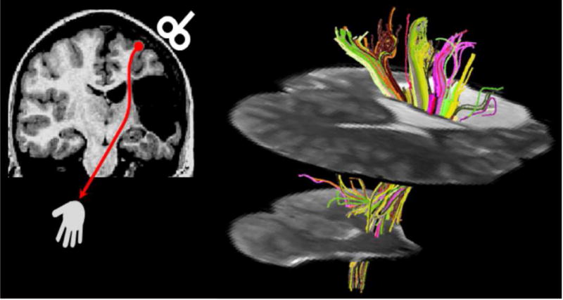
Diffusion tensor tractography in a patient with a cortico-subcortical infarction in the territory of the middle cerebral artery (modified from Staudt, 2010; reprinted with permission from John Wiley and Sons). Left: Coronal T1-weighted image depicting the lesion, leaving only a small bridge of preserved white matter between the lesion and the enlarged lateral ventricle. Transcranial magnetic stimulation (red) indicated preserved crossed cortico-spinal motor projections. Right: Diffusion tensor tractography visualizes the extensive connectivity mediated by this small bridge of preserved tissue (seed area for fiber tracking).