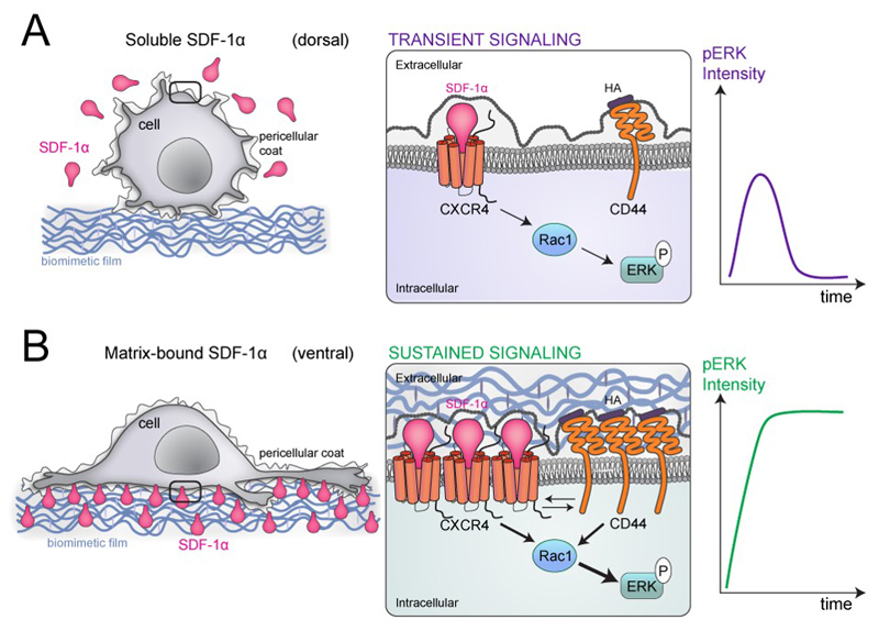
Figure 9.
Scheme summarizing the major differences in the response of MDA-MB-231 cancer cells to SDF-1α presented either as soluble cue (A) or as matrix-bound cue (B) for MDA-MB-231 breast cancer cells at different length scales from left to right. Left: at the cellular scale; middle at the plasma membrane/receptor scale and right: consequence in terms of phospho-ERK signaling. In the case of a soluble presentation (sSDF) (A), the local concentration of SDF-1α molecules is very low and SDF-1α is mostly presented at the dorsal side of the cells. As a consequence, cell adhesion is low and cells are round with only few protrusions. In this case, the signaling is transient since the CXCR4 and hyaluronan receptors (CD44 being the major one in MDA-MB231 cells) are only scarce and do not cooperate. SDF-1α-induced signaling via CXCR4 activates Rac1 and pERK but the intensity of the signal is small and its duration is transient (right scheme). In the case of matrix-bound SDF-1α (bSDF), the local concentration of SDF-1α in the polyelectrolyte film is locally very high so the cellular receptors (CXCR4), which are mostly localized at the basal side of the cancer cells, can bind to their SDF-1α ligands. This induces a localized receptor clustering (middle scheme) for CXCR4 as well as for the HA receptor. Indeed hyaluronan, the CD44 ligand, is provided by the pericellullar coat of cancer cells and by the polyelectrolyte film, which is made of HA. In this situation of spatial confinement at the ventral side of the cell, the two receptors CXCR4 (for SDF-1α) and CD44 (for HA) act in concert and activate Rac1 and the subsequent phosphorylation of ERK1/2. As a consequence, the intensity of the pERK signal is very high and it is sustained over a long time, for at least 16 h as shown in Figure 8B.

