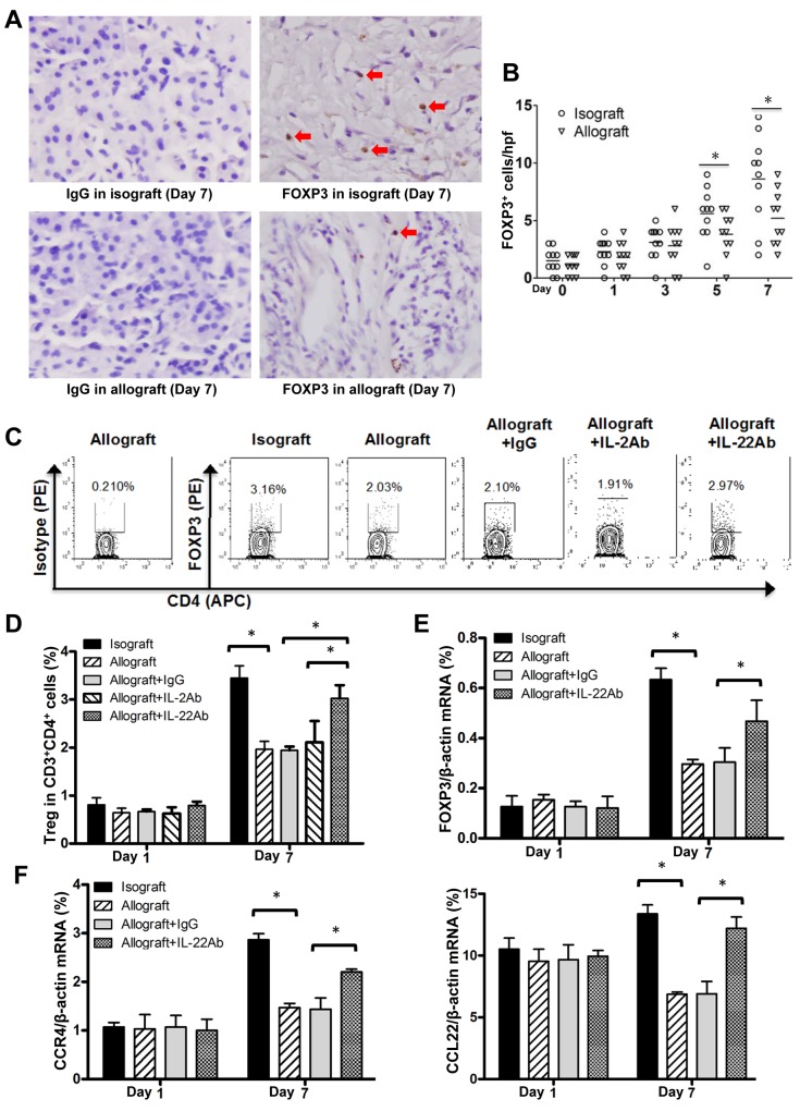
Figure 6. Decreased Treg cells at AR stage of allograft liver transplantation.
(A) IHC assay for Foxp3 positive cells in isografted or allografted liver tissues at 7 days postoperatively. Positive cells were indicated by red arrows. (B) Statistical analysis of liver portal areas Foxp3+ cell counts in IHC staining tissues. (C) The representative results of FCM assay for the frequency of Treg cells (CD3+CD4+Foxp3+) in liver lymphocyte of the indicated groups at the 7 day post transplantation. (D) The statistical analysis of Treg cell frequency in various groups at the indicated time points after liver transplantation (n=5). (E and F) Foxp3, CCR4 and CCL22 mRNA levels in liver tissues of recipients (n=5) in indicated groups at the indicated time points after liver transplantation. *p<0.05.

