Abstract
Purpose
This study is to develop a low-cost, easy-to-use, wide-field smartphone fundus video camera to enable affordable point-of-care examination and telemedicine.
Methods
The wide-field smartphone fundus camera is based on a unique design of miniaturized indirect ophthalmoscopy. For proof-of-concept prototype, we used a Samsung Galaxy S6 smartphone and all off-the-shelf components. A fiber coupled LED was used to deliver illumination light through a 1 mm micro mirror, which was conjugated to the subject pupil plane, for retinal illumination. A 60D ophthalmic lens was used to image the retina through the eye, and a plano-convex lens with 90 mm focal length was used to relay the retinal image to the smartphone camera.
Results
A totally wireless smartphone fundus camera was constructed, with a whole weight of 255 g. This device allowed both snapshot fundus photography and continuous video recording. 92° field of view (FOV) was achieved in single-shot images. Optic disc, macula, and retinal blood vasculatures can be clearly observed with image quality comparable to standard fundus camera.
Conclusion
Miniaturized indirect ophthalmoscopy enabled a low-cost, portable, wide-field smartphone fundus camera, which can foster telemedicine and clinical deployments of wide-field fundus photography for eye disease screening, diagnosis and treatment assessment.
Keywords: Fundus camera, fundus photography, field of view, illumination, imaging, smartphone, telemedicine, retina, retinal disease, eye disease
INTRODUCTION
Fundus examination is important for retinal disease screening, diagnosis, and treatment evaluation. In coordination with widely available internet technology, digital fundus photography has gained increasing interest for telemedicine examination of retinal diseases 1. Low-cost smartphone fundus cameras have been developed to explore affordable telemedicine of diabetic retinopathy (DR), age-related macular degeneration (AMD), retinopathy of premature (ROP), etc. Most of these smartphone fundus cameras employ the configuration of indirect ophthalmoscopy. By directly adopting binocular indirect ophthalmoscopy (BIO) lenses, these smartphone fundus cameras provide a low-cost solution for retinal examination. However, the BIO lenses are specially designed for head mounted BIO systems, which require long distance from the lens to the smartphone camera. Therefore, the BIO lens based smartphone fundus cameras are bulky, with a small field of view (FOV), typically < 45° in single-shot images. We report here a unique design of miniaturized indirect ophthalmoscopy to achieve 92° FOV in single-shot fundus image and to allow continuous video recording which opens the opportunity for real-time involvement of experienced ophthalmologists remotely.
MATERIALS AND METHODS
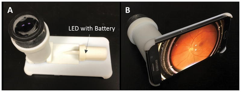
Figure 1 illustrates the optical layout of our custom-designed wide-field smartphone fundus video camera, and Fig. 2 shows representative photographs of our first lab prototype based on a Samsung Galaxy S6 smartphone. For proof-of-concept validation, we used all off-the-shelf components which were selected based on Opticstudio (Zemax) simulation. The framework of the smartphone adaptor was designed with 3D CAD simulation software, and was printed by a high-resolution 3D printer (Objet 30 Prime).
Figure 1.

(A) Optical layout of the smartphone fundus video camera. L1 is a 60D ophthalmic lens. L2 is relay lens with 90 mm focal length. L3 is the built-in camera lens. Solid light lines illustrate illumination light rays, and thin dashed lines show imaging light rays. (B) Schematic illustration of the optical fiber and micro mirror based light source. The center of the mirror is ~10 mm from the center of the camera sensor.
Figure 2.

Representative photographs of the smartphone fundus video camera.
Imaging system
As shown in Fig. 2A, the lens L1 is a 60D ophthalmic lens (Volk Optical Inc., V60C) to image the retina to the plane RI (dashed vertical line nearby the back focal plane of the lens L1 in Fig. 1A) between the lens L1 and lens L2 which is a plano-convex lens with 90 mm focal length (Edmund Optics, 67165). The focal length of the smartphone camera lens L3 is 4.3 mm. The retinal image RI is relayed to the smartphone camera sensor through the lenses L2 and L3. The Samsung Galaxy S6 smartphone consists of a 1/2.6″ camera sensor, with a frame resolution of 5312 × 2988 pixels. All images and videos were captured with dilated pupil. For capturing image and recording video, the stock camera application of the smartphone was used. To capture image iso was set to 100 and white-balance was set to daylight in manual imaging mode. Adjustable manual focusing slider was used for fine focusing and smartphone screen was filled by the retinal image by zoom function.
Illuminating system
A warm white LED (Thorlabs, MWWHD3) was used as the illumination light source. The LED power supply was integrated into the smartphone adaptor which allows a totally wireless fundus imager (Fig. 2A). A miniaturized light source is the kernel to meet the space limitation within the smartphone based portable device. The illumination light was collected with 1 mm diameter optical fiber. As illustrated in Fig. 1B, terminal part of the optical fiber was coupled with a 1 mm diameter 45° aluminum coated rod lens (Edmund Optics, 47–628). The mirror surface of the rod lens was relayed to the pupil of the subject through the lens L1 for retinal illumination.
Functional test
For evaluating the FOV and image resolution of our lab prototype device, normal subjects without eye diseases were recruited. This study was approved by the Institutional Review Board of the University of Illinois at Chicago and was in compliance with the ethical standards stated in the Declaration of Helsinki.
RESULTS
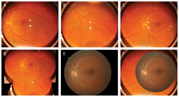
Figure 3A–C shows representative single-shot retinal images obtained by prototype device from one normal subject without eye disease. Figure 3D shows a montage of these three single-shot images in Fig. 3A–C. As shown in Fig. 3A–D, optic disc, macula, and retinal blood vasculatures can be clearly observed, with image quality comparable to standard fundus camera (Fig. 3E).
Figure 3.
(A–C) Representative single-shot images captured from a 41 years old age subject. The subject has no reported eye diseases. (D) Montage of single-shot images in Figs. 3A–C. (E) Representative fundus image from the same subject collected with a clinical fundus camera (Zeiss, Cirrus Photo 800), which has a single-shot FOV of 45° external angle, corresponding to 67.5°. (F) Overlap of images of Fig. 3C and Fig. 3E for FOV comparison.
A 6 s movie clip is provided as supplemental material to demonstrate the feasibility of continuous fundus video recording (see Video, Supplemental Digital Content, My Movie.mp4).
To quantify the FOV and spatial resolution of the prototype fundus camera, we took a picture of a resolution target placed at 1 m from the camera. By following the instructions defined by ISO 10940:2009 standard, a 30 μm central resolution was verified, and the horizontal FOV was calculated as 62° external angle, corresponding to 92° interior angle in single-shot images. External angle has been widely used to specify FOV in traditional fundus cameras, while interior angle is used in recently emerging wide-field fundus imagers, such as a Retcam and Optos. The estimated irradiance at the retina was 0.24 mW/cm2. According to the ISO 15004-2: 2007 standard, 11.5 hours continuous illumination is allowed for continuous video recording.
DISCUSSION
It is known that wide-field fundus photography is essential for screening, diagnosis and treatment evaluation of eye diseases such as diabetic retinopathy. However, the high equipment cost of existing devices is a limiting factor for clinical deployment of wide-field fundus photography, particularly in rural and underserved areas where both expensive instruments and skilled operators are not available. Low-cost smartphone fundus cameras promise convenient assessment of eye diseases at point-of-care environments, and may also enable affordable telemedicine screening to foster the access to medical cares in rural and underserved areas. Various smartphone-based ocular imaging techniques have been demonstrated in recent years 2–4. However, existing smartphone fundus cameras is limited by the small FOV in single-shot images. Using a donut-shaped trans-pupillary illumination as that in traditional fundus cameras, Maamari at al. combined crossed polarization technique with flashing light for smartphone based fundus camera 2. However, the FOV in single-shot images was still limited at 55°.
Moreover, the flashing light illumination excludes the potential of continuous video recording.
By employing trans-palpebral illumination, 152° FOV has been achieved in single-shot smartphone fundus image 5. However, clinical deployments of the trans-palpebral illumination based device is challenging due to the requirement of separate adjustment and optimization of imaging and illumination sub-systems.
Using all off-the-shelf components, we report here a wide-field smartphone fundus video camera that allows both snapshot imaging and video recording, with 92° FOV in single-shot images. The unparalleled FOV enables easy examination of retinal periphery that can be targeted by early stages of DR and other chorioretinal conditions. In coordination with widely available internet, the continuous video mode will enable real-time involvement of experienced ophthalmologists remotely, and will also allow montage data processing to readily increase the effective FOV beyond 180°.
Supplementary Material
Summary.
We developed a wide-field smartphone fundus camera based on a unique design of miniaturized indirect ophthalmoscopy. This is a totally wireless device to allow both snapshot fundus photography and continuous video recording, with 92° field of view (FOV) in single-shot images.
Acknowledgments
Funding: D.T. is supported by TUBITAK (The Scientific and Technological Research Council of Turkey). C.L. and X.Y. are supported by NIH R01 EY023522, NIH R01 EY024628, NIH R21 EY025760, NIH P30 EY001792, and unrestricted grant from Research to Prevent Blindness.
We thank Prof. Sait Egrilmez, Ege University School of Medicine, Izmir, Turkey for providing valuable suggestions on this project.
Footnotes
CONTRIBUTORS
D.T. contributed to system design, prototype construction, data acquisition and manuscript preparation; A.A. contributed to system design and image processing; M.K.E. contributed to data interpretation and manuscript preparation; C. L. contribute to optical design and construction; X.Y. supervised this project, and contributed to system design, data analysis, and manuscript preparation.
Proprietary interest: D.T. and X.Y. have pending patent application (US 62/546,830).
My Movie.mp4
References
- 1.Chin EK, Ventura BV, See KY, Seibles J, Park SS. Nonmydriatic Fundus Photography for Teleophthalmology Diabetic Retinopathy Screening in Rural and Urban Clinics. Telemedicine Journal and e-Health. 2014;20:102–108. doi: 10.1089/tmj.2013.0042. [DOI] [PMC free article] [PubMed] [Google Scholar]
- 2.Maamari RN, Keenan JD, Fletcher DA, Margolis TP. A mobile phone-based retinal camera for portable wide field imaging. The British journal of ophthalmology. 2014;98:438–441. doi: 10.1136/bjophthalmol-2013-303797. [DOI] [PubMed] [Google Scholar]
- 3.Suto S, Hiraoka T, Oshika T. Fluorescein fundus angiography with smartphone. Retina (Philadelphia, Pa) 2014;34:203–205. doi: 10.1097/IAE.0000000000000041. [DOI] [PubMed] [Google Scholar]
- 4.Kaya A. Ophthoselfie: Detailed Self-imaging of Cornea and Anterior Segment by Smartphone. Turkish journal of ophthalmology. 2017;47:130–132. doi: 10.4274/tjo.66743. [DOI] [PMC free article] [PubMed] [Google Scholar]
- 5.Toslak D, Thapa D, Chen Y, Erol MK, Paul Chan RV, Yao X. Trans-palpebral illumination: an approach for wide-angle fundus photography without the need for pupil dilation. Opt Lett. 2016;41:2688–2691. doi: 10.1364/OL.41.002688. [DOI] [PMC free article] [PubMed] [Google Scholar]
Associated Data
This section collects any data citations, data availability statements, or supplementary materials included in this article.


