Abstract
Purpose
Our case series aims to study the growing use of FDG PET/CT in diagnostic evaluation and follow up of IgG4-RD with emphasis on patients presenting with coronary artery involvement.
Methods
We conducted a search on the nuclear medicine and rheumatology service databases and identified patients with histologically proven IgG4-RD with FDG PET/CT performed at the Singapore General Hospital. The radiological, clinical, and laboratory findings of these patients were analyzed retrospectively.
Results
The series included ten male and two female patients. The commonest organ involved (five patients) was the pancreas. In three patients, coronary artery involvement manifested as soft tissue masses surrounding the arterial lumens. In these patients, histological diagnosis was established from alternative biopsy sites with abnormal metabolic activity on FDG PET/CT.
Correlation between laboratory and metabolic imaging findings was not statistically significant in our series.
Four patients had follow-up FDG PET/CT; three showed interval reduction in metabolic activity to baseline. One showed persistent abnormal metabolic activity before a rise in IgG4 levels. The metabolic imaging response was used to guide steroid dose.
Conclusions
FDG PET/CT is a useful tool in evaluation and follow-up of IgG4-RD, particularly in identifying alternative biopsy sites in patients who present with coronary artery involvement. Hypermetabolic coronary artery masses on FDG PET/CT should raise clinical suspicion of IgG4-RD. As the coronary artery masses may not show decrease in size after treatment, FDG PET/CT is also useful for metabolic response assessment.
Keywords: IgG4, Fluorodeoxyglucose, Pet/Ct, Coronary artery
Introduction
Immunoglobulin G4 (IgG4)-related disease (IgG4-RD) is a disorder that was first described in 2003, when extrapancreatic manifestations of disease were identified in patients with autoimmune pancreatitis [1].
The use of positron emission tomography with 2-deoxy-2-[fluorine-18]fluoro-D-glucose integrated with computed tomography (FDG PET/CT) for IgG4-RD has slowly been increasing [2]. With FDG PET/CT, whole-body surveys could be performed in a single sitting, disease could be detected early before clinical relapse, and physicians could assess treatment response before anatomical changes are discernable [3]. IgG4-RD can often be confused with other multi-system conditions such as lymphoma because of the myriad of organs that may be involved. Where the initially detected lesions are in locations with anatomically difficult access, a further potential strong point of FDG PET/CT would be the detection of alternative sites for histological confirmation of IgG4-RD.
We report a case series of patients with IgG4-RD who underwent FDG PET/CT at Singapore General Hospital (SGH). Interestingly, although our numbers are small, we had a relatively large proportion (three out of 12) of coronary artery involvement. We are adding a small case series to the growing body of evidence of coronary involvement in IgG4-RD [4–9].
Materials and Methods
We conducted a retrospective analysis of all patients at SGH with histologically proven IgG4-RD who had had an FDG PET/CT at any point in their clinical course. These patients were identified by searching the database at the Department of Nuclear Medicine and PET and the Department of Rheumatology of the Singapore General Hospital. The reports and images of the scans were accessed by authors 1, 3, 4, and 5 separately, and information on metabolic and morphologic abnormalities was recorded. If patients had repeated FDG PET/CT scans done, these were also recorded and the changes of a semi-quantitative measurement (SUVmax) were noted. The standardized uptake value (SUV), an index of FDG uptake in tissue, was computed as follows: SUV = region of interest activity/(injected radioactivity/body weight). Lesional SUVmax to liver background SUVmax ratios were also calculated. Liver background SUVmax (ranging from 3.0 to 4.0) was measured within a spherical volume of interest (diameter, 50 mm) positioned in an area of normal liver tissue in the right lobe).
Approval was granted by SGH’s institutional review board for this study and waiver of consent was obtained.
FDG PET/CT scans were performed using an LSO-based PET/CT scanner (GE Discovery 690, Wisconsin, USA) with 64-slice CT using 3D acquisition (FOV 70 cm diameter) and time of flight system with iterative reconstruction, according to guidelines set out by the Society of Nuclear Medicine and European Association of Nuclear Medicine [10, 11]. Scans were performed 60 to 110 min post injection of FDG.
Information recorded included demographic data, date of diagnosis, organs involved, and treatment information. Conventional radiologic imaging (ultrasonography [US], contrast-enhanced CT, and magnetic resonance imaging [MRI]) and laboratory results of patients were also recorded for correlation with FDG PET/CT results. Correlation between SUVmax and laboratory results were studied using Spearman’s rank correlation coefficient (rho).
Results
Demographics
A total of 12 patients (ten male and two female) were identified over a nine-year period. All patients were diagnosed to have IgG4-RD by the 2011 comprehensive diagnostic criteria (Table 1) [12].
Table 1.
Histological findings, location of biopsy, and correlative maximum serum IgG4 levels
| Patient no. | Maximum serum IgG4 levels (mg/dL) | Site of histology | IgG4 (HPF) | IgG4+/IgG+Ratio | Histology findings |
|---|---|---|---|---|---|
| 1 | 340 | Kidneys | >30 | >40% | Storiform fibrosis, lymphoplasmocytic infiltrate |
| 2 | 159 | Right iliac artery | >50 | 50% | Storiform fibrosis, lymphoplasmocytic infiltrate |
| 3 | 193 | Submandibular gland | >20 | 50% | Storiform fibrosis, lymphoplasmocytic infiltrate |
| 4 | 158 | Periaortic mass | >200 | >40% | Storiform fibrosis, lymphoplasmocytic infiltrate |
| 5 | 340 | Lacrimal gland and breast | >100 | >40% | Storiform fibrosis, lymphoplasmocytic infiltrate |
| 6 | 233 | Pancreas FNA | >30 | >40% | Fibrosis, lymphoplasmocytic infiltrate, phlebitis |
| 7 | 128 | Left lung mass | >50 | >40% | Storiform fibrosis, lymphoplasmocytic infiltrate |
| 8 | 340 | Cervical lymph node | >50 | 70% | Storiform fibrosis |
| 9 | 52 | Lacrimal gland | >100 | >40% | Storiform fibrosis, lymphoplasmocytic infiltrate |
| 10 | 285 | Pancreas | * | * | Storiform fibrosis |
| 11 | 285 | Pancreas | >50 | >40% | Storiform fibrosis, lymphoplasmocytic infiltrate |
| 12 | 126 | Pancreas | >30 | >40% | Storiform fibrosis, lymphoplasmocytic infiltrate |
FNA: fine needle aspirate
HPF: high powered field
*: figure not available
The median age at diagnosis was 58 years old (range 49 to 81). Eleven patients were of Chinese ethnicity, whilst one was Malay (Table 2). The time between initial scan (CT/MRI/FDG PET/CT) to subsequent FDG PET/CT ranged from 1 week to 4 years, with the median time frame being 4 months.
Table 2.
Patient demographics and organ involvement at initial FDG PET/CT
| Sex | Male | 10 |
| Female | 2 | |
| Race | Chinese | 11 |
| Malay | 1 | |
| Organs involved | Pancreas | 5 |
| Coronary vessels | 3 | |
| Salivary/lacrimal glands | 3 | |
| Lymph nodes | 3 | |
| Kidneys | 2 | |
| Breast | 1 | |
| Stomach | 1 | |
| Orbits | 1 | |
| Aorta | 1 | |
| Lung | 1 |
Scan Features
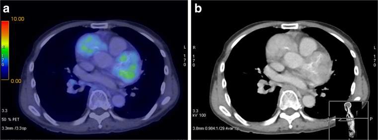
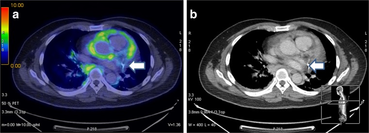
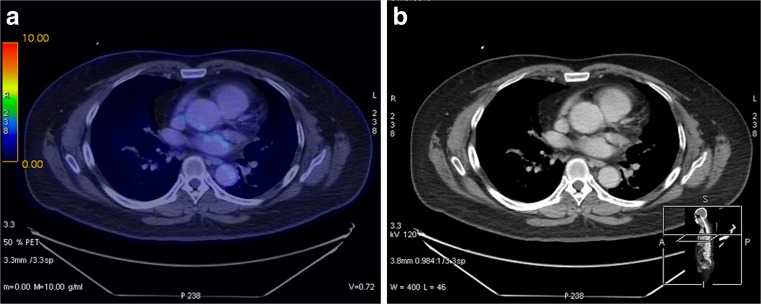
The commonest organs involved radiologically at time of presentation were the pancreas (five patients (42%)) followed by the coronary arteries, and the salivary/lacrimal glands (three patients (25%) respectively). Coronary artery involvement was part of a multi-system disease in the three patients included, but none had presenting symptoms related to the coronary arteries, for example acute coronary syndromes or dissections (Table 3). Coronary artery involvement manifested as hypermetabolic soft tissue masses surrounding the arterial lumens (Figs. 1, 2 and 3), an appearance that has very rarely been described in one of the key alternative diagnoses of lymphoma [13]. In all patients who had coronary artery involvement, the site of biopsy for initial diagnosis of IgG4-RD was remote from the coronary arteries (iliac vessel, kidney, and lip), but these biopsy sites were all shown to be FDG-avid on PET.
Table 3.
Presenting features, initial IgG4 levels, treatment, and follow up PET/CT findings
| Patient no. | Presenting features | IgG4 levels at the time of diagnosis (mg/dL) | Latest subsequent IgG4 levels (mg/dL) | Initial prednisolone dose or any other medication | Current prednisolone dose or any other medication | Follow-up scan findings |
|---|---|---|---|---|---|---|
| 1 | Loss of weight, fatigue | 269 | 340 (4 years post diagnosis) |
50 mg OM | 7.5 mg OM | Abnormal (4 years post diagnosis) |
| 2 | Paraproteinaemia, loss of weight, altered bowel habits | 22 | 159 (3 years post diagnosis) |
30 mg BD; Rituximab; Mycofenolate mofeteil 1 g BD |
5 mg OM | Normal (3 years post diagnosis) |
| 3 | Enlargement of salivary glands and loss of weight | 51 | 193 (1 year post diagnosis) |
80 mg OM | 5 mg OM; mycofenolate mofeteil 1 g BD | Normal (1 year post diagnosis) |
| 4 | Chest pain | 149 | 158 (1 year post diagnosis) |
30 mg OM; Methotrexate 20 mg once/week |
3 mg OM; methotrexate 15 mg once/week |
Normal (2 years post diagnosis) |
| 5 | Bilateral lacrimal gland enlargement | 269 | 340 (1 year post diagnosis) |
Nil | Nil | N/A |
| 6 | Obstructive jaundice | 233 | 340 (5 months post diagnosis) |
2 mg 4 times/week; Azathioprine 100 mg OM |
5 mg OM; azathioprine 150 mg OM |
N/A |
| 7 | Loss of weight | 128 | Nil | 5 mg OM | 5 mg OM | N/A |
| 8 | Increasing size of cervical nodes | 233 | 340 (4 months post diagnosis) |
40 mg OM | 5 mg OM | N/A |
| 9 | Orbital mass, left optic neuropathy, salivary gland enlargement | 52 | Nil | 60 mg OM | Nil | N/A |
| 10 | Obstructive jaundice, loss of weight | 340 | Nil | 30 mg OM | 20 mg OM | N/A |
| 11 | Obstructive jaundice | 285 | 224 (3 years post diagnosis) |
5 mg BD | 5 mg EOD | N/A |
| 12 | Pancreatitis | 126 | Nil | Nil | Nil | N/A |
OM: once in the morning
EOD: every other day
BD: twice daily
Fig. 1.
a, b: Patient 1. Hypermetabolic soft tissue masses are noted around the coronary vessels, predominantly the left anterior descending and left circumflex arteries
Fig. 2.
a, b: Patient 2. Hypermetabolic soft tissue masses surround all three coronary vessels
Fig. 3.
a, b: Patient 3. Arrows demonstrate FDG uptake with soft tissue masses around the coronary vessels
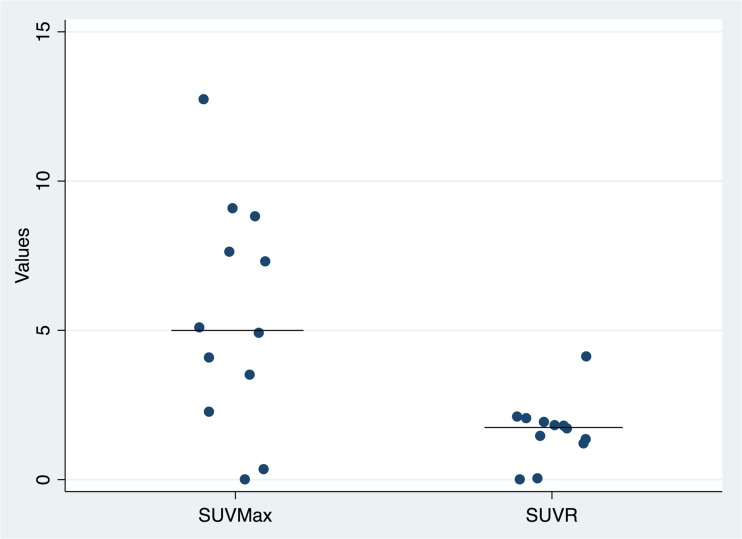
The median (range) SUVmax of lesions was 5.2 (2.7-12.8) in patients with positive FDG PET/CT. The median (range) ratio of lesional SUVmax to liver background SUVmax (SUVR) was 1.59 (0.90 to 4.27) (Fig. 4).
Fig. 4.
Dot graph of patients’ baseline SUVmax and SUVR (SUVmax of lesion: background liver SUVmax). The lines denote median values of both SUVmax and SUVR
Laboratory Features
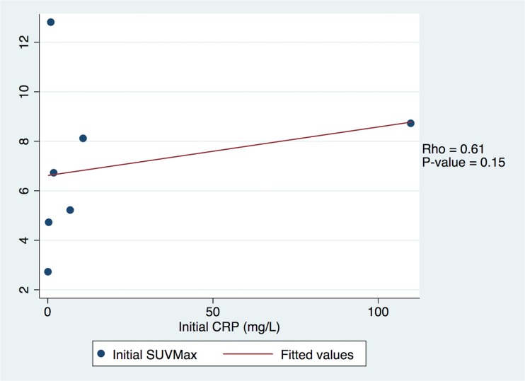
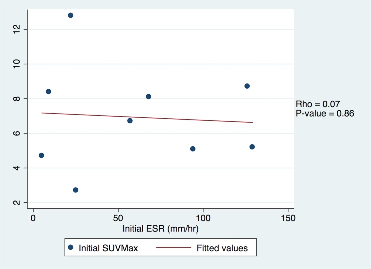
At initial diagnosis, all patients had IgG4 levels between 22 and >340 mg/dL, with median level of 191 mg/dL. There was no statistically significant correlation between IgG4 levels, CRP, ESR, and SUVmax (Figs. 5, 6 and 7) in our series.
Fig. 5.
Scatter plot of comparison of initial SUVMax to initial IgG4 levels showing no statistically significant relationship
Fig. 6.
Scatter plot of comparison of initial SUVMax to initial CRP levels showing no statistically significant relationship
Fig. 7.
Scatter plot of comparison of initial SUVMax to initial ESR levels showing no statistically significant relationship
For example, one patient had CRP level of 0.3 mg/L and ESR of 5 mm/h which was within the normal range, although his FDG PET/CT as well as IgG4 levels of 340 mg/dL were suggestive of active disease.
Follow-up
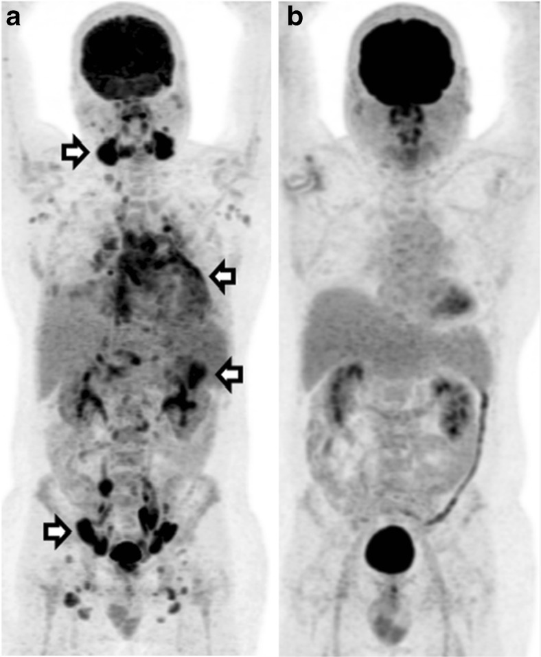
Patients were not on prednisolone at time of initial FDG PET/CT, but follow-up studies were done with ongoing treatment with immunosuppressants. Four patients had follow-up FDG PET/CT (Table 4), coincidentally including the three patients with coronary artery involvement. Three showed interval reduction in metabolic activity to background levels uniformly in all organs involved from a mean SUVmax of 6.9, with the shortest time being 3 months after treatment with oral prednisolone. In one of these patients, there was resolution of the coronary arterial lesions (Figs. 8 and 9). The fourth patient had been scanned multiple times with FDG PET/CT. However, after his repeat FDG PET/CT showed abnormal raised metabolic activity (SUVmax 4.2) in the coronary arteries and in the lymph nodes, an increase in steroid dose was guided by the metabolic imaging results. No flare-up of disease and no increase in prednisolone dosing were noted in the two years of follow-up in the three patients with good response on FDG PET/CT.
Table 4.
More detailed follow up findings (SUVmax, IgG4 levels, sites of involvement, and treatment regimen) of patients with repeated FDG PET/CT
| Initial FDG PET/CT SUVmax (organ) | SUVR | Other sites involved | Follow-up SUV Max |
SUVR | Duration from initial scan (months) | IgG4 at diagnosis (mg/dL) | Latest subsequent IgG4 levels (mg/dL) | Initial prednisolone dose or any other medication | Current prednisolone dose or any other medication |
|---|---|---|---|---|---|---|---|---|---|
| 5.1 (coronary vessels) | 1.59 | Lymph nodes kidneys |
4.2 | 1.2 | 48 | 269 | 340 (4 years) |
50 mg OM | 7.5 mg OM |
| 5.2 (coronary vessels) |
1.73 | Lymph nodes right iliac artery pericardium |
* | * | 38 | 22 | 159 (3 years) |
30 mg BD rituximab mycofenolate mofeteil 1 g BD |
5 mg OM |
| 8.7 (lymph nodes) |
2.49 | Lacrimal gland salivary gland pericardium Pancreas |
* | * | 11 | 51 | 193 (1 year) |
80 mg OM | 5 mg OM Mycofenolate mofeteil 1 g BD |
| 8.4 (periaortic mass) |
2.71 | Lacrimal gland lymph nodes |
* | * | 23 | 149 | 158 (1 year) |
30 mg OM methotrexate 20 mg once/week |
3 mg OM Methotrexate 15 mg once/week |
* denotes background levels of activity on FDG PET/CT
SUVR – SUVmax to liver SUVmax ratio
Fig. 8.
a, b: Post treatment follow up FDG-PET/CT in patient 3 which shows complete resolution of the FDG-avid soft tissue masses around the coronary vessels
Fig. 9.
a FDG-PET/CT maximal intensity projection image of patient 3 showing abnormal metabolic activity of the salivary glands, coronary arteries, left kidney, and lymph nodes in the mediastinum, axilla, abdomen, and pelvis. b Repeat FDG-PET/CT 1 year later after treatment with steroids and immunosuppressant showing complete metabolic resolution of previously involved sites
Discussion
There have been several retrospective [14] and prospective [15] studies evaluating the use of FDG PET/CT in IgG4-RD. To date, the use of FDG PET/CT to image the disease is still gaining traction.
We hope to add to the increasing body of evidence suggestive of benefit and highlight the increasing roles of FDG PET/CT in particular to guide biopsy site for diagnosis in patients where initial imaging showed difficult sites to biopsy (such as coronary arteries) as well as to evaluate disease response and recurrence.
Our study features a small case series of involvement of the coronary arteries. This may be related to the increasing use of CT coronary angiography or CT chest for other diagnostic purposes. These patients tend to have FDG-avid soft tissue nodular lesions or thickening surrounding all three of the coronary vessels. This is different from the usual pattern of aneurysms which tend to occur in involvement of the coronary vessels by other vasculitides [16].
The suspicion of IgG4-RD should be raised in patients with disease involvement of the coronary arteries with periarterial masses. Their clinical behavior tended to be similar to that of patients without coronary artery involvement, presenting without symptoms related to coronary artery involvement, and with radiological and biochemical improvement on prednisolone. There was no clear difference in terms of IgG4/IgG2/CRP/ESR levels between patients with and without coronary artery involvement. Interestingly, on follow up FDG PET/CT, the size of the soft tissue lesions in two out of three did not decrease dramatically as hoped. Rather, there was significant decrease in FDG uptake to that of background activity.
In our patients, we found that biopsy proven IgG4-RD shows SUVmax up to 12.8. Because there is still considerable overlap in terms of SUVmax with the majority of lymphoproliferative disorders, differentiation is not possible on the grounds of SUVmax alone although it can be suggested by the relatively lower levels and the pattern of uptake.
Potential weaknesses of this study are that it is a retrospective review of relatively small numbers of a fairly heterogeneous group of patients, and thus the strength of conclusions drawn from this may not be as robust as in other larger studies. However, given the relative rarity of the condition and in particular of coronary artery involvement, our study adds to the growing awareness and literature of the use of FDG PET/CT for evaluation of IgG4-RD.
Conclusion
In conclusion, the evidence for using FDG PET/CT in evaluating IgG4-RD is slowly growing. FDG PET/CT is a useful tool in evaluation and follow-up of IgG4-RD particularly in selecting alternative biopsy sites in patients who present with coronary artery involvement. Hypermetabolic coronary artery masses on FDG PET/CT should raise suspicion of IgG4-RD. As the coronary artery masses may not show decrease in size after treatment, FDG PET/CT is also useful for metabolic response assessment.
Acknowledgments
The authors wish to acknowledge the assistance of Mr. Mohamad Fairus bin Mohamad Zam for editing and formatting the PET/CT images, and Ms. Sheryl Quek from Singhealth Academy in editing and writing of this document.
Compliance with Ethical Standards
Conflict of Interest
Hian L Huang, Warren Fong, Wee M Peh, Kasat A Niraj and Winnie W Lam declare that they have no conflict of interest.
No research grant and funds were received for this study.
Ethical Approval
All procedures performed in studies involving human participants were in accordance with the ethical standards of the institutional and/or national research committee and with the 1964 Helsinki declaration and its later amendments or comparable ethical standards.
For this type of study formal consent is not required.
Informed Consent
The institutional review board of our institute approved this retrospective study, and the requirement to obtain informed consent was waived.
References
- 1.Lee TY, Kim MH, Park DH, Seo DW, Lee SK, Kim JS, et al. Utility of 18F-FDG PET/CT for differentiation of autoimmune pancreatitis with atypical pancreatic imaging findings from pancreatic cancer. Am J Roentgenol. 2009;193:343–348. doi: 10.2214/AJR.08.2297. [DOI] [PubMed] [Google Scholar]
- 2.Nakatani K, Nakamoto Y, Togashi K. Utility of FDG PET/CT in IgG4-related systemic disease. Clin Radiol. 2012;67:297–305. doi: 10.1016/j.crad.2011.10.011. [DOI] [PubMed] [Google Scholar]
- 3.Zhao Z, Wang Y, Guan Z, Jin J, Huang F, Zhu J. Utility of FDG PET/CT in the diagnosis of IgG4-related diseases. Clin Exp Rheum. 2016;1:119–125. [PubMed] [Google Scholar]
- 4.Hourai R, Miyamura M, Tasaki R, Iwata A, Takeda Y, Morita H, et al. A case of IgG4-related lymphadenopathy, pericarditis, coronary artery periarteritis and luminal stenosis. Heart Vessel. 2016;31:1709–1713. doi: 10.1007/s00380-016-0794-1. [DOI] [PubMed] [Google Scholar]
- 5.Keraliya AR, Murphy DJ, Aghayev A, Steigner ML. IgG4-Related disease with coronary arteritis. Circ Cardiovasc Imaging. 2016;9:e004583. doi: 10.1161/CIRCIMAGING.116.004583. [DOI] [PubMed] [Google Scholar]
- 6.Otani T, Matsumoto Y, Inoue K, Tanaka H, Hamada M. Morphological characterization of coronary arteries in immunoglobulin G4-related disease estimated by using computed tomography and magnetic resonance imaging. Int J Cardiol. 2016;215:111–113. doi: 10.1016/j.ijcard.2016.04.060. [DOI] [PubMed] [Google Scholar]
- 7.Delgado-García G, Sánchez-Salazar S, Rendón-Ramírez E, Castro-Medina M, Sáenz-Ibarra B. Barboza-Quintana Á et al myocardial ischemia as presenting manifestation of IgG4-related disease: a case-based review. Clin Rheumatol. 2016;35:2857–2864. doi: 10.1007/s10067-016-3292-z. [DOI] [PubMed] [Google Scholar]
- 8.Nishimura S, Amano M, Izumi C, Kuroda M, Yoshikawa Y. Takahashi Y, et al multiple coronary artery aneurysms and thoracic aortitis associated with IgG4-related disease. Intern Med. 2016;55:1605–1609. doi: 10.2169/internalmedicine.55.6314. [DOI] [PubMed] [Google Scholar]
- 9.Higashi H, Inaba S, Azuma T, Sumimoto T. Effects of steroid therapy for IgG4-related coronary periarteritis. Intern Med. 2016;55:1935–6. [DOI] [PubMed]
- 10.Delbeke D, Coleman RE, Guiberteau MJ, Brown ML, Royal HD. Siegel BA et al procedure guideline for tumor imaging with 18F-FDG PET/CT 1.0. J Nuc Med. 2006;47:885–895. [PubMed] [Google Scholar]
- 11.Boellaard R, Delgado-Bolton R, Oyen WJG, Giammarile F, Tatsch K, Eschner W, et al. FDG PET/CT: EANM procedure guidelines for tumour imaging: version 2.0. Eur J Nucl Med Mol Imaging. 2015;42:328. doi: 10.1007/s00259-014-2961-x. [DOI] [PMC free article] [PubMed] [Google Scholar]
- 12.Umehara H, Okazaki K, Masaki Y, Kawano M, Yamamoto M, Saeki T, et al. Comprehensive diagnostic criteria for IgG4-related disease (IgG4-RD), 2011. Mod Rheumatol. 2012;22:21–30. doi: 10.3109/s10165-011-0571-z. [DOI] [PubMed] [Google Scholar]
- 13.Yong Y, Lath N, Cheah F, Ng Y. Pictorial essay: uncommon causes of coronary artery encasement. J Cardiovasc Comput Tomogr. 2016;10(5):424–429. doi: 10.1016/j.jcct.2016.07.004. [DOI] [PubMed] [Google Scholar]
- 14.Ebbo M, Grados A, Guedj E, Gobert D, Colavolpe C, Zaidan M, et al. Usefulness of 2-[18F]-fluoro-2-deoxy-Dglucose-positron emission tomography/computed tomography for staging and evaluation of treatment response in IgG4-related disease: a retrospective multicenter study. Arthritis Care Res (Hoboken). 2014;66:86–96. [DOI] [PubMed]
- 15.Zhang J, Chen H, Ma Y, Xiao Y, Niu N, Wei L, et al. Characterizing IgG4-related disease with 18F-FDG PET/CT: a prospective cohort study. Eur J Nucl Med Mol Imaging. 2014;41:1624–1634. doi: 10.1007/s00259-014-2729-3. [DOI] [PMC free article] [PubMed] [Google Scholar]
- 16.Savage COS, Harper L, Cockwell P, Adu D, Howie AJ. Vasculitis BMJ. 2000;320(7245):1325–1328. doi: 10.1136/bmj.320.7245.1325. [DOI] [PMC free article] [PubMed] [Google Scholar]


