Correction to: Scientific Reports 10.1038/s41598-017-02270-6, published online 18 May 2017
The original version of this Article contained a typographical error in the title.
“In situ assembly of Ag nanoparticles (AgNPs) on porous silkworm cocoon-based would film: enhanced antimicrobial and wound healing activity”
now reads:
“In situ assembly of Ag nanoparticles (AgNPs) on porous silkworm cocoon-based wound film: enhanced antimicrobial and wound healing activity”
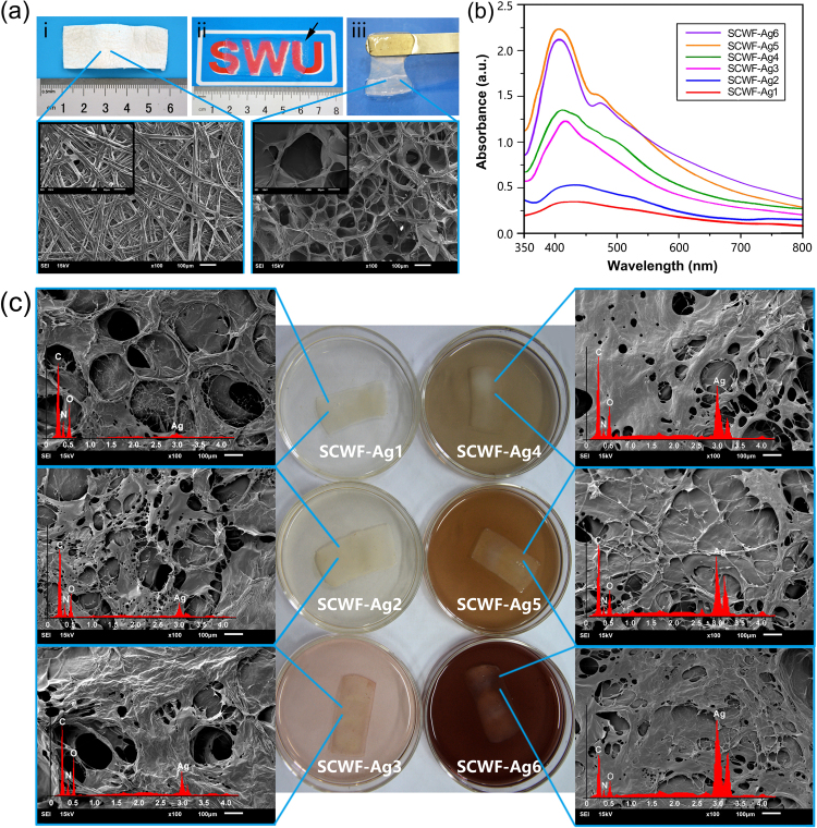
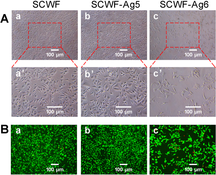
In addition, there were errors in Figure 2 and Figure 6. In Figure 2c, the micrograph for SCWF-Ag6 (right bottom) duplicated the micrograph for SCWF-Ag5 (right middle) and has now been replaced. In Figure 6A, panel c’ (SCWF-Ag6) duplicated panel a’ (SCWF) and has now been replaced.
Figure 2.
(a) Natural Bombyx mori cocoons (i); SCWF optical clarity and size (ii), wherein SCWF (black arrow) was placed on a piece of paper with one “SWU” symbol underneath. The gel (balanced on a metal spatula) was sufficiently elastic and flexible for easy handling (iii). The fibroin and sericin further dissolved to form a network that readily aggregated into transparent films, as shown in the scanning electron micrograph of lyophilized SCWF. (b) UV-vis absorption spectra of leaching aqueous dispersions of SCWF-Ag1–6 in deionized water. (c) Scanning electron micrographs of lyophilized SCWF-Ag1–6 with red curves of EDX and photographs of the respective SCWFs immersed in different concentration of AgNO3 aqueous solution after 4 h.
Figure 6.
(A) Growth observations of L929 cells treated with SCWF (a,a’), SCWF-Ag5 (b,b’), and SCWF-Ag6 (c,c’). (B) Calcein-AM/PI Double Stain Kit assay of L929 cells upon treatment with SCWF (a), SCWF-Ag5 (b) and SCWF-Ag6 (c). Live cells are stained by Calcein AM dye and produce an intense uniform green fluorescence (ex/em ~495 nm/~515 nm). Dead cells are stained by Calcein PI dye and emit bright red fluorescence (ex/em ~495 nm/~635 nm). The scale bar represents 100 μm.
These errors have now been corrected in the PDF and HTML versions of the Article
Footnotes
Kun Yu and Fei Lu contributed equally to this work.
Kun Yu and Fei Lu contributed equally to this work.
The original article can be found online at . 10.1038/s41598-017-02270-6.
The original article can be found online at https://doi.org/10.1038/s41598-017-02270-6.


